Introduction
Colorectal cancer (CRC) is the third most prevalent cancer diagnosed in both males and females and the second leading cause of cancer-related deaths worldwide, with an estimated of more than 900 000 deaths every year. A combination of hereditary and environmental risk factors such as smoking, addiction, excessive alcohol consumption, obesity and a high intake of red or processed meat play a significant role in developing CRC (Refs Reference Henrikson1–Reference Sung7). Currently, the primary treatment procedures for CRC are endoscopy, surgery, radiotherapy and chemotherapy, but despite the progress made in these areas, we still face many challenges (Ref. Reference Moore and Aulet8). For instance, radiotherapy and chemotherapy significantly increase the risk of subsequent CRC (Ref. Reference Allodji9). Therefore, novel therapeutic approaches are presently required to be discovered. MicroRNAs (miRNAs) are a family of small non-coding endogenous RNA molecules which regulate gene expression through binding to the complementary microRNA response elements (MREs) inside the mRNA non-translated regions or non-coding RNAs (Refs Reference Bartel10, Reference Frampton11). Consequently, miRNAs play a pivotal role in cell differentiation, proliferation and survival, and their differential expression patterns are involved in tumourigenesis (Refs Reference Rupaimoole12–Reference Zhang14). The activity of miRNAs is influenced by the presence of competing endogenous RNAs (ceRNAs), which vie for the miRNAs with shared MREs. These ceRNAs are circular RNAs (circRNAs), mRNAs, long non-coding RNAs (lncRNAs) and transcribed pseudogenes which act as miRNA sponges lowering the levels of available miRNAs to bind to the target mRNAs. Many of these diverse transcripts have proposed roles in human disease, including in various types of cancers (Ref. Reference Gao15). For example, Phosphatase and tensin homolog pseudogene 1 (PTENP1) is a transcribed pseudogene that regulates the mRNA and protein abundance of the tumour suppressor gene Phosphatase and tensin homolog (PTEN). It performs this in a miRNA-dependent way because of sharing of multiple conserved miRNA binding sites with PTEN. PTENP1 has a suppressive role in cell proliferation, and it would be selectively lost in human cancer (Ref. Reference Poliseno16).
In this review, we focused on the protein-coding and non-coding RNAs that function as ceRNAs in CRC. Furthermore, the ceRNAs’ potential roles as novel prognosis and diagnosis biomarkers were also investigated.
The ceRNA crosstalk in CRC
Dysregulated gene expression is noticeable in various types of cancers. miRNAs can positively or negatively regulate gene expression through modulation of transcriptional or translational programmes, but this miRNA-based regulation can be further altered by the amount of coding and non-coding endogenous transcripts which can bind the same miRNAs via their shared microRNA response elements (MREs) (Ref. Reference Bartel17). These transcripts, which have been proposed as ceRNAs since 2011, lead to indirect cross-coordination (Ref. Reference Salmena18). The conditions required for the creation of a ceRNA crosstalk mechanism are still unknown, but recent studies have shown that the ceRNA effectiveness is influenced by variable factors such as the relative abundance of ceRNA and target miRNAs, subcellular localisation and RNA binding proteins (RBPs). Disproportionate ceRNAs and their corresponding miRNA concentrations can clearly reduce the competition efficiency. Subcellular localisation determines the ceRNAs accessibility and RBPs are effective either directly by competition of ceRNAs for shared RBPs or indirectly by changing the secondary structures of ceRNAs (Refs Reference Mukherji19, Reference Tay20). Alteration in any of these factors may lead to various cancers through ceRNA network imbalance (Ref. Reference Cheng21).
Bioinformatics technology has recently revealed a growing number of ceRNAs that play a significant role in CRC. Therefore, studying and researching ceRNA networks can provide new insights into the pathogenesis and treatment of CRC.
The lncRNAs as ceRNAs
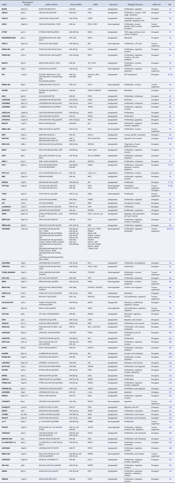
lncRNAs are non-coding RNAs with a length typically exceeding 200 nucleotides (Refs Reference Bach and Lee22–Reference Fazaeli25). Around 30 000 to 60 000 lncRNAs have been discovered in humans (Ref. Reference Ruan26). lncRNAs implicate in gene expression regulation and diverse biological processes (Ref. Reference Quinn and Chang27). They can influence the chromatin structure and function, the transcription of neighbouring and distant genes, and the mRNA stability, splicing and translation (Refs Reference Gil and Ulitsky28, Reference Mumbach29). Surprisingly, even some lncRNAs were translated into functional peptides (Ref. Reference Hartford and Lal30). Recent research has demonstrated that lncRNAs can potentially form the ceRNA networks through lncRNA–miRNA–mRNA interactions modulating the expression of target mRNAs. Thus, aberrantly expressed lncRNAs are often observed in various types of cancers, including CRC (Ref. Reference Spizzo31). For instance, lncRNA-metastasis-associated lung adenocarcinoma transcript 1 (MALAT1), was reported to be located in chromosome 11q13.1. It was demonstrated that MALAT1 is associated with cell migration, proliferation and invasion, tumour growth malignancy, metastasis and poor prognosis of CRC (Ref. Reference Zhao32). The role of MALAT1 in CRC was studied by Xu, Ji, Zheng, Yang and Fan, which was upregulated in primary CRC tissues and CRC cells including HCT116, SW620 and SW480 (Refs Reference Xu33–Reference Fan37). Hence, MALAT1 may also be defined as one of the oncogenic markers in CRC. It was confirmed that A-kinase anchoring protein 9 (AKAP-9) activation mediated MALAT1 by inducing the expression of phosphorylating serine and arginine-rich splicing factor 1 (SRSF1) and SRSF protein kinase 1 (SRPK1) could be involved in the CRC proliferation, tumour growth and metastasis (Ref. Reference Yang36). Furthermore, MALAT1 could bind directly to miR-508-5p and positively regulate RAB14, member RAS oncogene family (RAB14) expression, which suggested that MALAT1 functioned as a ceRNA for miR-508-5p (Ref. Reference Zhang38). Finally, MALAT1 induces CRC progression by affecting the miR-508-5p/RAB14 axis.
Small nucleolar RNA host gene 20 (SNHG20) which is located on chromosome 17q25.2 is highly expressed in CRC and is related to poor prognosis, advanced tumour, node and metastasis (TNM) stage and poor survival in patients with CRC (Ref. Reference Wang39). So, it was identified that the expression level of this lncRNA in 68 pairs of CRC and adjacent normal tissues was upregulated (Ref. Reference Wang39). In addition, it was reported that the unregulated expression of SNHG20 lncRNA contributes to the development and progression of CRC and a variety of other tumour types. SNHG20 acts as ceRNAs and sponges miR-495, hence SNHG20 modulates the expression of signal transducer and activator of transcription 3 (STAT3) following sponging miR-495 which lead to normally suppresses CRC cell proliferation, colony formation, migration and invasion (Ref. Reference Wang39). RP4 is a tumour suppressor lncRNA, which inhibits CRC cell proliferation and tumourigenesis through SH3 domain containing GRB2-like, endophilin B1 (SH3GLB1) gene expression regulation. SH3GLB1 expression inhibits the tumourigenesis in CRC by activating the autophagy-mediated cell death and suppressing the PI3K/Akt signalling. Liu et al. found that RP4 acts as ceRNA of SH3GLB1 and competitively binds to miR-7-5p. Thus, RP4 downregulation results in SH3GLB1 expression reduction observed in CRC (Ref. Reference Liu40). Hepatocellular carcinoma up-regulated lncRNA (HULC) is another lncRNA, which is highly expressed in CRC and significantly enhances CRC cell proliferation and metastasis through miR-613 sponging (Ref. Reference Dong41). miR-613 inhibits cell proliferation, migration and invasion, as well as the cell cycle arrest at the G1 phase by targeting Rhotekin (RTKN). Thus, HULC overexpression leads to RTKN overexpression in colon cancer tissues compared with nearby tissues (Ref. Reference Qu42).
The lncRNA-mediated competitive RNA crosstalk in CRC progression has been recognised and is summarised in Table 1.
Overview of lncRNAs as ceRNAs in CRC

The circRNAs as ceRNAs
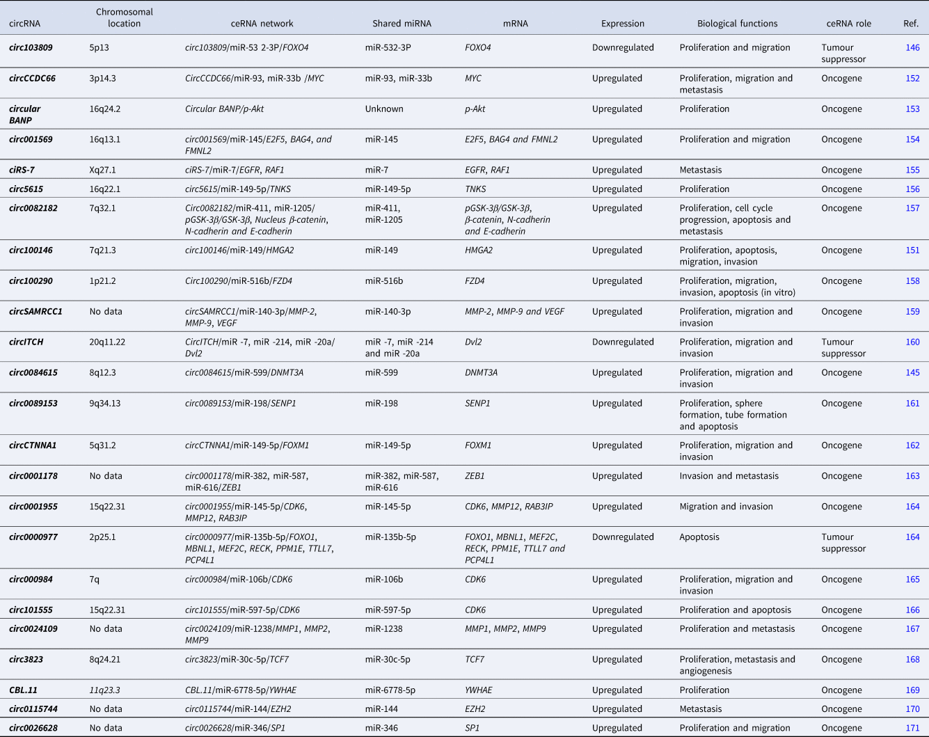
circRNAs are a large class of non-coding RNAs originally being generated as linear RNA transcripts but undergo a process called ‘back splicing’ in which a downstream splice donor site covalently links to an upstream splice acceptor site, forming a continuous covalently closed stable loop structure devoid of 5′ or 3′ polarities and poly-A tail (Refs Reference Holdt134, Reference Dragomir and Calin135). This structure makes circRNAs insensitive to exonucleases (Ref. Reference Suzuki136). CircRNAs were initially thought to be ‘junk’ generated by abnormal splicing events, except for the mouse testis-specific circRNA encoded from the sex-determining region Y (SRY) gene (Ref. Reference Capel137). In recent years, high-throughput RNA sequencing and bioinformatic algorithms have revealed the tissue-specific patterns of circRNA expression (Refs Reference Maass138, Reference Xia139). CircRNAs are primarily found in the cytoplasm and contain MREs capable of interacting with target miRNAs, thereby serving as ceRNAs and miRNA sponges (Ref. Reference Chen and Zhao140). They also participate in the transcription of their parental genes through interaction with RNA polymerase II (Ref. Reference Chen141). CircRNAs have been associated with many human diseases, including diabetes mellitus, neurological disorders, chronic inflammatory diseases, cardiovascular diseases and various types of cancer such as gastric cancer, bladder cancer and CRC (Refs Reference Li142–Reference Zhong144). One of the CRC-promoting circRNAs is circ0084615 that sponges miR-599 and repress its expression. DNA methyltransferase 3 alpha (DNMT3A) gene is a downstream target of miR-599. Zhang et al. showed that miR-599 inhibition increases DNMT3A mRNA and protein expression in HCT116 and SW480 cells. In other words, DNMT3A mRNA expression is negatively correlated with miR-599 expression and positively correlated with circ0084615 expression in CRC tissues. Depletion of circ0084615 prevents CRC cell proliferation, migration and invasion (Ref. Reference Zhang145). Circ103809 is another circRNA that normally sponges miR-532-3p, but its downregulation resulted in the high level of intracellular miR-532-3p leading to decreased expression of the Forkhead Box O4 (FOXO4) gene. This gene regulates many cellular pathways, and its downregulation has been reported recently in CRC (Ref. Reference Bian146). FOXO4 inhibits proliferation and migration via binding to a specific sequence of Adenomatous polyposis coli 2 (APC2) gene, which is an essential tumour suppressor in several cancers, including CRC, as a transcription factor and regulates the APC2 expression positively (Refs Reference van Es147–Reference Sun150).
Another example is circ100146 which its overexpression was reported in CRC patients. Liu et al. reported that circ100146 is upregulated in CRC patients. The higher expression level of circ100146 was also observed at CRC stages III and IV in comparison with CRC stages I and II. Circ100146 is a ceRNA that competitively binds to miR-149 and sponges it. This results in the overexpression of the High mobility group AT-hook 2 (HMGA2) gene, which has a binding sequence for miR-149. The abnormally increased expression of HMGA2 was also reported in CRC (Ref. Reference Liu151). Additional circRNAs are listed in Table 2.
Overview of circRNAs as ceRNAs in CRC

Pseudogenes as ceRNAs
There are approximately 11 000 pseudogenes scattered throughout the human genomes. Pseudogenes resemble the true active genes containing disruptive mutations or defects. Thus, they have been considered as nonfunctional ‘junk DNA’ sequences for decades, but certain functional pseudogenes have been discovered by sequencing technology advancement (Ref. Reference Chen172). This small fraction of pseudogenes possesses the ability to transcriptional or post-transcriptional regulation of parental or non-parental genes through the formation of small interfering RNAs (siRNAs) or a decrease in the concentration of miRNAs. Additionally, certain pseudogenes function as oncogenes and tumour suppressors. Therefore, they contribute to the pathogenesis of a wide variety of diseases such as cancer and CRC (Ref. Reference Sisu173). For instance, the dysregulated pseudogene Catenin Alpha 1 Pseudogene 1 (CTNNAP1) acts as a ceRNA that downregulates its cognate gene Catenin Alpha 1 (CTNNA1) expression through competition for the shared miRNA-141 (Ref. Reference Chen174). Additional pseudogenes implicated in CRC are listed in Table 3.
Overview of Pseudogenes as ceRNAs in CRC

The mRNAs as ceRNAs
Coding transcripts (mRNAs) exhibit the highest level of miRNA binding site conservation. Thus, they can function as ceRNAs as well as non-coding transcripts and regulate the expression levels of downstream mRNAs or serve as the miRNAs' sponges (Refs Reference Lee177, Reference Sumazin178). Tay et al. stated that several mRNAs can sponge miRNAs and regulate gene expression in multiple cancers (Ref. Reference Tay20). For instance, the PTEN ceRNA network includes several protein-coding transcripts such as CCR4-NOT transcription complex subunit 6 (CNOT6) and VAMP-associated protein A (VAPA) which regulate the tumour suppressor PTEN transcript (Ref. Reference Tay179). Although many mRNAs have been identified as ceRNA recently, there is still a need for more studies on the effects of mRNAs on miRNAs sponging in CRC.
The ceRNAs’ function in CRC signalling pathways and biological processes
Numerous ceRNAs have been shown to implicate the CRC cellular signalling pathways, such as Wnt/β-catenin, mitogen-activated protein kinase (MAPK), extracellular-signal-regulated kinase (ERK), transforming growth factor β (TGF-β) and epithelial–mesenchymal transition (EMT) signalling pathways. Additionally, ceRNAs are involved in biological processes, including tumour cell cycle and proliferation, apoptosis, invasion, migration and metastasis. Dysregulated ceRNAs in CRC tumour tissues may act as tumour oncogenes or tumour suppressors (Refs Reference Shan92, Reference Liu180, Reference Logan and Nusse181). We discussed validated ceRNAs and their biological roles in CRC signalling pathways in this review.
The ceRNAs and the signalling pathways involved in CRC cell proliferation
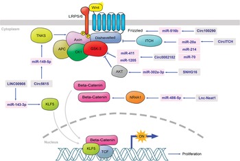
Cellular metabolism plays a pivotal role in all biological processes (Ref. Reference Zhu and Thompson182). Despite normal differentiated cells, cancer cells metabolism is primarily characterised by aerobic glycolysis to generate a significant amount of energy for cell activities (Ref. Reference Vander Heiden183). Moreover, the unlimited proliferation of tumour cells requires the rapidly increased synthesis of proteins and lipids. Various cell cycle proteins are also overexpressed in tumour cells resulted in cancer cell proliferation (Ref. Reference Otto and Sicinski184). Additionally, certain ceRNAs promote cell proliferation in all types of cancers including CRC by competitively binding to shared miRNA sequences and regulating mRNA synthesis and function. Some ceRNAs are reported in CRC with a direct effect on critical signalling pathways involved in cell proliferation, such as the Wnt/β-catenin pathway (Fig. 1). Signalling by the Wnt family of secreted and hydrophobic glycolipoproteins through β-catenin translocation to the nucleus and activation of targeted genes expression is a fundamental mechanism that regulates cell proliferation, cell polarity and cell fate determination during embryonic development and tissue homoeostasis (Ref. Reference Logan and Nusse181).
ceRNAs involved in Wnt/β-catenin signalling pathway: Circ5615 sponges miR-149-5p which causes TNKS overexpression and then Axin degradation. When the pathway is OFF, Axin phosphorylates β-catenin which leads to β-catenin degradation. As a result of Axin degradation, β-catenin accumulates in the cytoplasm, and the pathway continues. Therefore, the upregulation of circ5615 causes more proliferation by the upregulation of the target genes such as cyclin D1 gene. LINC00908 binds to miR-143-3p MRE leads KLF5 upregulation. This phenomenon facilitates the association of β-catenin with TCF increasing the transcriptional activity of TCF. KLF5 regulates cyclin D1 and CDK4 to trigger cell proliferation. Circ100290 sponges miR-516b inducing Frizzled protein gene upregulation and helps Wnt/β-catenin signalling pathway. CircITCH has adverse effects on miR-20a, miR-214 and miR-70, causing ITCH upregulation. Consequently, the Dishevelled (Dvl2) protein degrades and the Wnt/β-catenin signalling pathway would stop. Circ0082182 has a negative effect on GSK-3 by sponging miR-411 and miR-1205 and stops the pathway. GSK-3 phosphorylates β-catenin leading to β-catenin degradation. SNHG16 has the same action as circ0082182 on Wnt/β-catenin signalling pathway, but it sponges miR-302a-3p and upregulates AKT. As a result, it helps the c-Myc, ASCL2 and ETS2 genes expression. Lnc-NEAT1 binds to miR-486-5p and positively regulates NR4A1 leading to β-catenin accumulation in the cytoplasm. Consequently, the expression of c-Myc and cyclin D1 would be increased.

One of the ceRNAs involved in the Wnt/β-catenin pathway and overexpressed in CRC is an lncRNA called Nuclear Paraspeckle Assembly Transcript 1 (NEAT1). NEAT1 overexpression results in significant activation of the Wnt/β-catenin signalling pathway and upregulation of β-catenin, Cellular Myc (c-Myc) and cyclin D1 genes expression. NEAT1 modulates the expression of a transcription factor called Nuclear Receptor Subfamily 4 Group A Member 1 (NR4A1) by competitively targeting the shared miR-486-5p (Ref. Reference Liu180).
Circ-RNAs may also be involved in the Wnt/β-catenin signalling pathway. Ma et al. discovered that circ5615 stimulates the Wnt/β-catenin signalling pathway via Tankyrase (TNKS) expression regulation. TNKS has been shown to stabilise the β-catenin protein by promoting AXIN2 degradation. Circ5615 acts as a ceRNA of TNKS by binding to miR-149-5p. Therefore, circ5615 upregulation promotes TNKS overexpression and activation of the Wnt/β-catenin pathway. On the other hand, silencing circ5615 decreases the level of TNKS. TNKS has been implicated in a variety of functions, including cellular proliferation in CRC (Ref. Reference Ma156).
LINC00908 is another ceRNA that develops CRC cell proliferation through cell cycle promotion. Shan et al. reported that the percentage of cells in the G1 phase was increased while the percentage of cells in the S phase and the protein levels of cyclin D1, Cyclin Dependent Kinase 4 (CDK4), and Retinoblastoma protein (Rb) were significantly decreased by silencing LINC00908. LINC00908 can act as a sponge for miR-1433p, thereby promoting the expression of the Kruppel-like factor 5 (KLF5) gene, which is vital for cell proliferation (Refs Reference Guo78, Reference Shan92). snoRNA host gene 16 (SNHG16) is also a ceRNA that possesses significantly increased expression in CRC. Its expression is positively correlated with the expression of transcription factors regulated by the Wnt pathway including Achaete-Scute Family bHLH Transcription Factor 2 (ASCL2), ETS proto-oncogene 2, transcription factor (ETS2) and c-Myc, indicating that SNHG16 is regulated by the Wnt pathway (Ref. Reference Christensen185).
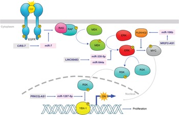
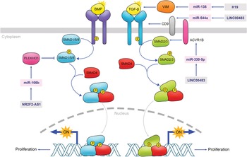
MAPK is another signalling pathway involved in cell proliferation, differentiation, migration and apoptosis. MAPK signalling is frequently overactivated in CRC cells and is thought to play a role in cellular hyperproliferation (Ref. Reference Yan186). The amount of LINC00483 was found to be increased when the MAPK signalling pathway was inhibited or growth arrest was followed by serum starvation, but the low level of LINC00483 expression was exhibited in CRC cells. Therefore, LINC00483 may have a tumour suppressor role. Competitively potential binding of LINC00483 to a variety of miRNAs, including miR-330-5p and miR-544a were predicted in CRC and it acts as a miRNAs sponge to modulate a set of multiple mRNAs (Ref. Reference Brex89) (Fig. 2).
ceRNAs involved in MAPK signalling pathway: CiRS-7 sponge's miR-7 leading RAF upregulation and facilitating MAPK signalling pathway. LINC00483 competitively binds to miR-330 and miR-544a MREs and has a negative effect on ERK and stops the pathway. NR2F2-AS1 sponges miR-106b increasing phosphorylated MYC and facilitating the MAPK pathway procedure. PRKCQAS1 binds to miR-1287-5p MRE and upregulates YBX-1 facilitating the MAPK signalling pathway.

The TGF-β signalling pathway is also dysregulated in cancer cells. TGF-β signalling pathway acts as a tumour suppressor leading to cell cycle arrest and apoptosis in early-stage cancer cells, but it has the potential to promote tumourigenesis and metastasis in late-stage tumours. Some ceRNAs such as Nuclear Receptor Subfamily 2 Group F Member 2 Antisense RNA 1 (NR2F2-AS1) inhibits the pathway. NR2F2-AS1 degrades SMAD5 via sponging miR-106b and upregulating Pleckstrin homology (PH)-domain-containing family O member 1 (PLEKHO1). Investigations have shown that miR-106b expression is significantly higher in CRC tissues than in normal colorectal tissues but PLEKHO1 is a potential CRC tumour suppressor which is downregulated in CRC tissues. On the other hand, some ceRNAs promote the TGF-β pathway. For instance, H19 competitively binds to miR-138 MRE and upregulates VIM. LINC00483 sponges miR-544a and helps the association of TGF-β 1 and 2. In addition, LINC00483 increases the ACVR1B expression, which facilitates SMAD1 and SMAD2 association, via sponging miR-330-5p (Refs Reference Colak and Ten Dijke187, Reference Akhurst and Hata188) (Fig. 3).
ceRNAs involved in TGF-β signalling pathway: NR2F2-AS1 can sponge miR-106b upregulating PLEKHO1. This causes SMAD5 degradation and stops the pathway. Therefore, the downregulation of NR2F2-AS1 causes more proliferation. H19 competitively binds to miR-138 MRE, which leads to VIM upregulation and promotes the TGF-β pathway. LINC00483 can sponge miR-544a and finally facilitates TGF-β 1 and 2 association (two domains of TGF-β receptor). Moreover, LINC00483 sponges miR-330-5p and have a positive effect on ACVR1B expression. ACVR1B facilitates SMAD1 and SMAD2 association and helps the procedure of the TGF-β signalling pathway.

The ceRNAs and signalling pathways involved in CRC cell apoptosis
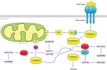
One of the most notable features of tumour cells is their ability to inhibit apoptosis (Ref. Reference Evan and Vousden189). Recent studies have demonstrated that ceRNAs can influence apoptosis in CRC (Fig. 4). Caspase-9 and caspase-3 are critical markers of the caspase-dependent pathway mediated by mitochondria. Shan et al. discovered that silencing LINC00908 induces apoptosis in CRC cells, with a consistently increasing rate of apoptosis and a significant increase in caspase-9 and caspase-3 cleavage (Ref. Reference Shan92).
ceRNAs involved in apoptosis signalling pathway: LINC00908 silencing effects on miR-143-3p resulted in KLF upregulation. Consequently, the caspase-9 would be increased and lead to apoptosis. Furthermore, KLF facilitates the conversion of procaspase-3 to caspase-3. Circ0082182 sponges miR-411 and miR-1205 and has a positive effect on caspase-9 in apoptosis signalling. EIF3J-AS1 inhibits YAP1 by affecting miR-3163. Therefore, it helps the conversion of procaspase-3 to caspase-3 leading to apoptosis.

Similarly, both NEAT1 silencing and NR4A1 inhibition induce apoptosis in CRC cells (Ref. Reference Liu180). Additionally, silencing circ0082182 increases pro-apoptotic caspase-3 and caspase-9 activity in CRC cells (Ref. Reference Liu157). It was represented by Liu et al. that the lncRNA Eukaryotic Translation Initiation Factor 3 Subunit J Antisense RNA 1 (EIF3J-AS1), which targets miR-3163, is vital for CRC cell apoptosis. They showed that silencing EIF3J-AS1 increases caspase-3 activity and the ratio of apoptotic cells. Yes1-associated transcriptional regulator (YAP1) is a downstream target of the tumour-suppressive Hippo pathway, and it has a negative correlation with miR-3163 and a positive correlation with EIF3J-AS1 in CRC. The results indicate that EIF3J-AS1 regulated YAP1 expression positively via miR-3163 (Ref. Reference Liu95).
It has been illustrated that Maternally Expressed Gene 3 (MEG3) expression is downregulated in human colorectal carcinoma tumour tissues and cells, correlated with increased miR-103a-3p and decreased Pyruvate Dehydrogenase E1 Subunit beta (PDHB). MEG3 can act as a sponge for miR-103a-3p and can also target PDHB. MEG3 restoration has the ability to induce apoptosis in CC and also promotes the expression of ER stress-related proteins such as 78-kDa glucose-regulated protein (GRP78), also known as Immunoglobulin Heavy Chain-Binding Protein (Bip), Activating transcription factor 6 (ATF6), CCAAT-Enhancer-Binding Protein Homologous Protein (CHOP), caspase-3 and caspase-9. As a result, it is capable of suppressing CC cancer (Ref. Reference Wang96).
The ceRNAs and signalling pathways involved in CRC cell invasion and migration
The EMT, which enables tumour cells to migrate and invade, is a frequent behaviour of tumour cells (Ref. Reference Pastushenko and Blanpain190). The TGF-β pathway, which promotes EMT in all types of tumours, is a critical and central pathway in this manner. TGF-β binds specifically to TGF-receptor (TGF-R) type 1 and 2 complexes. As a result of this, Mothers against decapentaplegic homolog 2 (SMAD2) and SMAD3 are phosphorylated. They then interact with SMAD4 and form complexes. Phosphorylated complexes of SMAD2, SMAD3 and SMAD4 induce EMT (Ref. Reference Akhurst and Hata188).
MIR22 host gene (MIR22HG) can interact with SMAD2 and regulate the TGF-β pathway's activity, inhibiting the interaction of SMAD2 and SMAD4 in the TGF pathway and thereby suppressing the EMT process (Ref. Reference Xu191).
Circ0082182 can lead to a miR-411 and miR-1205 sponge and activates the Wnt/β-catenin pathway, hence, inducing EMT in CRC cells. Circ0082182 expression decreases the levels of Phosphorylated Glycogen synthase kinase-3/Glycogen synthase kinase-3 (pGSK-3/GSK-3), nucleus β-catenin and N-cadherin while increasing the levels of E-cadherin. Thus, silencing circ0082182 inhibits the Wnt/β-catenin pathway and can inhibit EMT by increasing the expression of miR-411 and miR-1205 (Ref. Reference Liu157).
Besides, LINC00483 can participate in the invasion and migration of CRC cells. It is expressed at a low level in metastatic CRC when compared with primary tumours. Brex et al. used TGF-β1 and interleukin 6 (IL-6), two well-characterised inducers of EMT in CRC cells, to artificially induce EMT in CRC cells via distinct molecular cascades. They discovered that inducing EMT resulted in a decrease in LINC00483 expression. As a result, it inhibits EMT activation. Additionally, they discovered that Hepatocyte nuclear factor 4 (HNF4), a transcription factor, may play an oncogenic role in CRC by inhibiting the expression of LINC00483 (Ref. Reference Brex89).
NR2F2-AS1 is another ceRNA which participates in multiple signalling pathways. It is downregulated in CRC cells and promotes the migration and invasion abilities of CRC cells through the MAPK pathway, apart from being involved in CRC cell proliferation via the TGF-β pathway. The MAPK signalling pathway stimulates the epithelial to mesenchymal phenotypes transformation and MYC transcription factor activation, thereby inducing tumour migration and invasion. NR2F2-AS1 has a tumour-suppressive activity which inhibits the MAPK pathway by sponging for miR-106b and regulating the PLEKHO2 expression. It is also found that the expression level of miR-106b is negatively correlated with the NR2F2-AS1, PLEKHO2 and positively with the MYC expression level (Ref. Reference Liu192) (Fig. 2).
It has been reported that the lncRNA Protein Kinase C Theta Antisense RNA 1 (PRKCQ-AS1) can promote Y-box binding protein 1 (YBX1) mRNA and protein levels in CRC by sponging miR-1287-5p. YBX1 expression increases during CRC and is required for CRC malignancy via regulation of RAS/MEK/ERK signalling (Ref. Reference Cui88).
The ceRNAs as novel prognoses and diagnoses biomarkers for CRC
CRC is lethal cancer because its early stages are typically asymptomatic, and more than 60% of patients are diagnosed at a later stage. Stage IV CRC has a survival rate of 10% or less (Ref. Reference Sun193). As a result, a promising tumour biomarker may improve the survival rate of patients with CRC.
Tumour biomarkers are produced by cancer cells or other cells in the body in response to cancer and provide information about the disease's properties, such as the patient's survival, response to treatment, clinical status and aggressiveness. Tumour biomarkers can be used to aid in the diagnosis, prognosis and prediction of cancer. A promising tumour biomarker can be used to forecast future clinical characteristics such as the progression of the illness and its remission (Ref. Reference Goossens194). Proteins or other substances that cancer cells produce in greater quantities than normal cells have the potential to be used as tumour biomarkers (Ref. Reference Goossens194). However, research has demonstrated that non-coding RNAs and coding RNAs that act as ceRNAs in CRC progression can also be used as biomarkers.
For instance, Taurine upregulated gene 1 (TUG1) that sponges miR-600 plays a role in CRC metastasis, and it can be used as both a prognostic biomarker and a therapeutic target. Sun et al. exhibited an upregulation of this lncRNA in tumour tissue that was highly correlated with the patients' survival time (Ref. Reference Sun195). TUG1 overexpression was found to be oncogenic in vitro and increased colony formation, migration and metastatic potential in vivo. Additionally, they discovered that TUG1 could promote the expression of EMT-related genes (Ref. Reference Sun196).
Not only oncogenic but also tumour-suppressive ceRNAs can function as tumour biomarkers. Growth arrest-specific transcript 5 (GAS5) is a ceRNA sponging miR-222-3p (Ref. Reference Liu53). Yin et al. demonstrated that it is downregulated in human CRC tissues in comparison with adjacent normal lung tissues. Additionally, decreased GAS5 levels directly correlate with increased tumour size, low histological grade and advanced TNM stage. They demonstrated that patients with low GAS5 levels have a shorter survival time than those with high GAS5 levels. Additionally, the authors demonstrated that overexpression of GAS5 has the potential to inhibit cell proliferation (Ref. Reference Yin197).
Additionally, circRNAs have the potential to be used as tumour biomarkers. Because they lack a 5′ cap structure and a 3′ poly-A tail, circRNAs are resistant to exonucleases (Ref. Reference Suzuki136). In addition, they have a half-life of more than 48 h, in contrast to linear RNAs, which have an average half-life of 10 h (Ref. Reference Jeck and Sharpless198). Furthermore, circRNAs are stable in a variety of tissues and bodily fluids (Ref. Reference Zhang199). Zhang et al. observed that the expression level of circ0007534 is significantly high in CRC patients' plasma (Ref. Reference Zhang200). Circ0007534 can act as a ceRNA by sponging miR-761 (Ref. Reference Li201). Moreover, they demonstrated that a high level of the circ0007534 expression in patient plasma is associated with clinical progression, a metastatic phenotype, and poor differentiation in CRC. This circRNA has a high area under the curve (AUC) (0.780), sensitivity (0.92) and specificity (0.92) for discriminating CRC patients from healthy controls (0.522). They discovered that a high level of expression of the circ0007534 is associated with a poor prognosis in patients with CRC. As a result, circ0007534 may serve as a biomarker for CRC and may be associated with a poor prognosis (Ref. Reference Zhang200).
Another circRNA that can be used as a biomarker for CRC is cerebellar degeneration-related protein 1 antisense (CDR1-AS), also known as cirs-7, which acts as a mir-7 sponge. CDR1-AS expression is elevated in CRC tissue when compared with match normal mucosae. This is inversely proportional to lymph node metastasis, tumour size, T stage and poor overall survival (Ref. Reference Tang202). Weng et al. observed that CDR1-AS could be an independent risk factor for OS (Ref. Reference Weng155). As a result, CDR1-AS may serve as a prognostic biomarker for CRC and may be used as a therapeutic target in CRC patients.
Conclusions and perspectives
Recent research has been examining the functions of non-coding RNAs, coding RNAs and pseudogenes as tumour ceRNAs (Refs Reference Mukherji19–Reference Cheng21). It has been demonstrated that lncRNAs, circRNAs, pseudogenes and even mRNAs have the ability to regulate other mRNAs by acting as ceRNA to sponge miRNAs (Refs Reference Spizzo31, Reference Chen and Zhao140, Reference Sisu173). Presently, the advent of high-throughput sequencing and bioinformatics analysis technology has revealed that ceRNAs are implicated in a variety of biological processes, including the generation and development of CRC (Ref. Reference Cheng21). On the other hand, some problems need to be resolved because of the limited understanding of the ceRNA mechanisms (Ref. Reference Le203). Recent research indicates that dysregulated ceRNAs are essential in various biological processes associated with CRC, such as signalling pathways that respond to external stimuli, proliferation, apoptosis, invasion and migration (Refs Reference Shan92, Reference Liu180, Reference Akhurst and Hata188). We discussed current advances in the field of non-coding and protein-coding RNAs acting as ceRNAs in CRC, as well as validated ceRNAs engaged in CRC biological functions. Several ceRNAs are involved in other types of cancers. For instance, MIR22HG is involved in thyroid papillary carcinoma. This lncRNA displays low level of expression in thyroid papillary carcinoma whereas miR-24-3p is upregulated (Ref. Reference Xu191).
Additionally, we discussed the role of ceRNAs as novel prognostic and diagnostic biomarkers. CeRNAs are potential therapeutic targets not only for CRC, but also for other prevalent cancers, such as lung cancer, breast cancer and prostate cancer (Refs Reference Seo204–Reference Peng206). CeRNAs, such as some circRNAs, have distinct patterns of expression in tumour tissue and blood compared with normal tissue. Consequently, they are promising biomarkers and therapeutic targets for CRC (Refs Reference Suzuki136, Reference Jeck and Sharpless198, Reference Zhang199). Some ceRNAs are tumour suppressors in CRC, and the induction of the expression of ceRNAs in CRC tissue might have tumour-suppressive effects on CRC (Refs Reference Brex89, Reference Weng155, Reference Tang202). One of the best candidates for this antitumour procedure is circRNAs because of their high stability and long half-life (Refs Reference Suzuki136, Reference Jeck and Sharpless198). Exogenous circRNAs can be delivered via vectors containing DNA cassettes designed for circRNA expression or through transfection of purified in vitro-generated circRNAs (Ref. Reference Liu207). To this end, the introduction of engineered circRNAs into CRC cells or tissues as sponges for specific oncogenic miRNAs may represent a novel and efficient approach for future cancer therapy.
Although significant progress has been made in identifying and characterising ceRNAs, numerous critical unknown factors and limitations of research into the biological processes of ceRNAs remain to be addressed in future studies. For example, how ceRNAs perform their functions in cancer? ceRNAs are involved in cancer metastasis, cell proliferation, cell apoptosis and cell invasion and migration. However, very superficial research has been done in these areas. In addition, the major problem is how ceRNAs can be used in clinical practice. Recent studies have proposed that ceRNAs are promising diagnostic and prognostic biomarkers. Although this research suggests that particularly non-invasive biomarkers (blood samples) should be promoted for detection, completely non-invasive samples, such as urine, saliva and stool, should be considered in future studies.
In the end, we discussed that multiple signalling mechanisms are involved in the initiation and progression of ceRNA crosstalk in CRC, and our present understanding is insufficient. Thus, a better understanding of the mechanisms regulating ceRNA crosstalk in CRC and their related signalling pathways may aid in a better understanding of the mechanisms underlying the progression of CRC. The roles of pseudogenes acting as ceRNAs in cellular metabolic, molecular pathways in CRC are unknown. Pseudogenes were once thought to be non-coding RNAs that evolved as a result of gene mutations. Pseudogenes, far from being silent, have been proven to function as ceRNAs that influence tumour cell metabolism (Ref. Reference Jeck and Sharpless198).
Author contributions
MHA conceived the study and drafted the manuscript. Figures were prepared by ZZ. ZZ and MS revised the manuscript. MHA, ZZ and MS searched for relevant literature studies and provided the comments. All authors read and approved the final manuscript.
Financial support
The authors have no other relevant affiliations or financial involvement with any organisation or entity with a financial interest in or financial conflict with the subject matter or materials discussed in the manuscript apart from those disclosed. No writing assistance was utilised in the production of this manuscript.
Conflict of interest
The authors declare that they have no competing interests.