Introduction
The South American continent contains the greatest diversity of freshwater fish in the world (Birindeli and Sidlauskas Reference Birindelli and Sidlauskas2018). Within this diversity of fish, Brycon nattereri Günther, 1864, commonly known as pirapitinga, is a rare species, restricted to clean and well-preserved tributary channels (Galindo et al. Reference Galindo, Ferreira, Apolinário-Silva, Terra, Aprígio, Ota, Ohara, Almeida and Sofia2019). Due to being a species sensitive to environmental disturbances, it is considered endangered (Resende et al. Reference Resende, Silva, Pasa, Hilsdorf and Kavalco2021).
With the growing interest in preserving global biodiversity, particularly in freshwater environments, it is becoming increasingly evident that parasites are a fundamental part of this diversity. They have been one of the least studied organisms, despite playing important roles as integral components of food webs and ecosystem functions (Williams et al. Reference Williams, Faiad, Claar, French, Leslie, Oven, Guerra, Micheli, Zgliczynski, Haupt, Sandin and Wood2022). Monogeneans represent a portion of this parasitic diversity, with just over 5,500 described species worldwide, exhibiting a wide morphological variety in terms of hosts and microhabitats (Seidlová and Šimková Reference Seidlová, Benovics and Šimková2022).
Within the Class Monogenea, Dactylogyridae are the most studied, and the majority of their representatives are ectoparasites of characiform fish (Luque et al. Reference Luque, Pereira, Alves, Oliva and Timi2017). Monogeneans of the genus Tereancistrum Kritsky, Thatcher & Kayton, Reference Kritsky, Thatcher and Kayton1980 are parasites of Characiformes and have been recorded in three fish families: Anostomidae, Bryconidae, and Prochilodontidae. Only ten species have been described for this genus, the main characteristic of which is an accessory sclerite articulated to the ventral anchor: Tereancistrum kerri Kritsky, Thatcher & Kayton, Reference Kritsky, Thatcher and Kayton1980; T. parvus Kritsky, Thatcher & Kayton, Reference Kritsky, Thatcher and Kayton1980; T. ornatus Kritsky, Thatcher & Kayton, Reference Kritsky, Thatcher and Kayton1980; T. toksonum Lizama, Takemoto & Pavanelli, Reference Lizama, Takemoto and Pavanelli2004; T. curimba Lizama, Takemoto & Pavanelli, Reference Lizama, Takemoto and Pavanelli2004; T. pirassununguensis Cepeda, Ceccarelli & Luque, Reference Cepeda, Ceccarelli and Luque2012; T. arcuatus Cohen, Kohn & Boeger, Reference Cohen, Kohn and Boeger2012; T. paranaensis Karling, Lopes, Takemoto & Pavanelli, Reference Karling, Lopes, Takemoto and Pavanelli2014; T. flabellum Zago, Yamada, Franceschini, Bongiovani, Yamada & Silva, Reference Zago, Yamada, Franceschini, Bongiovani, Yamada and Silva2017; and T. takemotoi Leite, Pelegrini, Azevedo & Abdallah, Reference Leite, Pelegrini, Azevedo and Abdallah2020 (see the records of Tereancistrum spp. parasitizing Neotropical fish in Table 1).
List of Tereancistrum spp. species parasitizing the host families (Anostomidae, Prochilodontidae, and Bryconidae) of Neotropical fishes. All infection sites identified are gill filaments

The use of molecular markers, especially the mitochondrial COI region, has advanced our understanding of the taxonomy and evolution of fish parasites, such as monogeneans, which are highly diverse, especially in Neotropical freshwater ecosystems (Eiras et al. Reference Eiras, Takemoto, Pavanelli and Adriano2011; Hebert et al. Reference Hebert, Cywinska, Ball and DeWaard2003; Kentova et al. Reference Kmentová, Cruz-Laufer, Pariselle, Smeets, Artois and Vanhove2022; Luque et al. Reference Luque, Pereira, Alves, Oliva and Timi2017; Pereira et al. Reference Pereira, Castro, Vargas, Gomez and Oliveira2021). These markers not only help identify species but also reveal cryptic diversity and clarify taxonomic relationships (Cohen et al. Reference Cohen, Justo and Kohn2013). Despite their evolutionary value and increasing use in studies (Hasuike et al. Reference Hasuike, Scorsim, Arjona, Bellay, De Oliveira and Takemoto2024; Huang et al. Reference Huang, Meier, Todd and Chou2008; Neto and Domingues Reference Neto and Domingues2023; Yamada et al. Reference Yamada, Müller, Zago, Yamada, Ebert, Franceschini and Da Silva2023; Zago et al. Reference Zago, Yamada, Yamada, Franceschini, Bongiovani and Silva2020), the diversity of monogeneans remains largely unexplored, highlighting the importance of these tools in discovering new species, as in the case of Tereancistrum.
This study describes a new species of Tereancistrum and new record of Tereancistrum kerri parasitizing the gills of Brycon nattereri Günther, 1864, through the analysis of sclerotised structures and internal organs, as well as to molecularly characterize the specimens of T. kerri and T. campanum n. sp. by partial sequences of the mitochondrial cytochrome c oxidase subunit 1 (COI) gene region.
Material and methods
Host collections and study area
A total of four specimens of Brycon nattereri were collected in April 2023 using the gillnet method with meshes having openings of 12, 15, 25, 40, 50, and 80 mm between opposite nodes, according to Oliveira and Tejerina-Garro (Reference Oliveira and Tejerina-Garro2010), with modifications. Some of these specimens were deposited in the Coleção Ictiológica do Núcleo de Pesquisas em Limnologia, Ictiologia e Aquicultura (Nupélia-UEM) (NUP24315). The study was conducted on the Traíras River, one of the main tributaries of the Maranhão River, both of which belong to the Tocantins-Araguaia River Basin, located in the municipality of Niquelândia, State of Goiás, Brazil. Brycon nattereri was only collected at point P1 (14°39’20.4" S, 48°26’32.4" W). This study was approved by the Chico Mendes Institute for Biodiversity Conservation (ICMBio) (process n°. 02010.000260/01-73) and by the Biodiversity Authorization and Information System (SISBIO) (process n°. 71279-1). It was conducted under the principles adopted by the National Council for the Control of Animal Experimentation (CONCEA) and approved by the Ethics Committee for the Use of Animals at the Universidade Estadual de Goiás (n°. 003 - CEUA/UEG).
Parasitological processing
The fish were frozen, stored in polystyrene boxes containing gel ice, and transported to the Ichthyoparasitology Laboratory at the Núcleo de Pesquisas em Limnologia, Ictiologia e Aquicultura (Nupélia-UEM). The fish were examined in two different ways. First, for the morphological characterization of the parasites, the gills of the hosts were removed and fixed in a 5% formalin solution. The monogeneans were then triaged and transferred to 70% ethanol. Each monogenean specimen was mounted between a slide and coverslip containing Hoyer’s medium for the observation of sclerotized structures, while other specimens were stained with Gomori’s trichrome for the observation of internal organs, following the methodology of Eiras et al. (Reference Eiras, Takemoto and Pavanelli2006).
For molecular characterization, the hosts were triaged according to the methodology of Da Graça et al. (Reference Da Graca, Fabrin, Gasques, Prioli, Balbuena, Prioli and Takemoto2018), in which the parasites were collected fresh, without fixative reagents (alcohol or formaldehyde), and placed on a slide containing ultrapure water to confirm the morphotype and identify the main taxonomic characteristics, such as the shape of the accessory piece and the presence of the accessory sclerite on the haptor. Once the morphotypes had been confirmed and photographed, all the materials were transferred to 1.5-ml microtubes, each containing 20 μl of Milli-Q water for subsequent DNA extraction.
All measurements were expressed in micrometers (μm), presenting the mean followed by the range. A Nikon Eclipse e200 microscope equipped with a drawing tube and phase contrast light was used for the illustrations. Ecological terms (abundance, mean abundance, prevalence, and mean intensity) were used according to Bush et al. (Reference Bush, Lafferty, Lotz and Shostak1997). Holotypes and paratypes were deposited in the Coleção Helmintológica do Instituto Oswaldo Cruz, under register CHIOC - 40472a-f (a:Holotype and b-f: paratype).
Molecular characterization and delimitation
The DNeasy® Blood and Tissue Kit (QIAGEN®) was used to extract DNA from the parasites, following the protocol suggested by the manufacturer. A ProFlex™ 3×32-well thermal cycler was used to carry out the PCR. The DNA amplification reaction consisted of 1U of Taq DNA polymerase (5 U/μL, Invitrogen), Tris-KCL (20 mM Tris-HCl pH 8.4; 50 mM KCl), 1.87 mM MgCl₂, 0.1 mM of each dNTP, 4μM of each primer, DNA template (10ng), and Milli-Q water for a final volume of 23μL.
Specific primers for the mitochondrial region of cytochrome c oxidase subunit 1 (COI) were used to amplify the parasites’ DNA: COI_Mono_5: 5´TAATWGGTGGKTTTGGTAA-3´ and COI_Mono_3: 5´AATGCATMGGAAAAAAACA-3´ (Plaisance et al. Reference Plaisance, Rousset, Morand and Littlewood2008). The PCR products were checked on a 1% agarose gel, and the size of the fragments obtained was estimated using a marker of known molecular weight (100bp DNA Ladder, Invitrogen 0.5 μg/μL). The products obtained from DNA amplification were purified following the protocol described by Rosenthal et al. (Reference Rosenthal, Coutelle and Craxton1993). Sequencing was carried out by a private company on an Applied Biosystems® AB-3500 Genetic Analyser using the BigDye® Terminator kit.
The sequences obtained were edited and aligned using the BioEdit v7 software (Hall Reference Hall1999) and MEGA7 software (Kumar et al. Reference Kumar, Stecher and Tamura2016), respectively. MEGA7 software was used to obtain the p-distance and the gene tree using the maximum likelihood method and 1,000 bootstrap resampling. Only the sequences obtained in the present study of Tereancistrum kerri and Tereancistrum campanum n.sp. were used to understand the molecular relationship between congeneric species, since no sequences are available for the genus in the databases. Diaphorocleidus petrosusi (MF939755), a parasite of the Bryconidae family, was used as the outgroup (Supplementary material 1).
The same sequences were used to carry out molecular species delimitation tests. For this, we selected two molecular species delimitation methods that differ methodologically to compare the congruencies between them and access the best hypothesis that fits our data set: Assemble Species by Automatic Partition (ASAP, https://bioinfo.mnhn.fr/abi/public/asap/asapweb.html) (Puillandre et al. Reference Puillandre, Brouillet and Achaz2021) based on genetic distance; and the Poisson Tree Process (PTP), performed on the server https://species.h-its.org/ptp/ as a tree-based method (Zhang et al. Reference Zhang, Kapli, Pavlidis and Stamatakis2013).
Access to the genetic heritage of the specimens was authorized through the Sistema Eletrônico Nacional de Gestão do Patrimônio Genético e do Conhecimento Tradicional Associado (SisGen) under the number AB7CEFE, and the sequences obtained were deposited in GenBank: PQ474340- PQ474346 (Tereancistrum kerri) and PQ474347 (Tereancistrum campanum n.sp.).
Results
Taxonomic summary
Monogenea Van Beneden,1858.
Dactylogyridae Bychowsky, 1933
Tereancistrum Kritsty, Thatcher & Kayton, Reference Kritsky, Thatcher and Kayton1980
Tereancistrum campanum n. sp. (Figure 1)
Composite drawing A–H: A – Tereancistrum campanum n. sp. (ventral view); B – Male copulatory complex (ventral view); C – Accessory sclerite; D – Dorsal anchor; E – Ventral anchor; F – Dorsal bar; G – Ventral bar; H – Hooks.

Type host: Brycon nattereri
Type locality: Traíras river, Niquelândia- Goiás Brazil
Site of Infestation: Gill filaments.
Prevalence: 100%
Abundance: 23
Mean abundance: 5.7 ± 0.9
Mean Intensity: 5.7 ±0.9
Etymology: The specific name of the species refers to the bell shape of the accessory piece.
Another monogenea found: Tereancistrum kerri Kritsty, Thatcher & Kayton, Reference Kritsky, Thatcher and Kayton1980 (new host recorded).
Morphological description
Description: (based on 7 specimens clarified in Hoyer; 4 stained in Gomori trichrome). Body elongate 246 (192–312) long, 81 (46–127) wide, divided into cephalic region, trunk, short peduncle and haptor; cephalic region composed of two bilateral pairs of well-developed cephalic lobes, cephalic glands indistinct. Two pairs of eyes, posterior larger than the anterior, accessory granules present or absent (some may have 3 pairs of ocelli, anterior inconspicuous or small; there may be accessory granules distributed around the ocelli). Pharynx spherical 12 (11–14) in diameter; esophagus short, bifurcated into two intestinal caeca; caeca confluent, posterior to gonads. Haptor subhexagonal 62 (44–78) long, 85 (58–120) wide. Ventral anchor 42 (39–44) long, with broadly curved shaft and point, developed roots, small fold on base; tip of superficial root reflexed; base 15 (12–17) wide. Accessory sclerite associated to ventral anchor 34 (25–44) long, 5 (4–6) wide, with a spatulate termination. Two haptoral muscles attached to the accessory sclerite. Dorsal anchor 33 (24–39) long, with well-developed base and roots, shaft bent proximally, point attenuated to fine recurved tip; base 13 (12–14) wide. Ventral bar 54 (41–69) long and 5 (4–6) wide in an ‘open V-shaped’ shape with slightly expanded ends. Dorsal bar 23 (20–29) long, 4 (3–5) wide in a ‘U’ shape. Seven pairs of hooks, 19 (17–20) long, similar in size, shape, Dactylogyridae distribution; each with depressed thumb, recurved point, inflated proximal portion of shank, filamentous hook loop approximately 1/2 shank length. Gonads intercecal, overlapping. Ovary elongate 23 (20–24) long, 9 (9–10) wide. Testis 31 (24–35) long, 11 (11–12) wide, posterior to ovary. The copulatory complex is composed of a male copulatory organ with 2 1/2 clockwise rings 121 (107–132) long. The accessory piece robust not connected to the base of the male copulatory organ 22 (18–29) long, 15 (12–20) wide, comprising 2 subunits; the proximal part composed a thin sclerotized thread serving as guide for de MCO, distal region ‘bell’ shaped ornamented; oval prostatic reservoir. Vaginal pore dextral is composed of a thin elongated tube slightly sclerotized, vaginal opening funnel-shaped. Vitelline granules are distributed throughout the trunk, except in the cephalic region, reproductive organs, peduncle and haptor.
Remarks: The new species is included in the genus Tereancistrum due to the presence of the accessory sclerite connected to the ventral anchor. Tereancistrum campanum n. sp. can be easily distinguished from other species by the shape of its robust, bell-shaped accessory piece. When compared to other species, only the structures present in the haptor (anchors, bars, and hooks) of T. kerri and T. arcuatus are similar to those in T. campanum n. sp., but they can easily be differentiated by the male copulatory complex. T. kerri has a simple tapered tube with a small base, while the new species has 2 1/2 clockwise rings. We compared the new species with T. arcuatus, and both species have similar anchors and bars, but Tereancistrum campanum n. sp. has a male copulatory organ with 2 1/2 clockwise rings, while T. arcuatus has a ‘J’-shaped copulatory organ.
When compared with T. curimba, T. toksonun, T. ornatus, T. pirassununguensis, and T. takemotoi, all of these species have a ‘Y’-shaped dorsal bar, which can be easily differentiated from Tereancistrum campanum n. sp., which has an open ‘V’-shaped dorsal bar. In comparison with T. paranaensis, T. parvus, and T. flabellum, the morphology of the anchors, the shape of the bars, and the male copulatory complex show significant differences when compared to the species described in this study. T. parvus has a ‘V’-shaped dorsal bar, while Tereancistrum campanum n. sp. has an open ‘V’-shaped dorsal bar. When compared to T. flabellum, there is a difference in the base of the male copulatory organ, which is shaped like a ‘shell’, differing from the species described in this study. T. paranaensis has a male copulatory organ with a thin accessory piece, while, in contrast, Tereancistrum campanum n. sp. has a robust and ornate accessory piece.
Molecular characterization and delimitation
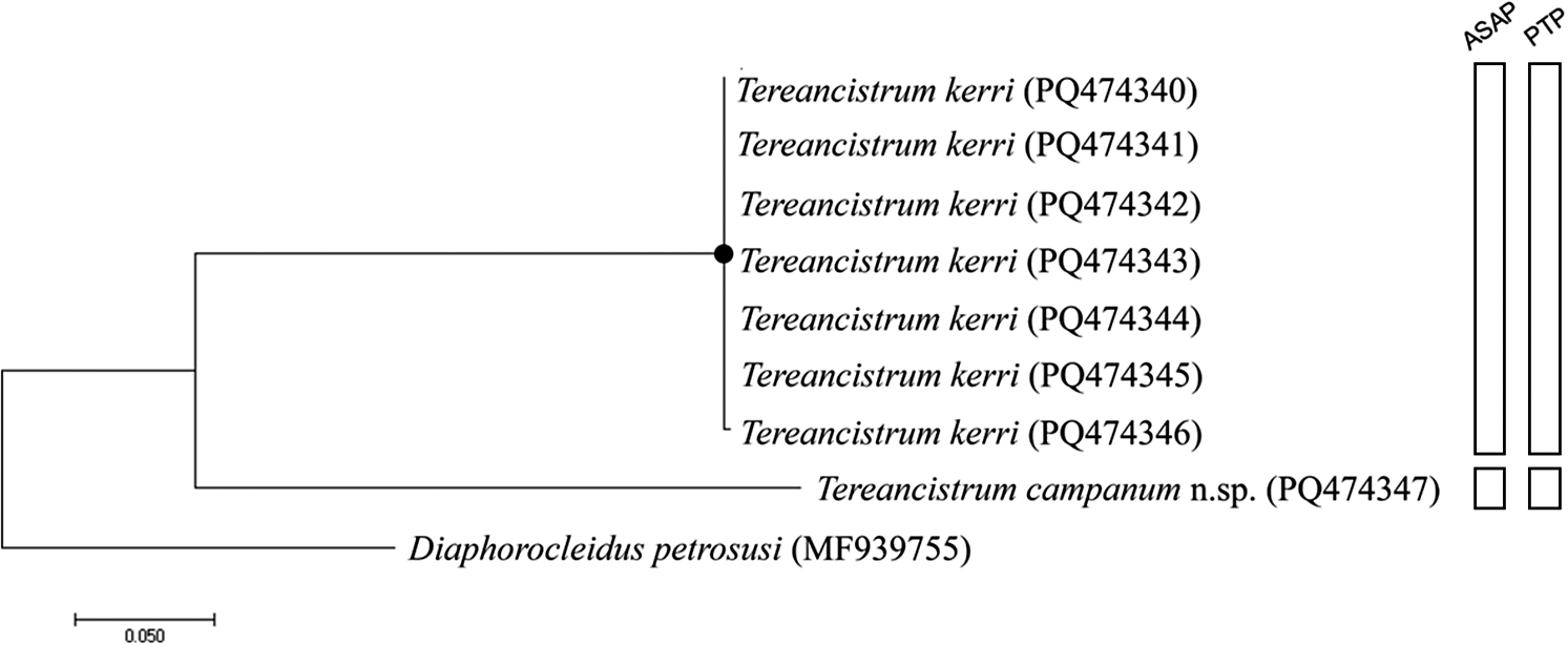
After editing and alignment, partial sequences of the COI region, with 615 base pairs (bp), were obtained for Tereancistrum campanum n.sp. and Tereancistrum kerri. Considering the specimens identified morphologically as T. kerri, all sequences were identical to each other, except for specimen TK4; the species showed a distance ranging from 25.4% to 22.6% with the species described here (Supplementary material 1).
Based on the gene tree for COI using the HKY+I nucleotide substitution model (Figure 2), specimens morphologically identified as T. kerri were grouped in the same cluster, while the specimen identified as T. campanum n. sp. formed a separate group. In both species delimitation tests (ASAP and PTP), T. campanum n. sp. was recognized as a distinct Molecular Operational Taxonomic Unit (MOTU).
Gene tree constructed using the maximum likelihood method with 1,000 bootstrap resamplings for the COI molecular marker. Diaphorocleidus petrosusi was used as an outgroup, and the nucleotide substitution model used was HKY+I. Support values ≥99 are highlighted with circles. The Molecular Operational Taxonomic Units (MOTUs) delimited by the ASAP and PTP methods are represented in the bars on the right-hand side of the gene tree.

Discussion
This study increases the number of currently valid species in the genus Tereancistrum to eleven, with species parasitizing three fish families: Anostomidae, Bryconidae, and Prochilodontidae. Among these species, T. parvus, T. flabellum, and T. paranaensis are primarily associated with the Anostomidae family, while T. toksonum, T. curimba, T. pirassununguensis, and T. takemotoi are commonly found in Prochilodontidae. Finally, T. kerri, T. arcuatus, and T. campanum n. sp. are exclusively found in Bryconidae (Cohen et al. Reference Cohen, Justo and Kohn2013; Leite et al. Reference Leite, Pelegrini, Azevedo and Abdallah2020; Zago et al. Reference Zago, Yamada, Franceschini, Bongiovani, Yamada and Silva2017).
Based on COI mtDNA sequences and the species delimitation tests employed, we determined that the specimen identified morphologically as T. campanum n. sp. represents a Molecular Operational Taxonomic Unit (MOTU) distinct from other congeneric specimens, showing a genetic distance ranging from 25.4% to 22.6% (Supplementary material 1), a level of divergence commonly found between different monogenean species (Hasuike et al. Reference Hasuike, Scorsim, Arjona, Bellay, De Oliveira and Takemoto2024; Neto and Domingues Reference Neto and Domingues2023; Zago et al. Reference Zago, Yamada, Yamada, Franceschini, Bongiovani and Silva2020).
Although T. kerri, T. arcuatus, and T. campanum n. sp. parasitize Bryconidae and share morphological similarities, specifically regarding their haptoral structures, the sequence data obtained for T. campanum n. sp. confirms that this specimen refers to a distinct species from T. kerri. However, the absence of available sequences for T. arcuatus in the literature, and consequently in the public databases, prevented us from performing a molecular comparison that could further clarify the relationship between the species that parasitize Bryconidae.
This study contributes to the understanding of the parasitic fauna in the Tocantins-Araguaia River Basin in Goiás State, Brazil, expanding the known geographical distribution of the genus Tereancistrum and enhancing our knowledge of parasitic diversity in Brazilian freshwater fish. However, the lack of available sequences for other species of the genus in public databases limits our ability to perform a more robust phylogenetic analysis for the entire genus.
Supplementary material
The supplementary material for this article can be found at http://doi.org/10.1017/S0022149X25000148.
Data availability
The datasets generated and analyzed during the study are available in the GenBank repository under the following accession numbers: PQ474340-PQ474346 (Tereancistrum kerri) and PQ474347 (Tereancistrum campanum n.sp.)
Code availability
Not applicable.
Acknowledgements
The authors are grateful to Capes and CNPq for funding the study, to the Núcleo de Pesquisa em Limnologia Ictiologia e Aquicultura - NUPÉLIA-UEM for providing the infrastructure, and to Legado verdes do Cerrado-Reserva Votorantim for all their support and help during the collections.
Author contribution
All authors contributed to the study’s conception and design. Material preparation and data collection were carried out by WTH, SB, and RMT. Molecular analyses were performed by BS, ISA, and AVO. The drawings have been made by WTH. All authors contributed to the writing of the manuscript. All authors read and approved the final manuscript.
Financial support
This work was supported by Conselho Nacional do Desenvolvimento Científico e Tecnológico.
Competing interest
The authors declare that they have no conflict of interest.
Consent to participate
Not applicable.
Consent for publication
Not applicable.
Ethical standards
All applicable guidelines for the care and use of animals were followed.