MY BACKDOOR DEVELOPMENT OF AN INTEREST IN MALARIA
In hindsight, the first 2 papers I published in Parasitology (Clark et al. Reference Clark, Cox and Allison1977a, Reference Clark, Richmond, Wills and Allisonb), extracts from my Ph.D. thesis, were central to the path my work was to take for the next 30 years. This was towards arguing that tumour necrosis factor (TNF) and other similar host-origin soluble mediators, none of which had yet been described, were central to immunity and disease pathogenesis in infectious diseases, including malaria. Rather than simply review the science, which has been amply covered in recent years (Clark and Cowden, Reference Clark and Cowden2003; Clark et al. Reference Clark, Alleva, Mills and Cowden2004, Reference Clark, Budd, Alleva and Cowden2006, Reference Clark and Chaudhri2008a), this contribution tells, for those who may well do something similar, and equally unexpected, as their career develops, how the story unfolded. The journey lacked overall guidance, a starting hypothesis, and pre-determined milestones. Neither did I have relevant research experience, merely success in solving a cattle disease in inland Australia that had been a long-standing mystery. Thought to be infectious, it turned out to be a plant poisoning with a fascinating pathophysiological action that explained why only cattle are affected (Clark, Reference Clark1973). This depended on having lived in that country for years, and having the confidence of the locals.
My new direction was simply driven by curiosity. During a basic macrophage function-based Ph.D., the purpose of going to London in 1972, I did not expect to call on my experience of examining Babesia bovis-infected blood smears (and thus the ability to recognize a drug-killed parasite inside a red cell when I saw one) nor my working knowledge of the gross and histological pathology of cattle that had died from infection with this organism. But without this background, a rare opportunity nowadays, when most stay continuously in the education system, I suspect I would have achieved little.
Once acclimatized, my days soon filled with cytotoxicity assays in which mouse macrophages killed chicken red cells coated with minute amounts of antibody (Sanderson et al. Reference Sanderson, Clark and Taylor1975). We used these as targets simply because they had much lower background levels of Cr51 release than did mouse red cells, and the vague future prospect was that this might tell us something about tumour immunology. How much more like reality, I thought, to use red cells from the same species as the macrophages, and while about it, why not Babesia-infected red cells, since I imagined a career back in cattle babesiosis. But was there a mouse Babesia? Gunnel Huldt, a visiting Swedish toxoplasma researcher, knew Alister Voller, who put me on to Frank Cox at Kings College on The Strand, and I came back on the Underground to the MRC laboratory at Northwick Park Hospital in Harrow carrying a cage of Babesia microti-infected mice. The consequences of this trip have informed all my work since. In a move thought natural by all at the time, but less easy to accomplish now that research is more directed, it led to me ditching my nascent interest in tumour immunology.
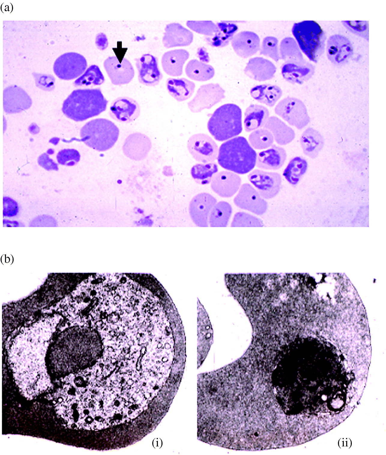
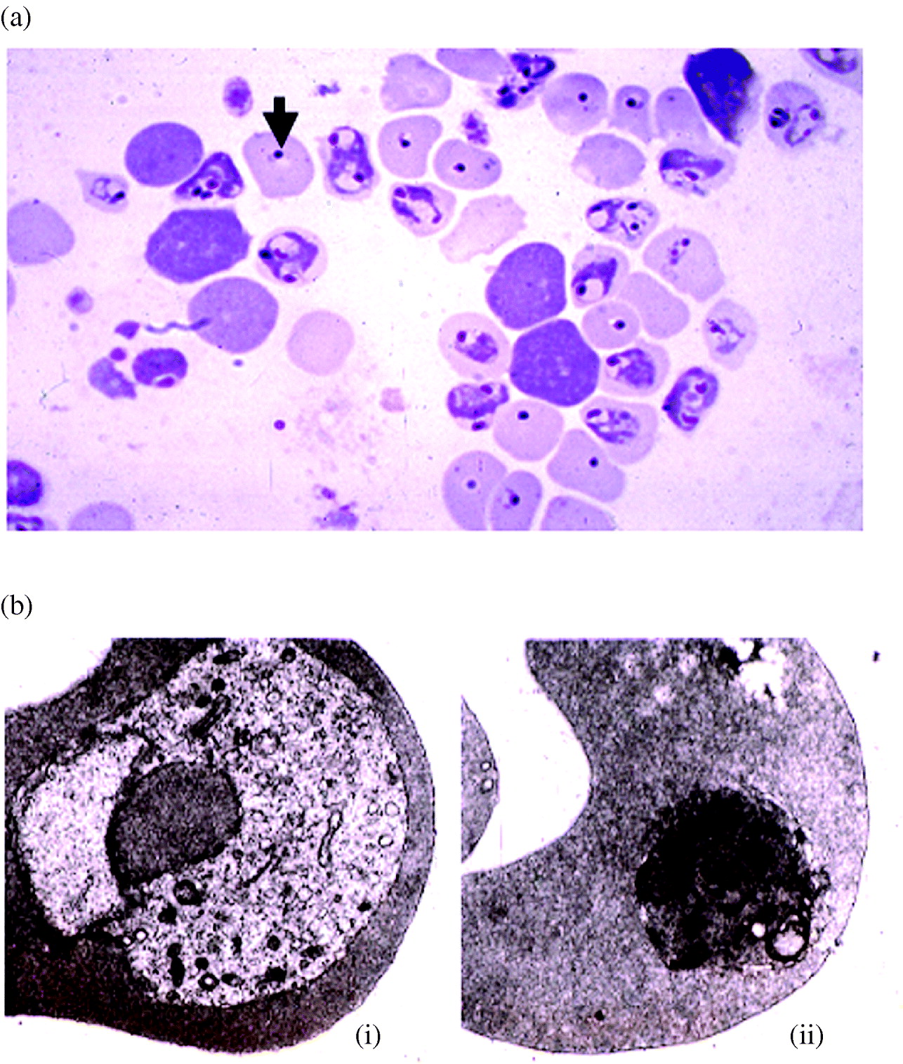
From my experience with reading post-drug blood smears of cattle babesiosis it seemed obvious to me, once I passaged these parasites, that mice routinely kill B. microti inside circulating red cells, at the time the mice themselves became slightly ill, and that this had to be the reason it was not a lethal parasite. Electron microscopy confirmed their intra-erythrocytic location (arguably they were attached and could not enter) and that they were now what we would call apoptotic (Clark et al. Reference Clark, Richmond, Wills and Allison1975). I first assumed everyone knew this, and probably why it happened. My next impulse, through sheer curiosity, was to wonder whether this might also happen to malaria parasites, closely related but a much bigger fish. Earlier cross-protection experiments (Cox, Reference Cox1970) and later electron microscopy (Fig. 1) convinced me that this was probably so. This potentially made nonsense of the then current antibody-based mechanism of immunity against these organisms. Hence Ted Wills, Joan Richmond and I took their electron micrographs to Laurie Bannister, at Guys Hospital, for his opinion. He and Geoff Butcher encouraged us that this phenomenon was worth pursuing because they had observed it when they challenged dually infected rhesus monkeys (Butcher et al. Reference Butcher, Bannister and Mitchell1976). Unexpectedly, Plasmodium coatneyi died en masse inside red cells when monkeys previously immunized against Plasmodium knowlesi were challenged with this parasite (Butcher et al. Reference Butcher, Mitchell and Cohen1978). In their view this could explain why some monkeys had high levels of this antibody yet unexpectedly failed to resist a challenge infection (Butcher et al. Reference Butcher, Mitchell and Cohen1978), while others with little or no anti-merozoite antibody were unexpectedly immune (Langhorne et al. Reference Langhorne, Butcher, Mitchell and Cohen1979). Clearly, some powerful influence beyond specific antibody directed against erythrocytic forms, the then popular dogma, was reproducibly occurring. We published another update of the data, logic and implications in Parasitology (Clark et al. Reference Clark, Richmond, Wills and Allison1977b). Belatedly, what was termed the cell-mediated approach to malaria vaccination eventually became a mainstream research direction (Good et al. Reference Good, Xu, Wykes and Engwerda2005).
Many aberrant forms of Plasmodium chabaudi (see arrow) immediately post-peak parasitaemia in (a) light microscopy of smear of circulating blood that proved, by electron microscopy (b), to be apoptotic parasites inside erythrocytes. Micrographs of healthy (i) and apoptotic (ii) parasite shown. Their presence can be missed if smears are not made frequently, since they are quickly removed by the spleen. This period is extended on splenectomized mice. Prior BCG prematurely caused this same degeneration in Babesia and Plasmodium spp. in mice. Adapted, with permission, from Cytokines and Growth Factors Reviews (2007) 18, 335.
ARRIVING AT THE POSSIBILITY OF TNF INVOLVEMENT IN PARASITE KILLING
The work might then have stalled had not Jean-Louis Virelizier, a virologist colleague, suggested we see if a newly-described soluble mediator, Type II interferon, (Salvin et al. Reference Salvin, Ribi, Granger and Youngner1975) later termed immune interferon, and then interferon-gamma, and generated in mice infected with BCG (the Bacillus Calmette-Guérin strain of Mycobacterium tuberculosis) plus Old Tuberculin, was behind this parasite death. Accordingly, he kindly gave me some BCG-infected mice, and several weeks later, following Salvin's protocol, I infected them with 106B. microti. This was dramatically protective (Clark et al. Reference Clark, Allison and Cox1976), with parasites not even becoming patent (showing on smears). Indeed, if big enough doses (109) were given to create an instant parasitaemia, organisms could be seen dead inside circulating red cells about 12 h after injection. The effect was indeed profound, since it also protected totally against massive doses (109) of its lethal cousin, Babesia rhodaini, a parasite so virulent that infection with only 1 parasite is enough to kill a mouse. Again, parasites died inside circulating red cells. These BCG-protected mice, whether infected with 106 or 109Babesia parasites, proved to possess an immunity as strong and durable as that seen in previously infected mice, in the absence of detectable antibody. Moreover, BCG protected reasonably well against the predictable mouse malarias (Clark et al. Reference Clark, Allison and Cox1976), those that cross-protect with mouse babesias (Cox, Reference Cox1970).
When Corynebacterium parvum proved to be just as protective (Clark et al. Reference Clark, Cox and Allison1977a), (also a Parasitology paper), it became evident that the host response to babesiosis had fundamental similarities to that induced by tumours (Old et al. Reference Old, Clarke and Benacerraf1959; Halpern et al. Reference Halpern, Biozzi, Stiffel and Mouton1966), macrophage-dwelling bacteria (Dubos and Schaedler, Reference Dubos and Schaedler1957; Howard et al. Reference Howard, Biozzi, Halpern, Stiffel and Mouton1959; Collins and Scott, Reference Collins and Scott1974) and tissue-dwelling protozoa (Swartzberg et al. Reference Swartzberg, Krahenbuhl and Remington1975; Smrkovski and Larson, Reference Smrkovski and Larson1977). Protection against these organisms by BCG and C. parvum had always been assumed to operate by enhancing their phagocytosis, but our parasites were dying inside circulating erythrocytes, implicating some unidentified circulating soluble factor(s). This raised the idea of the same novel soluble agent(s), perhaps arising from activated macrophages, being involved in the immunity against all these other organisms as well.
Looking for clues on the nature of this non-antibody soluble agent(s), we noted that BCG and other protectants (reviewed by (Clark and Cowden, Reference Clark and Cowden2003)) not only shared the ability to protect mice against tumours, but also made them extremely susceptible to lipopolysaccharide (LPS), i.e. bacterial endotoxin (Pirsch et al. Reference Pirsch, Mika and van der Maaten1957; Abernathy et al. Reference Abernathy, Bradley and Spink1958; Suter et al. Reference Suter, Ullman and Hoffman1958; Clark, Reference Clark1979). Indeed, we could reliably predict which previously untested agents would sensitize mice to LPS, and how well, by the degree to which they protected them against these blood parasites. It was most timely that LPS had recently been realised not be directly harmful, but to act indirectly through undefined mediators it induced host cells to release (Berry et al. Reference Berry, Moore, Goodrum, Couch and Schlessinger1977). Hence we joined in the fashionable task of wondering what soluble factor from macrophages might kill tumour cells, except our focus was parasite killing.
EXTENDING TNF INVOLVEMENT TO DISEASE PATHOLOGY
This was when we began to consider whether these soluble factors might be involved in the pathology of malaria and babesiosis. The idea evolved from the concept that, since mouse Plasmodium and Babesia spp. also protected against themselves and each other (Cox, Reference Cox1970), it was logical that, like BCG, they too should sensitize mice to LPS. In the process of establishing this we injected LPS into mice during the early stages of malarial infection, while they were apparently normal but very LPS-sensitive, and found that within a few hours we had serendipitously caused the rapid onset of the usual biochemical and tissue pathology that these parasites, alone, normally cause at higher densities (Clark, Reference Clark1978). The more advanced were infections with fatal strains of haemoprotozoa, the more minute the dose of LPS required to mimic the range of adverse changes normally observed only in terminal infections. If we waited even longer the same changes occurred without the assistance of LPS (Clark, Reference Clark1978). This implied that undisturbed malaria infection could duplicate the consequences, in terms of releasing harmful soluble factors, of LPS injection. Therefore these mediators logically were major contributors to these pathological changes in malaria disease as well as those observed in diseases caused by the bacteria that produce LPS.
In the process of trying to link this, in mediator terms, with the protective effect of BCG and C. parvum, Jean-Louis Virelizier and I sounded out several contacts of his in Paris. In particular Ion Gressor at Villejuif was an open and friendly source of advice, but we found no practical leads. After considering several possibilities, including immune interferon, we read a PNAS paper by a Sloan-Kettering group in New York on a mediator they had termed tumour necrosis factor (TNF) (Carswell et al. Reference Carswell, Old, Kassel, Green, Fiore and Williamson1975). Whereas we had been wondering about how BCG and C. parvum protected against haemoprotozoa, they were chasing the protective effect of these agents against experimental tumours. Like our unknown mediator, theirs was triggered by LPS in mice primed several weeks earlier with BCG or C. parvum, and, like ours, it killed rapidly dividing cells, theirs tumours, our haemoprotozoa. I visited their lab, and soon I began working with them. Betsy Richards (E. A. Carswell, first author of the first TNF paper), proved to be open and honest, and swiftly became a trusted collaborator, friend, and confidant.
At about this stage of the thought processes I was obliged to return to Australia because of a scholarship commitment. Lack of local interest in these ideas (except from Carolyn Geczy in Sydney, also with overseas cytokine experience) ultimately proved to be an advantage, since it forced me to maintain and expand my international perspective through travel, and my contact with confidantes in the UK, France and the US continued to be as strong as when I had lived in London.
During these first Canberra years I first came across the ideas of Brian Maegraith, an Australian who had stayed on in the UK since the 1930s. In those years no one wanted to talk about how host-origin mediators might explain systemic infectious disease. I did, however, find a group in the US, the Shock Society, who were interested in linking such mediators with the pathology of traumatic shock. I became a regular at their meetings, and found we had much in common. When I professed an interest in malaria they told me of a Brian Maegraith, and how, during the Second World War, he had equated the anuria of crush syndrome and falciparum malaria (Maegraith and Findlay, Reference Maegraith and Findlay1944). So I searched for anything else he might have written on malaria. Ignorance of his existence, monograph, or text-book, shows just how outside the malarial disease world I then was, and how heretical my ideas must have seemed within it. Nevertheless, at our first meeting in May 1978, Brian Maegraith was prepared to listen, and we soon recognized that my TNF approach to malarial disease was entirely consistent with the original approach he had begun decades earlier. He acquainted me with his earlier sepsis work in Oxford (Maegraith, Reference Maegraith1938), and how malaria, when he encountered it while an army pathologist in Sierra Leone, in his terms fitted it like a glove. For a decade we, and often Alec Fletcher, enthusiastically talked work in Liverpool every 6 months or so, and I was honoured to be asked to contribute to Brian's 80th birthday Festschrift (Clark, Reference Clark1987b).
During this period I also had the benefit of much interaction in Canberra with Michael Boyle, then in private practice, who had extensive practical experience of ‘Pyrexias of Unknown Origin’ in army hospitals in Vietnam. He broadened my horizons by making me aware that if my story fitted malaria and sepsis it must surely fit rickettsial and viral diseases as well. He also had friends at the Walter Reed from that era who could not do enough for me once they realised I knew Michael Boyle. This was an essential education.
In 1978 I wrote a Lancet Hypothesis article on the involvement of TNF in the host response to malaria and babesiosis (Clark, Reference Clark1978). The first collaborative experiments with the Sloan-Kettering group showed that malaria infection could take the place of BCG or C. parvum as a primer for TNF and interleukin-1 (IL-1) generation (Clark et al. Reference Clark, Virelizier, Carswell and Wood1981). Next, serum containing TNF proved to inhibit in vivo multiplication of Plasmodium vinckei (Clark et al. Reference Clark, Virelizier, Carswell and Wood1981). While we predicted, from the LPS experiments described above (Clark, Reference Clark1978), that malaria would provide its own trigger for TNF release, we could not detect TNF in terminal mouse malaria serum using the L cell assay of the time. Once we realised that these assays could be made more sensitive by adding actinomycin D we had no difficulty in detecting it in such serum (Clark and Chaudhri, Reference Clark and Chaudhri1988b). Unfortunately the advent of ELISA assays was still some years off.
In 1981 we therefore put forward the idea (discussed with Brian Maegraith, Frank Cox, Betsy Richards, Michael Boyle and others since 1978) that, as well as protecting the host through causing parasites to die inside circulating red cells, TNF and associated mediators, including IL-1 (we referred to it by an old name, lymphocyte activating factor (LAF)) could also harm the host when produced excessively. This, we reasoned, would explain the disease, including fever, hypoglycaemia, bone marrow depression, coagulopathy, hypergammaglobulinaemia, hypotension and rise in serum levels of acute-phase reactants seen in malaria and bacterial sepsis (Clark et al. Reference Clark, Virelizier, Carswell and Wood1981; Clark, Reference Clark1982b). Some 40 key background papers that demonstrate the development of the idea of TNF being much more than a tumour killer (Clark and Cowden, Reference Clark and Cowden2003). In broad terms, this approach had led us to the then novel concept that the same soluble mediator(s), of host origin, including and perhaps led by TNF, had a previously unsuspected dual set of functions in disease in general – governing a new type of protective response against infectious agents, and also causing disease pathology when generated excessively (Clark, Reference Clark2007). There are now >70 000 PubMed hits on TNF alone, the majority on its roles in physiology and disease mechanisms. Moreover, work from the field of rheumatoid arthritis, where anti-TNF agents are the largest success in applied biotechnology, has demonstrated that TNF is a master regulator within the inflammatory cytokine cascades (Charles et al. Reference Charles, Elliott, Davis, Potter, Kalden, Antoni, Breedveld, Smolen, Eberl, deWoody, Feldmann and Maini1999).
In these papers (Clark et al. Reference Clark, Virelizier, Carswell and Wood1981; Clark, Reference Clark1982b), the second being the record of an invited talk at an international sepsis meeting in Heidelberg in late 1981, I predicted that the long-elusive malarial toxin, much written about 100 years ago (Ross, Reference Ross1911) as the probable cause of malarial illness, would prove to be functionally similar to LPS, the only trigger for TNF then known. We argued that the ‘toxin’, like LPS, would not be directly harmful, but act through releasing TNF, and therefore could be defined by its ability to do this in vitro. This model led to proposals that phosphatidylinositol (Taverne et al. Reference Taverne, Bate and Playfair1989; Bate et al. Reference Bate, Taverne, Bootsma, Mason, Skalko, Gregoriadis and Playfair1992) and later glycosylphosphatidylinositol (Schofield and Hackett, Reference Schofield and Hackett1993) were the malarial toxin that triggers the harmful pro-inflammatory cytokine cascade.
RATIONALIZING INNATE DIFFERENCES IN PARASITE LOAD REQUIRED FOR ONSET OF ILLNESS
I had wondered, when starting to work with B. microti, why mice never showed illness below 60% parasitaemia, whereas from my experience cattle infected with B. bovis, just like (I found out later) people with P. falciparum or P. vivax, need so few organisms to make a previously unexposed host ill that the diagnostic routine is to search hard on a thick smear. Mouse and human comparison of infections with B. microti, and Saimiri and human comparisons with P. falciparum, indicated the phenomenon was a host, not parasite, characteristic. Hence in 1980 I decided to see whether these cytokine ideas explained the relative parasite densities required for onset of illness in previously unexposed hosts infected with Plasmodium, Babesia, and related species. A combination of experiments and reading made me appreciate a novel correlation extending from lizards, through birds, to various mammals, including man. Namely, innate tolerance to endotoxin, which makes animals and people ill through releasing cytokines such as TNF, correlates nicely with innate tolerance to haemoprotozoa (Clark, Reference Clark1982a). Moreover, I had noted, acquired human malarial tolerance correlates with LPS tolerance (Rubenstein et al. Reference Rubenstein, Mulholland, Jeffery and Wolff1965). More than anything beforehand, these patterns convinced me I was on the right track, albeit no one ever asked for a reprint! Neither, however, has anyone ever put up an alternative theory to explain the phenomenon, which is everyday experience to parasitologists examining blood smears from hosts as diverse as reptiles, birds, and man. I now appreciate that the pattern also applies to trypanosomiasis.
THE ADVENT OF rTNF
Like other purified, but not yet cloned, mediators, cytokines such as TNF were not easy to obtain before the mid-1980s, and the literature stayed small until they were available in recombinant form. Over the period from the publication of our key arguments in 1981 and 1982, until Bruce Beutler in Dallas unofficially provided me with some rTNF in 1986, we were forced to concentrate on putative down-stream mediators of cascades initiated by TNF, such as oxygen-derived free radicals. Once we had enough rTNF to be able to use it in vivo we showed it would kill mouse malaria parasites in vivo (Clark et al. Reference Clark, Hunt, Butcher and Cowden1987b), mimic mouse malaria pathology in general (Clark et al. Reference Clark, Cowden, Butcher and Hunt1987a), and in particular the dyserythropoiesis and erythrophagocytosis (Clark and Chaudhri, 1988 b) as well as foetal loss (Clark and Chaudhri, Reference Clark and Chaudhri1988a). In principle this was all entirely consistent with what I had predicted 7 years earlier, and observed at the Dana Farber Institute at Harvard, during a visit in 1987 to see the side-effects of rTNF therapy in tumour patients. From my perspective, they were inadvertently reproducing malarial disease (Spriggs et al. Reference Spriggs, Sherman, Michie, Arthur, Imamura, Wilmore, Frei and Kufe1988; Bauer et al. Reference Bauer, Cate, Barzeger, Spriggs, Sherman and Rosenberg1989). As reviewed by Clark et al. (Reference Clark, Alleva, Mills and Cowden2004), the parallels were quite uncanny. At about the same time various groups, including ourselves, correlated the level of TNF in falciparum malarial serum with disease severity (Kern et al. Reference Kern, Hemmer, Van Damme, Gruss and Dietrich1989; Butcher et al. Reference Butcher, Garland, Adjukiewicz and Clark1990; Kwiatkowski et al. Reference Kwiatkowski, Hill, Sambou, Twumasi, Castracane, Manogue, Cerami, Brewster and Greenwood1990).
The term cachectin, for a time commonly seen in the parasitological literature (Beutler and Cerami, Reference Beutler and Cerami1987), warrants explanation here. Inevitably, the sequencing technology applied to TNF was also being used at the same time to define the molecular nature of other chemically purified cytokines. The pleotropic properties of TNF meant a number of researchers found that a serum fraction they had named and functionally defined proved to be identical to TNF. One example, an LPS-induced macrophage product that came to light in 1982, as yet in an unnamed form (Kawakami et al. Reference Kawakami, Pekala, Lane and Cerami1982), through trying to understand weight loss in trypanosomiasis. Its defining characteristic was an ability to inhibit the enzyme lipoprotein lipase, and 3 years later (a decade after TNF) it was purified (Beutler et al. Reference Beutler, Mahoney, Le, Pekala and Cerami1985b) and termed cachectin. Like many other soluble mediators it proved, on sequencing (Beutler et al. Reference Beutler, Greenwald, Hulmes, Chang, Pan, Mathison, Ulevitch and Cerami1985a), to be identical to a previously sequenced (Aggarwal et al. Reference Aggarwal, Kohr, Hass, Moffat, Spencer, Henzel, Bringman, Nedwin, Goeddel and Harkins1985) molecule that had been termed TNF 10 years earlier (Carswell et al. Reference Carswell, Old, Kassel, Green, Fiore and Williamson1975). In due course the term cachectin dropped out of currency. Other researchers referred to their molecules as TNF as soon as they were sequenced (Takeda et al. Reference Takeda, Iwamoto, Sugimoto, Takuma, Kawatani, Noda, Masaki, Morise, Arimura and Konno1986), which was less confusing.
TNF AWARENESS EXPANDING TO OTHER INFECTIOUS DISEASES
The idea of TNF (and of course, functionally-related cytokines) causing pathology in infectious disease had a smooth transition from babesiosis and malaria into being implicated in the diseases caused by various other infectious agents. Examples are Mycobacterium spp. (Rook et al. Reference Rook, Taverne, Leveton and Steele1987), Leishmania spp. (Raziuddin et al. Reference Raziuddin, Abdalla, el Awad and al Janadi1994), Toxoplasma gondii (Arsenijevic et al. Reference Arsenijevic, Girardier, Seydoux, Chang and Dulloo1997), Salmonella spp. (Bhutta et al. Reference Bhutta, Mansoorali and Hussain1997), Brucella abortus (Ahmed et al. Reference Ahmed, Al Matrouk, Martinez, Oishi, Rotimi and Nagatake1999), and Listeria monocytogenes (Nakane et al. Reference Nakane, Yamada, Hasegawa, Mizuki, Mizuki, Sasaki and Miura1999). A parallel literature exists documenting the involvement of this cytokine in killing these infectious agents (Clark et al. Reference Clark, Alleva, Budd and Cowden2008a). In due course we (Clark and Cowden, Reference Clark and Cowden1989) had extended the logic of this approach to understanding viral diseases, and 15 years later (Clark et al. Reference Clark, Alleva, Mills and Cowden2004) noted how the literature on the diseases caused by Marburg, Ebola, Lassa, Junin and influenza viruses had caught up with this prediction. Since then we have proposed a new influenza treatment based on these principles (Budd et al. Reference Budd, Alleva, Alsharifi, Koskinen, Smythe, Mullbacher, Wood and Clark2007). Selling the idea of excess inflammatory cytokines being central to these above diseases is much easier than for falciparum malaria, since they have no sequestering parasitized red cells to have generated an alternative explanation.
FURTHER EXPANSION OF INFLAMMATORY CYTOKINES INTO UNDERSTANDING MALARIA
To be a serious contender as the primary cause of disease in malaria, as well as in other systemic infectious diseases, the cytokine theory (Clark et al. Reference Clark, Virelizier, Carswell and Wood1981; Clark, Reference Clark1987a; Clark and Rockett, Reference Clark and Rockett1994) has had to have answers to the traditional view of falciparum malaria disease, which still sees it as a systemic hypoxia caused by vascular obstruction by sequestered parasitized red cells (MacPherson et al. Reference MacPherson, Warrell, White, Looareesuwan and Warrell1985; Fairhurst and Wellems, Reference Fairhurst and Wellems2006; Maier et al. Reference Maier, Rug, O'Neill, Brown, Chakravorty, Szestak, Chesson, Wu, Hughes, Coppel, Newbold, Beeson, Craig, Crabb and Cowman2008). After all, changes such as hypoglycaemia, hyperlactataemia and metabolic acidosis, all conventionally indicating the host being forced towards anaerobic respiration, as well as neurological symptoms that could be roughly equated to the effects of cerebral vascular obstruction if one avoids reading the stroke literature too closely, are all present in severe malaria. To engage with this viewpoint, invitations to write a number of reviews that focus on malaria, but also summarize the wider literature on roles of inflammatory cytokines in disease pathophysiology (Clark and Cowden, Reference Clark and Cowden2003; Clark et al. Reference Clark, Alleva, Mills and Cowden2004, Reference Clark, Budd, Alleva and Cowden2006, Reference Clark, Alleva, Budd and Cowden2008a; Clark and Griffiths, Reference Clark, Griffiths, Schroten and Wirth2007) have been accepted. The fundamental question addressed by these reviews is whether falciparum malaria has a unique pathophysiology, or our argument that it is similar in principle to that of the diseases with which it is clinically confusable. We are not aware of recent reviews in favour of the opposite case.
These reviews are sufficiently recent for their content not to need reiterating here, but several points warrant attention. As reviewed (Clark et al. Reference Clark, Budd, Alleva and Cowden2006), of particular relevance are the cytokine-induced changes that mimic hypoxia by inducing the heterodynamic protein, hypoxia-inducible factor-1 (HIF-1), the most thoroughly studied hypoxia-responsive transcription factor. It has more than 40 target genes, and tunes the products of these genes to set the body up for coping with hypoxic conditions. Hence a degree of mimicry of hypoxia is inevitable when these cytokines are increased. Another way in which the effects of hypoxia are reproduced in a range of severe infectious diseases is through the capacity of inflammatory cytokines to render the mitochondrial respiratory chain dysfunctional. In 1954 Brian Maegraith published electron micrographs of mitochondria from experimental malaria tissues, and discussed the possibility of functional hypoxia in malaria arising from interference with oxygen acceptance by host cells (Maegraith, Reference Maegraith1954). Later his group wrote of serum factors producing changes consistent with this (Riley and Maegraith, Reference Riley and Maegraith1962). Updates of the way in which the inflammatory cytokine literature in this area has progressed are available in our reviews (Clark and Cowden, Reference Clark and Cowden2003; Clark et al. Reference Clark, Budd, Alleva and Cowden2006, Reference Clark, Alleva, Budd and Cowden2008a). Key names in the literature are Fink (Fink, Reference Fink2001), Crouser (Crouser, Reference Crouser2004), and Singer (Brealey et al. Reference Brealey, Karyampudi, Jacques, Novelli, Stidwill, Taylor, Smolenski and Singer2004).
Since these reviews were written another established mediator of inflammatory disease, angiopoietin-2, which promotes cytokine production and vascular leakage, has become part of the migration across from the literatures of human sepsis (Giuliano et al. Reference Giuliano, Lahni, Harmon, Wong, Doughty, Carcillo, Zingarelli, Sukhatme, Parikh and Wheeler2007) and trauma (Giamarellos-Bourboulis et al. Reference Giamarellos-Bourboulis, Kanellakopoulou, Pelekanou, Tsaganos and Kotzampassi2008) to that of falciparum malaria (Yeo et al. Reference Yeo, Lampah, Gitawati, Tjitra, Kenangalem, Piera, Price, Duffull, Celermajer and Anstey2008). While further strengthening the case for the inflammatory basis of these diseases, and consistent with focussed sequestration being a secondary consequence of this, these data also add a rationale for a tendency for vascular leakage in all 3 of these states (sepsis, trauma and malaria), and remind us of the multifactorial nature of inflammation. In particular, this mediator can be expected to affect lung function. Consequent arguments for angiopoietin-1 being a treatment for the acute lung injury of experimental sepsis (Witzenbichler et al. Reference Witzenbichler, Westermann, Knueppel, Schultheiss and Tschope2005; Xu et al. Reference Xu, Qu, Cao, Sai, Chen, He and Yu2008) have obvious implications for the same lesion in vivax and falciparum malaria (Anstey et al. Reference Anstey, Jacups, Cain, Pearson, Ziesing, Fisher, Currie, Marks and Maguire2002).
TNF AND CEREBRAL MALARIA
Many still regard primary vascular blockage from sequestered parasitized red cells as the primary reason for the onset and maintenance of the neurological changes in falciparum malaria (Taylor et al. Reference Taylor, Fu, Carr, Whitten, Mueller, Fosiko, Lewallen, Liomba and Molyneux2004; Maier et al. Reference Maier, Rug, O'Neill, Brown, Chakravorty, Szestak, Chesson, Wu, Hughes, Coppel, Newbold, Beeson, Craig, Crabb and Cowman2008). However, 2 recent vivax papers (Genton et al. Reference Genton, D'Acremont, Rare, Baea, Reeder, Alpers and Muller2008; Tjitra et al. Reference Tjitra, Anstey, Sugiarto, Warikar, Kenangalem, Karyana, Lampah and Price2008) might well re-open this debate. In brief, these 2 large studies, from groups working independently in different parts of New Guinea, found that the generally acknowledged non-sequestering parasite, P. vivax, causes encephalopathy, including coma, and a fatal outcome, to the same degree as does P. falciparum. Obviously, this is a challenge to the primary vascular blockage school of thought. In contrast, our proposal (Clark et al. Reference Clark, Budd, Alleva and Cowden2006) that the cerebral pattern of sequestration frequently seen at autopsy in falciparum malaria arises, as a secondary cytokine-induced event, at sites where endothelial thrombomodulin expression is innately low (Bajaj et al. Reference Bajaj, Kuppuswamy, Manepalli and Bajaj1999), but is not necessary for impaired neurological function, serious harm or fatality, has no difficulty with these new vivax data. As discussed below, to me the future of understanding the neurological changes of human malaria infections and similar conditions resides in the literature on cytokine production and function within the central nervous system. This includes a deeper comprehension of their normal physiological roles as neurotransmitters, and how their perturbation affects function. As discussed below, this literature at present is dominated by TNF.
FUTURE DIRECTION OF TNF AND DISEASE PATHOPHYSIOLOGY
TNF and the brain
Neurophysiologists appreciate that TNF is normally generated within various brain cells, and has key roles in regulating normal synaptic functions such as overall strength (Beattie et al. Reference Beattie, Stellwagen, Morishita, Bresnahan, Ha, Von Zastrow, Beattie and Malenka2002), transmission via glutamate expression (Pickering et al. Reference Pickering, Cumiskey and O'Connor2005), trafficking of AMPA receptors (Ferguson et al. Reference Ferguson, Christensen, Gensel, Miller, Sun, Beattie, Bresnahan and Beattie2008), homeostatic synaptic scaling (Stellwagen and Malenka, Reference Stellwagen and Malenka2006), long-term potentiation (Cumiskey et al. Reference Cumiskey, Butler, Moynagh and O'Connor2007), and maintaining normal background levels of neurogenesis (Bernardino et al. Reference Bernardino, Agasse, Silva, Ferreira, Grade and Malva2008). It also regulates neuronal type-1 inositol trisphosphate receptors (IP3R), which are central to neuronal Ca2+ homeostasis, and thus the ionic signalling cascades on which their normal function depends (Park et al. Reference Park, Yule and Bowers2008). In addition, synaptic nitric oxide (NO) integrates synaptic firing in a globally organized fashion (Steinert et al. Reference Steinert, Kopp Scheinpflug, Baker, Challiss, Mistry, Haustein, Griffin, Tong, Graham and Forsythe2008). As we have argued (Clark et al. Reference Clark, Rockett and Cowden1992), this mechanism (NO from iNOS induced by TNF) would readily account for reduced synaptic function in cerebral malaria.
As noted above, for many years high serum TNF levels have been associated with severity of severe falciparum malaria, in many minds more or less a synonym for cerebral malaria. However more recently it has been demonstrated that elevated CSF, but not serum, levels of TNF were associated with cerebral malaria, as well as subsequent neurological and cognitive morbidity (John et al. Reference John, Panoskaltsis Mortari, Opoka, Park, Orchard, Jurek, Idro, Byarugaba and Boivin2008). This is consistent with a recently developed concept of the central nervous system recognizing inflammation in the rest of the body (Hopkins, Reference Hopkins2007). Much literature is extending this idea, which seems to be a very plausible link between severe malaria and the persistent neurological deficiencies often reported (Murphy and Breman, Reference Murphy and Breman2001). A recent experimental example is colonic inflammation in the rat inducing cells in the hippocampus to generate TNF (Riazi et al. Reference Riazi, Galic, Kuzmiski, Ho, Sharkey and Pittman2008). This was functionally significant, since infusion of TNF antibody into the cerebral ventricles prevented the seizures associated with gut inflammation, but not the degree of gut inflammation itself. TNF generation in the rest of the brain is yet to be examined in this context. These principles might well prove to apply to all systemic inflammatory states that have associated encephalopathies lacking frank inflammation within the blood-brain barrier, such as falciparum and vivax malaria, sepsis and influenza. In my view this area of research is set to expand, conceivably, at a practical level, led by an approach being used in Alzheimer's disease (Tobinick et al. Reference Tobinick, Gross, Weinberger and Cohen2006). TNF-induced mitochondrial dysfunction (above) has also been reported in a model of sepsis encephalopathy (d'Avila et al. Reference d'Avila, Santiago, Amancio, Galina, Oliveira and Bozza2008), and therefore plausibly occurs in cerebral malaria. The field of sickness behaviour opens another window of awareness for understanding how TNF generates parasitic diseases (Clark et al. Reference Clark, Budd and Alleva2008b).
Mitochondrial biogenesis as well as dysfunction is controlled by cytokines such as TNF
As we have reviewed (Clark et al. Reference Clark, Alleva, Budd and Cowden2008a), cytokine-induced mitochondrial dysfunction is entirely consistent with systemic inflammation in malaria mimicking primary hypoxia. This mimicry provides plausible, but we think misleading, indirect evidence for sequestration of parasitized red cells being the primary event in the disease caused by falciparum malaria. But this is only one side of the effects of these cytokines, including TNF, through the nitric oxide and oxygen radicals they generate, on mitochondria. To see the bigger picture, and thus realise the context of any small window, one should also appreciate the physiological roles of these mediators in increasing mitochondrial biogenesis, i.e. generation of new mitochondria, during the recovery phase of infections (Suliman et al. Reference Suliman, Welty Wolf, Carraway, Tatro and Piantadosi2004, Reference Suliman, Welty Wolf, Carraway, Schwartz, Hollingsworth and Piantadosi2005; Nisoli and Carruba, Reference Nisoli and Carruba2006; Haden et al. Reference Haden, Suliman, Carraway, Welty Wolf, Ali, Shitara, Yonekawa and Piantadosi2007). This particular big picture is, in turn, part of a larger one throughout the body, in which low levels of TNF enhance proliferation of a cell or organelle that higher levels destroy – see thymocytes (Ranges et al. Reference Ranges, Zlotnik, Espevik, Dinarello, Cerami and Palladino1988; Hernandez et al. Reference Hernandez Caselles and Stutman1993), hepatocytes (Bour et al. Reference Bour, Ward, Cornman and Isom1996; Diehl and Rai, Reference Diehl and Rai1996), haematogenesis (Clark and Chaudhri, Reference Clark and Chaudhri1988b; Rebel et al. Reference Rebel, Hartnett, Hill, Lazo Kallanian, Ferrara and Sieff1999), and neurogenesis (Bernardino et al. Reference Bernardino, Agasse, Silva, Ferreira, Grade and Malva2008). Its actions on mitochondria simply follow this pattern, as part of the organism's homeostasis. To me, this homeostasis seems to be the fundamental physiological role of TNF, and disease occurs when it moves to either end of the spectrum.
Why should a mitochondrion be susceptible to cytokines such as TNF anyway?
The short answer, I suspect, is because TNF, when in high concentrations, mistakes it for something related to a Rickettsia. This contains the kernel of a fascinating project for an enterprising investigator. The two main ingredients are to be familiar with the work of Margulis on symbiogenesis, which regenerated awareness of the concept of microbial origin of organelles (Margulis and Chapman, Reference Margulis and Chapman1998) and that of Emelyanov on a common origin of Rickettsiae and mitochondria (Emelyanov, Reference Emelyanov2001). One must also appreciate that, like many other intracellular organisms, Rickettsiae are susceptible to TNF, or rather the NO it generates by inducing iNOS (Turco and Winkler, Reference Turco and Winkler1993).
Conclusion
In conclusion, I trust that the experiences and literature summarized in this commentary will encourage young investigators to look for opportunities to conduct their own investigator-driven research, as soon as possible in their careers, and persist with it, changing fields as their imagination demands. If pursued with luck and enthusiasm, one's own investigator-driven research can be much more rewarding, both to the field and the individual, than the alternative of being part of a larger group with a pre-determined research direction. Certainly, the experience of introducing a host-origin mediator, TNF, at the time worked on by just one laboratory in tumour biology, into the worlds of response against invading organisms and disease pathogenesis, and persisting long enough to see both aspects prosper, has been a great joy, as well as providing a strong sense of purpose.
The author owes much gratitude to many colleagues, particularly (in chronological order) Frank Cox, Jean-Louis Virelizier, Betsy Richards (E. A. Carswell), Brian Maegraith, Michael Boyle, Bill Cowden, Kirk Rockett and Melissa Awburn for advice and encouragement, and to the Australian National Health and Medical Research Council for the renewable-on-merit Fellowship that has made this work possible.