Introduction
Over the past few years, study of the Al-rich assemblage of minerals from the Gap Garonne, Var, France has yielded two new species (forêtite, Mills et al., Reference Mills, Kampf, McDonald, Favreau and Chiappero2012; bariopharmacoalumite, Mills et al., Reference Mills, Rumsey, Favreau, Spratt, Raudsepp and Dini2011), new polytypes (e.g. bariopharmacoalumite-Q2a2b2c, Grey et al., Reference Grey, Mumme, Price, Mills, Macrae and Favreau2014) and exceptionally large crystals have been used to help solve the structures of minerals in combination with high-brilliance synchrotron radiation (e.g. cyanotrichite, Mills et al., Reference Mills, Christy, Colombo and Price2015; camerolaite, Mills et al., Reference Mills, Christy, Favreau and Galea-Clolus2017). Exceptional crystals of the hydrated copper aluminium arsenate ceruleite have been discovered at the mine and, in combination with synchrotron radiation, have been used to elucidate the structure of the mineral first described by Dufet in 1900.
Occurrence
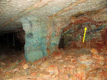
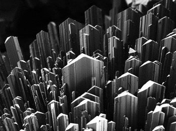
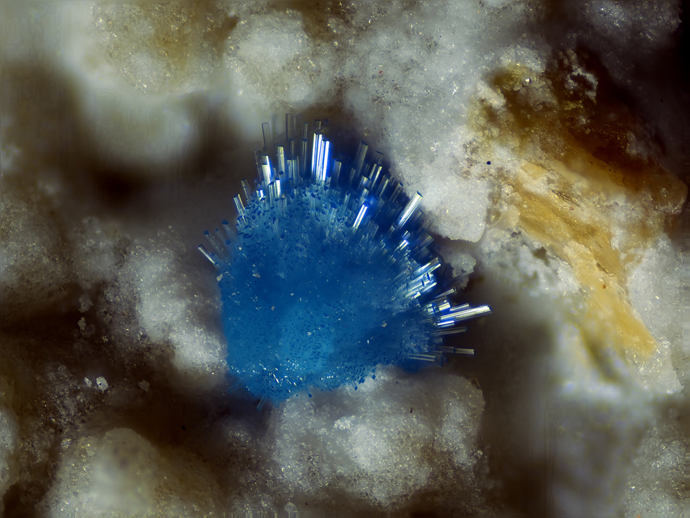
The specimen studied (GF6566; now registered in the collections of Museums Victoria, specimen number M53971) was collected in the early 1990's in Hall D of the South mine, Gap Garonne. The mineralized face, parallel to the mine wall, was located near the top of the conglomerate layer, at the level of the mine roof (Fig. 1). It was lined with mansfieldite‒scorodite in botryoidal crusts or clusters of brownish distinct crystals. Ceruleite occurs as isolated sky blue balls up to 1 mm in diameter. Individual crystals may reach ~100 µm × 20 µm × 5 µm, which are of by far the best quality for the species so far reported (Fig. 2). Their lustre is glassy, owing to the thickness of the constituent needles, contrasting with the usual velvety appearance of the mineral at the mine. Sometimes, thick transparent needles protrude from the balls (Fig. 3), creating the aspect of a naval mine. Other minerals collected in the vicinity include: bariopharmacoalumite, forêtite and olivenite. The occurrence of bariopharmacoalumite, forêtite and mansfieldite/scorodite illustrates the unusual extent of incorporation of Al into the secondary arsenates of this mineral assemblage.
Copper (+aluminium) rich mineralization in Hall D, South mine, Cap Garonne. The location of the ceruleite mineralization is arrowed.

Secondary electron microscopy photograph of ceruleite blades showing prominent striations on the {001} face. Field of view = 100 µm across.

Cluster of ceruleite crystals on mansfieldite. Field of view = 1.3 mm across.

Experimental
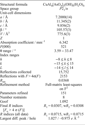
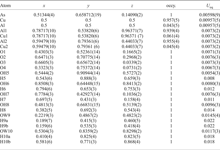
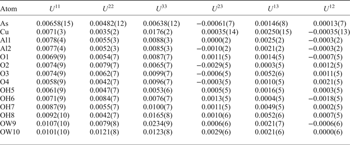
Several crystals were examined by qualitative energy-dispersive spectroscopy, which showed the presence of only Cu, Al and As as major cations, consistent with the major constituents for ceruleite. A 20 µm × 1 µm × 1 µm blade of ceruleite was mounted on a nylon loop to minimize diffraction from the sample mount for an X-ray data collection on the micro-focus macromolecular beam line MX2 of the Australian Synchrotron. Data were collected at 100 K using an ADSC Quantum 315r detector and monochromatic radiation with a wavelength of 0.71080 Å. A φ scan was employed with frame widths of 1° and a counting time per frame of 1 s. The data was integrated in P  $\bar 1$ using XDS (Kabsch, Reference Kabsch2010). XPREP (Bruker, 2001) gave a space group of P2/n consistent with the E-statistics, |E 2–1| = 0.897, which indicated a centrosymmetric space group. The absorption correction was carried out with SADABS (Bruker, 2001), giving 2166 unique reflections with an R int of 0.0368. A partial model was found using SHELXT (Sheldrick, Reference Sheldrick2015), which gave an R 1 of 0.080. Missing atoms were then located via subsequent difference-Fourier syntheses during the refinement, performed using SHELXL-97 (Sheldrick, Reference Sheldrick2008). All hydrogen atoms were located via calculation of the bond-valence sums on the oxygen atoms, consideration of non-bonded O···O distances which might indicate the presence of hydrogen bonds, and careful inspection of the difference-Fourier maps. Hydrogen atoms were softly constrained to a distance of 0.90(3) Å and a U eq 1.5× that of its corresponding oxygen atom. Details concerning data collection and structure refinement are provided in Table 1. Fractional coordinates and atomic displacement parameters are provided in, respectively, Table 2 and Table 3.
Crystal data and structure refinement for ceruleite.

Atomic coordinates and equivalent isotropic displacement parameters and site-occupancy factors for ceruleite.

Anisotropic displacement parameters for ceruleite.

Crystal structure
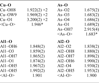
The crystal structure of ceruleite contains Cu in square planar coordination with 2 × OH8 at 1.922 Å and 2 × OW9 at 1.972 Å. Additional potential ligands 2 × O1 are in positions appropriate to complete a very elongated octahedron around the Cu atom, but at a distance (3.200 Å) so long that any interaction is very weak (0.016 valence units, using the bond-valence parameters of Brese and O'Keeffe, Reference Brese and O'Keeffe1991). Two symmetrically distinct Al atoms are both surrounded by 2 × O2– and 4 × OH– in rather regular octahedra with Al–O = 1.838–1.992 Å. Scattering factors suggest that there is ~5% substitution of Cu at Al sites and vice versa, as shown by the occupancy data in Table 2. Arsenic is surrounded by a tetrahedron of 4 × O2– at 1.675–1.689 Å. Interestingly, a fifth potential ligand (OH7) is situated beyond the centre of one face of the tetrahedron, but again at a distance so long (2.915 Å) that it cannot be considered significantly bonded (0.045 vu). Bond distances for the non-hydrogen cations are shown in Table 4.
Bond distances in Å for Cu, Al and As in ceruleite.

*Mean of four shortest bonds only
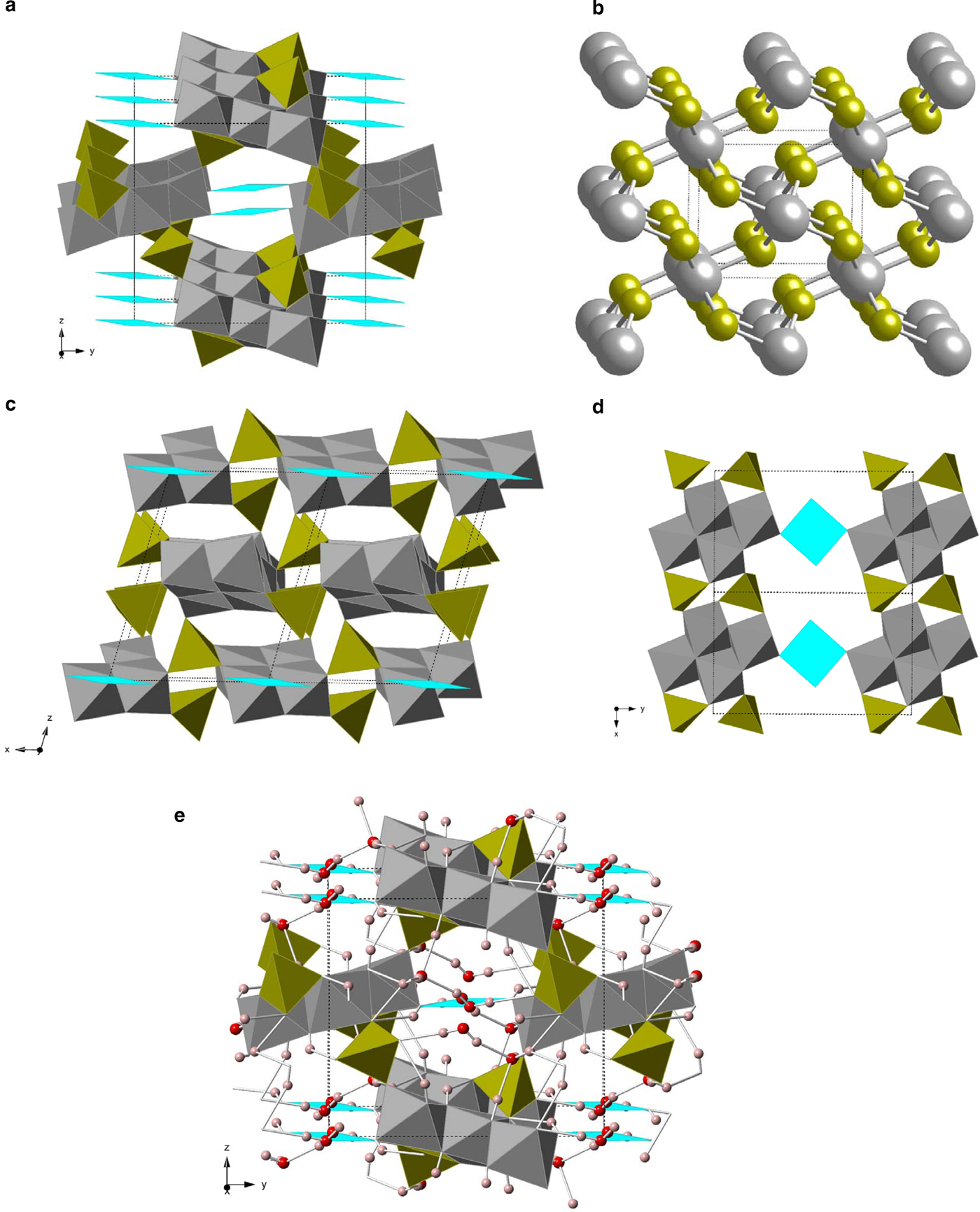
The Al(O,OH)6 octahedra share edges to form rhombus-shaped tetramers. AsO4 tetrahedra share two corners with one such rhombus and the other two corners with each of two other rhombi, linking them into a very open mesoporous framework (Fig. 4a). If the Al4 rhombi are considered as single units, then each As links three rhombi and each rhombus links six AsO4 tetrahedra, with a topology similar to that of Fe and S atoms in marcasite, FeS2 (Fig. 4b,c). The densest linkage of rhombi and tetrahedra is along the a direction, where Al–Al–As 3-rings and Al–As–Al–As 4-rings link rhombi into ribbons that run || a and are flattened on approximately (001) (Fig. 4d). This structural anisotropy presumably accounts for the fibrous-bladed habit of the crystals. There are large channels || a between these ribbons, but trans hydroxide corners of Cu(OH)2(H2O)2 squares lie in the channels and link Al4 rhombi along || b. The H2O molecules are also in the channels, with OW9 bound to Cu but OW10 held in place only by hydrogen bonds (Fig. 4e). Other hydrogen bonds decorate the sides of the channels but do not play an essential role in holding the framework together. As far as we can tell, there are no strong topological similarities to any other structures reported for arsenate, phosphate or sulfate minerals, or for that matter, any compound in the Inorganic Crystal Structure Database.
(a) Polyhedral framework of ceruleite structure, viewed nearly parallel to a. Al(O,OH)6 octahedra are grey, AsO4 tetrahedra are olive and Cu(OH,H2O)4 squares are cyan. (b) Structure of marcasite (FeS2) shown in ball-and-stick format using the same colour scheme as (a), showing topological equivalence between the network of Al4 tetramers and As in ceruleite, and Fe and S in marcasite. (c) Framework viewed nearly parallel to b, emphasizing 6:3 coordination pattern of Al4 tetramers and As tetrahedra. (d) Layer of framework between 0.25 < z < 0.75, showing dense Al4As2 ribbons running || a, connected by Cu squares. (e) Complete ceruleite structure in the same orientation as (a), with H atoms shown as small pink spheres and O atoms of H2O as larger red spheres.

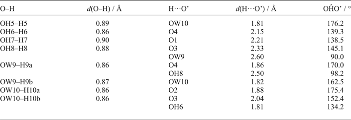
As noted above, bond valences calculated for the structure excluding hydrogen were used to identify O2–, OH– and H2O species. Non-bonded O…O distances were then used to identify probable hydrogen bonds, and electron density residua corresponding to H atoms were found close to all the positions where they would be predicted from the geometry of the other ligands around the associated oxygen atom(s). All primary O–H distances were in the range 0.85–0.90 Å, and the H–O–H bond angles for the two H2O molecules were 111(4)° and 102(4)° for OW9 and OW10 respectively. After a final stage of refinement, the hydrogen bond geometries were as shown in Table 5, acknowledging that all parameters have rather large attendant uncertainties (e.g. 0.02–0.03 Å for O–H distances).
Hydrogen bond geometry for ceruleite.

There were no H···O distances in the 2.60–2.69 Å range, but several more at 2.69–2.80 Å, which have calculated bond valences <0.03 valence units (vu) and are interpreted to be non-bonded. Hence, a cut-off distance of 2.6 Å was used for H···O bonds. Every hydrogen atom was found to form one strong hydrogen bond with H···O = 1.80–2.33 Å. For H8, H9a and H10b, a second weak hydrogen bond also occurred, with H···O >2.5 Å. The short H-bonds showed a range of O–H···O angles of 138–177°, from strongly bent to nearly linear, while the long bonds made much smaller angles of 90–134° with the primary O–H bond. Inclusion of the H bonds of Table 5 gave all O atoms a coordination number (CN) of 4, with cations arranged in an approximate tetrahedron, except for O2 (CN = 3, plane triangular) and OH8 and OW9 (CN = 3 + 1, with weak H···O links at > 2.5 Å located almost coplanar with the other ligands).
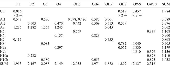
Bond valences were calculated from interatomic distances using the parameters of Brese and O'Keeffe (Reference Brese and O'Keeffe1991) except for H–O bonds, where the parameterization of Yu et al. (Reference Yu, Xue and Ratajczak2006) was used, with r 0 = 0.79 Å for primary bonds of length <1.05 Å, but r 0 = 1.409 Å otherwise. Table 6 shows that bond-valence sums on the large cations Cu, Al and As are all within 3% of their ideal values. The spread of bond-valence sums for oxygen atoms is broader, but all are still within 9% of 2 vu except for OW10, which is somewhat overbonded at ~2.3 vu. This is probably due to errors in the locations of donor hydrogen atoms H5 and H9b. Small displacements of those would weaken their hydrogen bonds to OW10, reducing the overbonding on that oxygen atom and also on H5 and H9b themselves (cf. Table 6). Even bond-valence sums for the hydrogen atoms are all acceptably close to ideal at 0.90–1.18 vu.
Bond valences for ceruleite (in valence units).

Discussion
Dufet (Reference Dufet1900) first reported ceruleite from the Emma Luisa gold mine, Guanaco, Antofagasta province, Antofagasta region, Chile, and gave a formula CuAl4(AsO4)2(OH)8·4–4.5H2O, which was later revised to Cu2Al7(AsO4)4(OH)13·11.5H2O by Schmetzer et al. (Reference Schmetzer, Berdesinski, Bank and Kroužek1976) on the basis of their wet-chemical analysis. It is interesting to note that the formula corresponding to the structure determination of the present study represents a revision back to the simpler stoichiometry CuAl4(AsO4)2(OH)8·4H2O, confirming the accuracy of the original analysis by Dufet (Reference Dufet1900).
Acknowledgements
Pierre Clolus is thanked for taking the colour photo and Vincent Bourgoin (Association Jean Wyart, Université Pierre et Marie Curie, Paris) for the SEM photo.