Introduction
Noonan syndrome is an autosomal dominant RASopathy with an estimated prevalence of 1 in 1,000–2,500 live births, caused by germline gain-of-function variants in genes encoding components of the Renin-Angiotensin System/Mitogen-Activated Protein Kinase (RAS/MAPK) signalling pathway. Reference Romano, Allanson and Dahlgren1,Reference Roberts, Allanson, Tartaglia and Gelb2 To date, pathogenic variants in at least thirteen genes have been identified as causative, with approximately 50% of Noonan syndrome cases attributable to gain-of-function mutations in the PTPN11 gene, which encodes the protein tyrosine phosphatase SHP2. RAF1 variants are among the more common genetic causes, accounting for approximately 5% of Noonan syndrome cases, and are disproportionately associated with severe cardiac disease, particularly hypertrophic cardiomyopathy. Approximately 60–80% of cases arise from de novo mutations. Reference Tartaglia, Gelb and Zenker3,Reference Bhambhani and Muenke4
The clinical phenotype of Noonan syndrome is broad, encompassing distinctive craniofacial features, postnatal short stature, webbed neck, chest wall deformities, congenital cardiac defects, lymphatic anomalies, renal malformations, variable neurodevelopmental involvement, and an increased predisposition to malignancy. Reference Roberts, Allanson, Tartaglia and Gelb2 Cardiovascular disease is the dominant source of morbidity and early mortality. Hypertrophic cardiomyopathy occurs in 22–29% of Noonan syndrome patients overall but carries a substantially higher prevalence in specific genotypes. Reference Hickey, Mehta and Elmi5 Overall survival in Noonan syndrome-associated hypertrophic cardiomyopathy is approximately 71% at three years, with a further phase of late mortality commencing after 10 years of age. Reference Hickey, Mehta and Elmi5 In infants under six months of age presenting with congestive heart failure, one-year survival falls to as low as 31%. Reference Wilkinson, Lowe and Salbert6
Genotype-phenotype correlations in Noonan syndrome are most pronounced for cardiac disease. Hypertrophic cardiomyopathy is present in approximately 75% of patients with pathogenic RAF1 variants—the highest rate among all Noonan syndrome-associated genes—and in approximately 56% of those with RIT1 variants. Reference Aoki, Niihori, Inoue and Matsubara7 The RAF1 p. Ser257Leu variant, identified in Case 2 of this report, is among the most frequently reported pathogenic variants in Noonan syndrome-associated hypertrophic cardiomyopathy and has been shown experimentally to confer increased kinase activity and elevated Mitogen-Activated Protein Kinase Kinase (MEK/MAP2K) and Extracellular Signal-Regulated Kinase (ERK) phosphorylation. Reference Aoki, Niihori, Inoue and Matsubara7,Reference Razzaque, Nishizawa and Komoike8 In contrast, PTPN11 mutations account for the majority of Noonan syndrome cases but are associated with hypertrophic cardiomyopathy in only 18–30% of patients. Nonetheless, severe hypertrophic obstructive cardiomyopathy does arise in the context of PTPN11 mutations and, when present, portends a clinically challenging course. Reference Gelb, Roberts and Tartaglia9
Conventional management of Noonan syndrome-associated hypertrophic obstructive cardiomyopathy relies on beta-blockers, calcium-channel blockers, disopyramide, and surgical septal myectomy in refractory cases. Reference Elliott, Anastasakis and Borger10 These approaches target the haemodynamic consequences of disease rather than its molecular basis, and a meaningful proportion of patients retain severe obstruction despite optimised medical and surgical management, representing a significant and unmet therapeutic need.
Trametinib is a highly selective, reversible allosteric inhibitor of MEK1/2, approved by the FDA for adult oncologic indications. In 2019, Andelfinger et al. first reported successful off-label use of trametinib in two infants with RIT1-related Noonan syndrome and severe early-onset hypertrophic obstructive cardiomyopathy, demonstrating marked regression of cardiac hypertrophy. Reference Andelfinger, Marquis and Raboisson11 Subsequently, a systematic review of 17 published cases across multiple Noonan syndrome genotypes reported short-term haemodynamic improvement in all patients treated with trametinib, with no deaths attributable directly to the drug. Reference De Brouchoven, Lorand and Bofferding12 Notably, RAF1-related Noonan syndrome—associated with the most severe hypertrophic cardiomyopathy phenotype—has been reported to respond to trametinib in several case series, including the seminal report by Meisner et al. (2021), in which biventricular hypertrophic cardiomyopathy and multifocal atrial tachycardia resolved with trametinib in a patient carrying the same RAF1 p. Ser257Leu variant as Case 2 in this report. Reference Meisner, Bradley and Russell13
Herein, we describe two 10-year-old patients with Noonan syndrome-associated hypertrophic obstructive cardiomyopathy—one carrying a confirmed PTPN11 variant and one carrying a confirmed RAF1 variant—who achieved rapid and significant haemodynamic improvement following initiation of trametinib. These cases provide complementary genotype-specific evidence supporting the clinical utility of mitogen-activated protein kinase kinase inhibition in Noonan syndrome-associated hypertrophic obstructive cardiomyopathy.
Case reports
Case 1 (PTPN11 c.1528C>G; p. Gln510Glu)
A 10-year-old male with genetically confirmed Noonan syndrome (PTPN11 c.1528C>G; p. Gln510Glu, identified by next-generation sequencing) was referred for evaluation of progressive exertional chest pain. His past medical history was notable for severe hypertrophic obstructive cardiomyopathy, pulmonary valve dysplasia, and prior right ventricular outflow tract obstruction. There was no family history of Noonan syndrome or cardiac disease. In 2017, he underwent complex cardiac surgery at a tertiary referral centre, comprising left ventricular septal myectomy, resection of right ventricular outflow tract muscle bundles, fenestration of intracavitary chordae, fenestration of the anterior papillary muscle of the mitral valve, and pulmonary valve commissurotomy. Prior to this admission, he was maintained on atenolol 20 mg twice daily.
On presentation, the patient was haemodynamically stable with normal vital signs. Physical examination revealed dysmorphic features consistent with Noonan syndrome, including low-set ears, widely spaced eyes, and a short webbed neck. Cardiovascular examination revealed a harsh systolic ejection murmur loudest at the left sternal border, with a palpable thrill. Respiratory, abdominal, and peripheral perfusion examinations were unremarkable. Laboratory investigations, including full blood count, renal function, liver enzymes, and cardiac biomarkers, were within normal limits. NT-proBNP measured 2,600 pg/mL prior to initiation of trametinib.
Twelve-lead electrocardiography demonstrated corrected QT interval (QTc) prolongation (QTc 510 ms) in addition to features of left ventricular hypertrophy. Given this finding and the known risk of corrected QT interval prolongation with atenolol in susceptible patients, beta-blocker therapy was transitioned from atenolol to nadolol, which offers comparable rate-limiting and anti-obstructive efficacy with a more favourable corrected QT interval profile.
Echocardiographic assessment demonstrated biventricular hypertrophy, predominantly involving the left ventricle, with marked interventricular septal thickening (IVSd approximately 21 mm) and a posterior wall thickness of 23 mm. A severe dynamic mid-cavitary left ventricular outflow tract obstruction was present at the subaortic level, associated with systolic anterior motion of the mitral valve, generating a peak gradient of approximately 130 mmHg and a mean gradient of 80 mmHg (Figure 1). The anterior mitral valve leaflet demonstrated prolapse with mild-to-moderate mitral regurgitation. The aortic valve was structurally normal with no regurgitation. The right ventricular outflow tract was patent with no residual obstruction. The pulmonary valve was dysplastic with severe free pulmonary regurgitation, consistent with prior commissurotomy. Left ventricular systolic function was preserved (ejection fraction approximately 75%). No pulmonary hypertension or pericardial effusion was identified. These findings were concordant with prior echocardiographic studies, confirming the absence of interval progression.
Case 1: Pre-treatment echocardiogram at day 1 demonstrating severe LVOT obstruction with SAM of the mitral valve (parasternal long-axis and 4-chamber views). Peak LVOT gradient ∼130 mmHg.

Figure 1. Long description
Panel A: A grayscale echocardiogram image showing a vertical cross-section of the heart. The image displays various heart structures with different shades of gray. Panel B: A color Doppler echocardiogram image showing a vertical cross-section of the heart. The image includes color-coded areas indicating blood flow patterns, with red and blue regions representing different flow directions. Panel C: A grayscale echocardiogram image showing another vertical cross-section of the heart from a different angle. The image displays various heart structures with different shades of gray. Panel D: A color Doppler echocardiogram image showing a vertical cross-section of the heart from a different angle. The image includes color-coded areas indicating blood flow patterns, with red and blue regions representing different flow directions.
In view of the severity of left ventricular outflow tract obstruction and the underlying gain-of-function PTPN11 variant conferring constitutive hyperactivation of the renin-angiotensin system/mitogen-activated protein kinase pathway, a decision was made to initiate targeted mitogen-activated protein kinase kinase inhibitor therapy. After multidisciplinary discussion and obtaining informed consent from the patient’s guardians, trametinib was commenced at a dose of 0.025 mg/kg/day. The medication was well tolerated throughout, with no observed adverse effects, including dermatologic, gastrointestinal, ophthalmologic, or haemodynamic complications. Following initiation of trametinib, the corrected QT interval was monitored serially and remained within normal limits (QTc 430 ms), with no further prolongation attributable to the combination of nadolol and trametinib.
A follow-up echocardiogram performed three days after initiation of trametinib demonstrated a significant and rapid haemodynamic response, with reduction of the left ventricular outflow tract peak gradient from approximately 130 mmHg to 40 mmHg (Figure 2). Repeat laboratory investigations remained within normal limits. NT-proBNP at follow-up measured 933 pg/mL at two-month follow-up, with interval regression of left ventricular wall thickness on echocardiography (interventricular septal thickness 19 mm, decreased from 21 mm at baseline; posterior wall thickness 21 mm, decreased from 23 mm at baseline) with persistence of the reduced left ventricular outflow tract gradient at approximately 40 mmHg (mean 28 mmHg) and no interval deterioration in biventricular function or valvular abnormalities.
Case 1: Post-treatment echocardiogram at day 3 demonstrating reduction of LVOT gradient to ∼40 mmHg (parasternal long-axis and 4-chamber views).

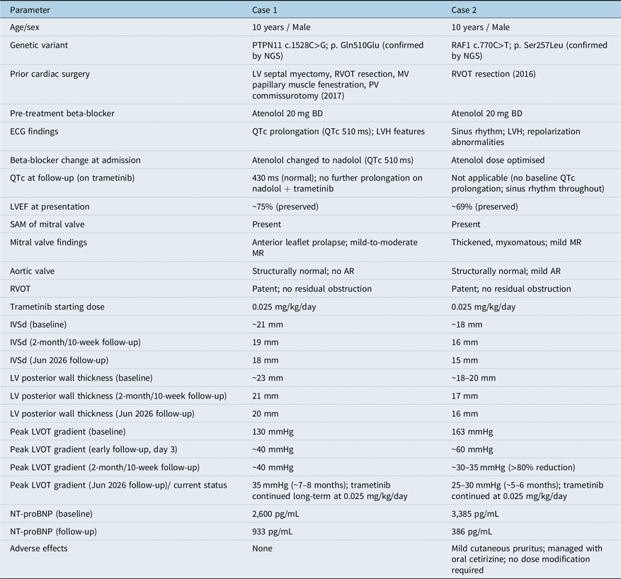
The patient has remained on continuous trametinib therapy at 0.025 mg/kg/day without interruption or dose adjustment. At the most recent follow-up in June 2026, approximately seven to eight months after treatment initiation, repeat echocardiography demonstrated further interval improvement. The left ventricular outflow tract peak gradient had decreased further to approximately 35 mmHg, and structural regression of hypertrophy was evident, with interventricular septal thickness reduced to 18 mm and posterior wall thickness reduced to 20 mm. Systolic anterior motion of the mitral valve was no longer evident, and mitral regurgitation had decreased to trace-to-mild in severity. The pulmonary valve remained dysplastic with severe free pulmonary regurgitation, unchanged from baseline and consistent with the patient’s prior surgical commissurotomy. Left ventricular systolic function remained preserved (ejection fraction approximately 70%), and there was no pericardial effusion or pulmonary hypertension. NT-proBNP at this follow-up had decreased further to 410 pg/mL. The corrected QT interval remained within normal limits on nadolol. No adverse effects have been observed with continued therapy (Table 1).
Summary of clinical, echocardiographic, and treatment characteristics

Table 1. Long description
A table comparing clinical, echocardiographic, and treatment characteristics of two cases. The table has 21 rows and 3 columns. Column headers are Case 1 and Case 2. Row labels include Age/sex, Genetic variant, Prior cardiac surgery, Pre-treatment beta-blocker, ECG findings, Beta-blocker change at admission, QTc at follow-up (on trametinib), LVEF at presentation, SAM of mitral valve, Mitral valve findings, Aortic valve, RVOT, Trametinib starting dose, IVSd (baseline), IVSd (2-month/10-week follow-up), IVSd (Jun 2026 follow-up), LV posterior wall thickness (baseline), LV posterior wall thickness (2-month/10-week follow-up), LV posterior wall thickness (Jun 2026 follow-up), Peak LVOT gradient (baseline), Peak LVOT gradient (early follow-up, day 3), Peak LVOT gradient (2-month/10-week follow-up), Peak LVOT gradient (Jun 2026 follow-up)/current status, NT-proBNP (baseline), NT-proBNP (follow-up), Adverse effects. Row 1: Age/sex, 10 years / Male, 10 years / Male. Row 2: Genetic variant, PTPN11 c.1528CG; p. Gln510Glu (confirmed by NGS), RAF1 c.770CT; p. Ser257Leu (confirmed by NGS). Row 3: Prior cardiac surgery, LV septal myectomy, RVOT resection, MV papillary muscle fenestration, PV commissurotomy (2017), RVOT resection (2016). Row 4: Pre-treatment beta-blocker, Atenolol 20mg BD, Atenolol 20mg BD. Row 5: ECG findings, QTc prolongation (QTc 510ms); LVH features, Sinus rhythm; LVH; repolarization abnormalities. Row 6: Beta-blocker change at admission, Atenolol changed to nadolol (QTc 510ms), Atenolol dose optimised. Row 7: QTc at follow-up (on trametinib), 430ms (normal); no further prolongation on nadolol trametinib, Not applicable (no baseline QTc prolongation; sinus rhythm throughout). Row 8: LVEF at presentation, 75% (preserved), 69% (preserved). Row 9: SAM of mitral valve, Present, Present. Row 10: Mitral valve findings, Anterior leaflet prolapse; mild-to-moderate MR, Thickened, myxomatous; mild MR. Row 11: Aortic valve, Structurally normal; no AR, Structurally normal; mild AR. Row 12: RVOT, Patent; no residual obstruction, Patent; no residual obstruction. Row 13: Trametinib starting dose, 0.025 mg/kg/day, 0.025 mg/kg/day. Row 14: IVSd (baseline), 21 mm, 18 mm. Row 15: IVSd (2-month/10-week follow-up), 19 mm, 16 mm. Row 16: IVSd (Jun 2026 follow-up), 18 mm, 15 mm. Row 17: LV posterior wall thickness (baseline), 23 mm, 18-20 mm. Row 18: LV posterior wall thickness (2-month/10-week follow-up), 21 mm, 17 mm. Row 19: LV posterior wall thickness (Jun 2026 follow-up), 20 mm, 16 mm. Row 20: Peak LVOT gradient (baseline), 130 mmHg, 163 mmHg. Row 21: Peak LVOT gradient (early follow-up, day 3), 40 mmHg, 60 mmHg. Row 22: Peak LVOT gradient (2-month/10-week follow-up), 40 mmHg, 30-35 mmHg (80% reduction). Row 23: Peak LVOT gradient (Jun 2026 follow-up)/current status, 35 mmHg (7-8 months); trametinib continued long-term at 0.025 mg/kg/day, 25-30 mmHg (5-6 months); trametinib continued at 0.025 mg/kg/day. Row 24: NT-proBNP (baseline), 2,600 pg/mL, 3,385 pg/mL. Row 25: NT-proBNP (follow-up), 933 pg/mL, 386 pg/mL. Row 26: Adverse effects, None, Mild cutaneous pruritus; managed with oral cetirizine; no dose modification required.
IVSd = interventricular septal thickness in diastole; LVOT = left ventricular outflow tract; LVEF = left ventricular ejection fraction; SAM = systolic anterior motion; MR = mitral regurgitation; AR = aortic regurgitation; PR/PI = pulmonary regurgitation/insufficiency; RVOT = right ventricular outflow tract; PA = pulmonary artery; PV = pulmonary valve; MV = mitral valve; LVH = left ventricular hypertrophy; NGS = next-generation sequencing; BD = twice daily; OD = once daily; QTc = corrected QT interval.
Case 2 (RAF1 c.770C>T; p. Ser257Leu)
A 10-year-old male with genetically confirmed Noonan syndrome (heterozygous RAF1 c.770C>T; p.(Ser257Leu)) identified by next-generation sequencing presented to the emergency department with complaints of palpitations and exertional chest pain. He carried an established diagnosis of Noonan syndrome and severe hypertrophic obstructive cardiomyopathy, for which he had undergone surgical resection of right ventricular outflow tract obstruction in 2016 at a tertiary cardiac centre. He had been maintained on atenolol 20 mg twice daily prior to this presentation. There was no reported family history of Noonan syndrome or cardiac disease; however, parental genetic testing to confirm de novo status of the RAF1 variant was not performed.
On presentation, the patient was haemodynamically stable. Physical examination demonstrated dysmorphic features consistent with Noonan syndrome, including hypertelorism, low-set ears, and a short neck. Auscultation revealed a loud systolic murmur consistent with left ventricular outflow tract obstruction. There were no signs of heart failure, and peripheral perfusion was intact. Laboratory investigations, including cardiac biomarkers, were unremarkable. NT-proBNP measured 3,385 pg/mL prior to initiation of trametinib.
Twelve-lead electrocardiography demonstrated sinus rhythm with features of left ventricular hypertrophy and repolarisation abnormalities. Echocardiographic assessment revealed severe diffuse left ventricular hypertrophy with a prominent mid-cavitary obstruction situated approximately 2.5–3 cm below the aortic annulus, with turbulent flow on colour Doppler imaging. The peak left ventricular outflow tract gradient measured 163 mmHg with a mean gradient of 73 mmHg. Interventricular septal thickness was approximately 18 mm in diastole and 24 mm at peak systole; posterior wall thickness ranged from 18 to 20 mm. Systolic anterior motion of the mitral valve was present (Figure 3). The mitral valve was thickened and myxomatous with accessory tissue; the annulus measured 24 mm with no stenosis and mild mitral regurgitation. The aortic valve was tricuspid with an annulus of 20 mm and mild aortic regurgitation. The previously operated right ventricular outflow tract was widely patent with no residual obstruction; the pulmonary valve annulus measured 22 mm with mild pulmonary insufficiency and a mean pulmonary artery pressure of 12 mmHg, with no significant pulmonary hypertension. Left ventricular systolic function was preserved (ejection fraction approximately 69%). No pericardial effusion was identified.
Case 2: Pre-treatment echocardiogram at day 1 demonstrating severe LVOT obstruction with SAM of the mitral valve (parasternal long-axis and 4-chamber views). Peak LVOT gradient 163 mmHg.

Given the severity of left ventricular outflow tract obstruction in the context of prior right ventricular outflow tract surgery and ongoing beta-blocker therapy, targeted mitogen-activated protein kinase kinase inhibitor therapy was initiated following multidisciplinary review and informed consent. Atenolol dosage was optimised, and trametinib was commenced at 0.025 mg/kg/day. The medication was well tolerated throughout; however, the patient developed mild cutaneous pruritus that resolved with oral cetirizine.
A follow-up echocardiogram performed three days after initiation of trametinib demonstrated a significant and rapid haemodynamic response, with a reduction of the left ventricular outflow tract peak gradient decreased from 163 mmHg to approximately 60 mmHg, with a reduction in mean gradient to 36–40 mmHg (Figure 4). With continued therapy, further improvement was observed, with the peak gradient declining to approximately 30–35 mmHg in later follow ups—a greater than 80% reduction from baseline over approximately ten weeks. Echocardiography at this ten-week assessment also demonstrated interval regression of left ventricular wall thickness (interventricular septal thickness 16 mm in diastole, decreased from 18 mm at baseline; posterior wall thickness 17 mm, decreased from 18 to 20 mm at baseline). NT-proBNP at follow-up measured 386 pg/mL.
Case 2: Post-treatment echocardiogram at day 3 demonstrating a reduction of LVOT gradient to ∼30–35 mmHg (parasternal long-axis and 4-chamber views).

Figure 4. Long description
Panel A: A grayscale echocardiogram image showing a parasternal long-axis view of the heart. The image displays various heart structures, including the left ventricle and left atrium. Panel B: A color Doppler echocardiogram image showing the same parasternal long-axis view with color flow mapping. The colors red and blue indicate blood flow direction and velocity. Panel C: A grayscale echocardiogram image showing a 4-chamber view of the heart. This view displays the left and right ventricles, as well as the left and right atria. Panel D: A color Doppler echocardiogram image showing the 4-chamber view with color flow mapping, highlighting blood flow through the heart chambers.
The patient has remained on continuous trametinib therapy at 0.025 mg/kg/day without interruption. At the most recent follow-up in June 2026, approximately five to six months after treatment initiation, echocardiography demonstrated further interval improvement. The left ventricular outflow tract peak gradient had decreased further to approximately 25–30 mmHg, and structural regression of hypertrophy was evident, with interventricular septal thickness reduced to 15 mm in diastole and posterior wall thickness reduced to 16 mm. The mitral valve remained mildly thickened with trace-to-mild mitral regurgitation, and mild aortic regurgitation persisted unchanged. Left ventricular systolic function remained preserved (ejection fraction approximately 68%), with no pulmonary hypertension and no pericardial effusion. NT-proBNP at this follow-up had decreased further to 210 pg/mL. The cutaneous pruritus noted earlier in treatment had fully resolved, and no further adverse effects have been observed with continued therapy (Table 1).
Discussion
The two cases presented herein add meaningfully to the growing literature on trametinib as a targeted disease-modifying therapy for Noonan syndrome-associated hypertrophic obstructive cardiomyopathy. Together, they cover two of the most clinically important Noonan syndrome genotypes—PTPN11 and RAF1—in older paediatric patients with established, surgically treated hypertrophic obstructive cardiomyopathy who had failed conventional medical management. The rapid and substantial haemodynamic improvement observed in both cases, in the absence of adverse effects, strengthens the case for mitogen-activated protein kinase kinase inhibition as a viable therapeutic strategy across the molecular spectrum of Noonan syndrome-associated hypertrophic obstructive cardiomyopathy.
Molecular basis of Noonan syndrome-associated hypertrophic obstructive cardiomyopathy: PTPN11 and RAF1
Both PTPN11 and RAF1 encode proteins that function within the renin-angiotensin system/mitogen-activated protein kinase signalling cascade, and gain-of-function variants in either gene result in constitutive downstream MEK-ERK hyperactivation—the shared molecular driver of cardiomyocyte hypertrophy in Noonan syndrome.
PTPN11 encodes SHP2, a non-receptor protein tyrosine phosphatase that acts upstream of Renin–Angiotensin System (RAS) as a positive regulator of Mitogen-Activated Protein Kinase (MAPK) signalling by dephosphorylating inhibitory tyrosine residues on renin–angiotensin system effectors, thereby amplifying downstream mitogen-activated protein kinase kinase and extracellular signal-regulated kinase activity. Reference Leegaard, Gregersen, Nielsen, Bjerre and Handrup14 Gain-of-function PTPN11 variants—such as p. Gln510Glu in Case 1—result in constitutive SHP2 activation, sustaining elevated phospho-ERK levels in cardiomyocytes. Although hypertrophic cardiomyopathy is less prevalent in PTPN11-related Noonan syndrome (18–30%) than in RAF1-related disease, the severity of obstruction can be comparable when it does occur, as demonstrated by Case 1’s peak left ventricular outflow tract gradient of 130 mmHg despite prior surgical myectomy. The PTPN11 c.1528C>G; p. Gln510Glu variant identified in Case 1 maps to the protein tyrosine phosphatase domain of SHP2, distinct from the N-SH2 and C-SH2 domains in which the majority of Noonan syndrome-associated PTPN11 variants cluster. Protein tyrosine phosphatase-domain variants are less frequently reported than N-SH2 variants but have been shown to similarly relieve the autoinhibited conformation of SHP2, producing constitutive catalytic activation and sustained downstream MEK-ERK signalling. Reference Aoki, Niihori, Inoue and Matsubara7 The p. Gln510Glu variant itself has been reported only sporadically in the Noonan syndrome literature and has not, to our knowledge, been subjected to dedicated functional validation; its classification as gain-of-function is therefore inferred from its domain location and from the clinical phenotype observed in this patient, rather than confirmed by in vitro kinase assay. This represents a limitation in the molecular characterisation of Case 1 and highlights a need for functional studies of less common PTPN11 variants identified in Noonan syndrome-associated hypertrophic obstructive cardiomyopathy.
RAF1 encodes Raf-1 proto-oncogene serine/threonine kinase (MAP3K), which functions directly downstream of renin–angiotensin system and phosphorylates MEK1/2, the immediate upstream activators of ERK1/2. Reference Aoki, Niihori, Inoue and Matsubara7,Reference Razzaque, Nishizawa and Komoike8 The RAF1 c.770C>T; p. Ser257Leu variant identified in Case 2 is a recurrent, well-characterised pathogenic variant clustered in the CR2 domain of RAF1, which has been demonstrated to produce elevated kinase activity, increased mitogen-activated protein kinase kinase and extracellular signal-regulated kinase phosphorylation, and altered downstream signalling in cardiomyocytes consistent with cardiac hypertrophy. Reference Aoki, Niihori, Inoue and Matsubara7,Reference Razzaque, Nishizawa and Komoike8 This variant was first reported by Razzaque et al. in 2007 in four of thirty Noonan syndrome patients, two of whom had severe hypertrophic cardiomyopathy, and has since been documented in numerous unrelated individuals with Noonan syndrome-associated hypertrophic cardiomyopathy in multiple cohorts. Reference Razzaque, Nishizawa and Komoike8 Importantly, hypertrophic cardiomyopathy is present in approximately 95% of patients with pathogenic RAF1 variants—the highest gene-specific hypertrophic cardiomyopathy prevalence in Noonan syndrome—underscoring the direct pathogenic role of RAF1 in myocardial hypertrophy. Reference Aoki, Niihori, Inoue and Matsubara7
Preclinical evidence directly supports mitogen-activated protein kinase kinase inhibition as a targeted approach for RAF1-mediated hypertrophic cardiomyopathy. Wu et al. demonstrated in a knock-in mouse model of Noonan syndrome carrying the Raf1 L613V variant that postnatal MEK-ERK inhibition ameliorated both the cardiac hypertrophy and morphological phenotype of the disease. Reference Wu, Simpson and Hong15 In vitro studies using induced pluripotent stem cell-derived cardiomyocytes from patients with Noonan syndrome-associated gain-of-function variants confirmed that individual pathogenic variants are both necessary and sufficient to induce cardiomyocyte hypertrophy and that pharmacological mitogen-activated protein kinase kinase inhibition attenuates this effect. Reference Gelb, Roberts and Tartaglia9 These converging preclinical data provide the biological rationale for trametinib’s efficacy in both PTPN11- and RAF1-related Noonan syndrome-associated hypertrophic obstructive cardiomyopathy.
RAF1-specific clinical evidence for trametinib
The RAF1 genotype in Case 2 is particularly well-represented in the published trametinib literature, providing a strong reference framework for the response observed. In the systematic review by De Brouchoven et al. (2025), two of the seventeen reviewed cases carried the RAF1 c.770C>T; p. Ser257Leu variant—precisely the same variant identified in Case 2 of the present report. Reference De Brouchoven, Lorand and Bofferding12 One of these patients, reported by Meisner et al. (2021), was a 20-week-old infant with biventricular hypertrophic cardiomyopathy, pulmonary valve stenosis, and multifocal atrial tachycardia; trametinib at 0.025 mg/kg/day resulted in complete resolution of multifocal atrial tachycardia within 72 hours and significant improvement of biventricular hypertrophic cardiomyopathy over six months. Reference Meisner, Bradley and Russell13 A second RAF1 p. Ser257Leu patient reported by Mussa et al. (2021) was a preterm neonate with severe biventricular obstructive hypertrophic cardiomyopathy who showed transient improvement in cardiac function and reduction in septal thickness on trametinib before dying of pulmonary capillary hemangiomatosis—a cause of death considered unrelated to trametinib. Reference De Brouchoven, Lorand and Bofferding12
Case 2 extends these observations to an older child with established RAF1-related hypertrophic obstructive cardiomyopathy who had already undergone surgical right ventricular outflow tract intervention and remained on optimised beta-blocker therapy, yet carried a severely elevated peak left ventricular outflow tract gradient of 163 mmHg. The greater than 80% gradient reduction achieved with trametinib over ten weeks in this patient—without dose escalation or adverse effects—represents one of the most substantial reported haemodynamic responses to mitogen-activated protein kinase kinase inhibition in the RAF1 genotypic subgroup and in older paediatric patients.
PTPN11-specific clinical evidence and comparison
While RAF1-related hypertrophic obstructive cardiomyopathy has a well-established response profile to trametinib, evidence for PTPN11-related disease is more limited. Pascarella et al. (2024) described a neonate with PTPN11 p. Phe285Ser-related Noonan syndrome who exhibited reduction in left ventricular posterior wall thickness after two months of trametinib, with no recurrence of chylothorax at 18-month follow-up. Reference Pascarella, Limongelli and De Falco16 Nakano et al. (2022) documented reduction in left ventricular mass and NT-proBNP in a child carrying the same variant. Reference Nakano, Rankin and Annam17 Case 1 extends the PTPN11-specific evidence base by demonstrating a rapid and dramatic reduction in left ventricular outflow tract gradient—from 130 to 40 mmHg within three days—in a 10-year-old with post-surgical hypertrophic obstructive cardiomyopathy, a patient profile not previously described.
The speed of gradient reduction in Case 1 is particularly striking and warrants mechanistic consideration. Structural regression of myocardial hypertrophy is generally a slow process, typically unfolding over months to years. Reference Andelfinger, Marquis and Raboisson11,Reference De Brouchoven, Lorand and Bofferding12 A gradient reduction of this magnitude within three days is more consistent with a functional effect of mitogen-activated protein kinase kinase inhibition on left ventricular outflow tract dynamics—specifically, reduction of dynamic outflow obstruction through decreased myocardial hypercontractility and improved diastolic relaxation—rather than a structural change. This is supported by evidence that extracellular signal-regulated kinase signalling modulates intracellular calcium handling and myofilament calcium sensitivity, both of which contribute to the dynamic nature of left ventricular outflow tract obstruction in hypertrophic cardiomyopathy. Reference Chaput and Andelfinger18
Dosing, treatment duration, and monitoring
Both patients received trametinib at approximately 0.025 mg/kg/day, consistent with the most commonly used starting dose in the paediatric Noonan syndrome literature Reference De Brouchoven, Lorand and Bofferding12 and with recommendations from paediatric oncology phase I studies. Reference Geoerger, Moertel and Whitlock19 This dosage is lower than that used in oncologic indications—a mechanistically justified distinction, as germline RASopathy mutations generally confer a lower magnitude of pathway hyperactivation than somatic cancer mutations, and the therapeutic goal is normalisation rather than ablation of mitogen-activated protein kinase signalling. Reference De Brouchoven, Lorand and Bofferding12,Reference Chaput and Andelfinger18 In both cases, no dose escalation was required, suggesting that 0.025 mg/kg/day may be an appropriate starting and maintenance dose for hypertrophic obstructive cardiomyopathy in this age group.
The optimal treatment duration for Noonan syndrome-associated hypertrophic obstructive cardiomyopathy remains undefined. Attempts at treatment cessation have been associated with haemodynamic relapse in some reported cases, including recurrence of elevated left ventricular outflow tract gradients and rising NT-proBNP levels necessitating re-treatment. Reference Leegaard, Gregersen, Nielsen, Bjerre and Handrup14,Reference Pascarella, Limongelli and De Falco16 Wolf et al. (2024) reported in a retrospective cohort of 30 infants that, following extended trametinib treatment, sustained cardiac and clinical improvement was maintained after treatment weaning in five patients without relapse. Reference Wolf, Zenker and Boleti20 Based on current evidence, a pragmatic approach involves continuing therapy until sustained cardiac remodelling is documented, typically 3 months to 2 years, followed by gradual weaning with close echocardiographic and biomarker surveillance.
Both patients in this series demonstrate a clinically important pattern of continued improvement over time. With continued, uninterrupted trametinib therapy, both patients demonstrated progressive improvement beyond the initially documented response. In Case 1, the left ventricular outflow tract peak gradient decreased further from 40 mmHg at two months to approximately 35 mmHg at seven to eight months, with interventricular septal thickness decreasing from 19 to 18 mm and posterior wall thickness from 21 to 20 mm over the same interval. In Case 2, the gradient decreased further from 30 to 35 mmHg at ten weeks to approximately 25–30 mmHg at five to six months, with interventricular septal thickness decreasing from 16 to 15 mm and posterior wall thickness from 17 to 16 mm over the same interval. In both cases, the haemodynamic improvement observed acutely within days of treatment initiation was therefore followed by a second, slower phase of incremental functional and structural improvement over subsequent months, consistent with the two-phase model proposed below.
This biphasic pattern of response is, in our view, one of the more clinically informative observations of this report. It suggests that trametinib’s haemodynamic benefit in Noonan syndrome-associated hypertrophic obstructive cardiomyopathy arises through two distinct, temporally separated mechanisms: an early functional effect, evident within days and most plausibly mediated by extracellular signal-regulated kinase-dependent modulation of myocardial contractility and calcium handling (as discussed in Section PTPN11-Specific Clinical Evidence and Comparison), followed by a slower, structural effect over subsequent months as sustained MEK-ERK inhibition permits gradual regression of myocardial hypertrophy itself. The continued incremental gains seen in both patients between the early and extended follow-up assessments are consistent with ongoing structural remodelling rather than a plateaued functional ceiling. This has direct implications for clinical practice and the design of future studies: the full benefit of trametinib in Noonan syndrome-associated hypertrophic obstructive cardiomyopathy may not be apparent from early follow-up alone, and continued therapy over a period of months should be anticipated to yield further gains in both gradient reduction and structural regression, supporting its use as a sustained, disease-modifying therapy rather than a short-course intervention.
Safety and tolerability
Trametinib was well tolerated in both patients. Case 1 experienced no adverse effects at any point during the follow-up period, while Case 2 developed mild, self-limiting cutaneous pruritus that resolved with oral antihistamine therapy and did not require dose modification. This is consistent with the published Noonan syndrome literature, in which non-critical adverse events—most commonly cutaneous toxicity (eczema, acneiform rash), diarrhoea, and gastrointestinal symptoms—were reported in 7 of 17 reviewed cases, predominantly at doses ≥ 0.025 mg/kg/day, and led to dose reduction or discontinuation in only a minority. Reference De Brouchoven, Lorand and Bofferding12 Adverse effects in Noonan syndrome patients receiving trametinib appear less severe than those observed in adult oncology patients, attributed to the lower doses used, the absence of combination chemotherapy, and the fundamental difference in therapeutic intent: in RASopathies, mitogen-activated protein kinase kinase inhibition aims to normalise signalling rather than ablate it. Reference De Brouchoven, Lorand and Bofferding12,Reference Chaput and Andelfinger18
Three deaths were reported across the published Noonan syndrome trametinib literature; none were considered directly attributable to drug toxicity. Reference De Brouchoven, Lorand and Bofferding12 Nonetheless, given the off-label nature of trametinib in this population, structured monitoring is advisable throughout treatment, incorporating serial echocardiography, NT-proBNP measurement, skin and ophthalmologic examination, and hepatic and haematological bloodwork, as outlined in the protocol proposed by Nakano et al. Reference Nakano, Rankin and Annam17
Clinical implications
Both patients in this series had undergone prior surgical right ventricular outflow tract intervention and were receiving optimised beta-blocker therapy, yet continued to carry severe, symptomatic left ventricular outflow tract obstruction. This illustrates the persistence of haemodynamically significant residual disease after current standard-of-care strategies and the clinical imperative for molecularly targeted approaches. The response to trametinib in both cases—spanning two distinct genotypes, two different mechanisms of renin-angiotensin system/mitogen-activated protein kinase pathway activation, and different clinical presentations—suggests that mitogen-activated protein kinase kinase inhibition is effective regardless of the specific upstream molecular lesion, as long as it converges on mitogen-activated protein kinase kinase hyperactivation.
The fact that Case 2’s RAF1 genetic result was confirmed only after treatment initiation also highlights an emerging clinical reality: in patients with a strong clinical Noonan syndrome diagnosis and life-limiting hypertrophic obstructive cardiomyopathy, it may be appropriate to initiate trametinib while genetic results are pending, particularly when conventional management has been exhausted. However, we strongly advocate that comprehensive molecular genetic testing be completed in all such patients, both to validate the diagnosis, inform prognosis, and allow genotype-specific therapeutic guidance.
The evidence base for trametinib in Noonan syndrome, while expanding, remains predominantly derived from uncontrolled case reports and series subject to publication bias. Prospective, multicentre clinical trials with standardised dosing protocols, objective haemodynamic endpoints, biomarker assessment, and structured long-term follow-up are urgently required. International registries represent a critical interim measure for systematically accumulating real-world safety and efficacy data across the Noonan syndrome genotypic spectrum.
This report has several limitations that warrant explicit acknowledgement. First, although follow-up has now extended to seven to eight months in Case 1 and five to six months in Case 2 with continued structural and functional improvement, this remains short relative to the months-to-years timeframe over which Noonan syndrome-associated hypertrophic obstructive cardiomyopathy and its treatment typically evolve, and longer-term data will be needed to confirm whether the observed gains are maintained or further extended. Second, both cases are derived from a single-patient, retrospective, off-label treatment context without a control group, and causal attribution of haemodynamic and structural change to trametinib—rather than to natural disease fluctuation or other unmeasured factors—rests on temporal association and biological plausibility rather than controlled comparison. Third, the PTPN11 p. Gln510Glu variant in Case 1 lacks dedicated functional validation in the literature, introducing uncertainty into its molecular classification as gain-of-function. Finally, as a two-patient case report, these findings cannot be generalised to the broader Noonan syndrome population, and prospective multicentre data are required before firm conclusions can be drawn about optimal treatment duration, weaning strategy, or long-term safety.
Conclusion
We describe two paediatric patients with Noonan syndrome-associated severe hypertrophic obstructive cardiomyopathy—one harbouring a PTPN11 c.1528C>G; p. Gln510Glu variant and one harbouring a RAF1 c.770C>T; p. Ser257Leu variant—who achieved rapid and clinically significant haemodynamic improvement following initiation of trametinib, with further incremental functional and structural gains observed over subsequent months of continued therapy. Both patients had failed conventional pharmacological management and retained severe left ventricular outflow tract obstruction despite prior surgical intervention. Trametinib was well tolerated with no adverse effects in one case, with a mild rash in the second.
These cases provide complementary, genotype-specific evidence supporting the clinical utility of mitogen-activated protein kinase kinase inhibition across two mechanistically distinct Noonan syndrome genotypes. The shared therapeutic response to trametinib in PTPN11- and RAF1-related hypertrophic obstructive cardiomyopathy supports a pathway-level rather than gene-specific rationale for mitogen-activated protein kinase kinase inhibitor therapy in Noonan syndrome—reinforcing the concept of trametinib as a genotype-agnostic precision therapeutic for this condition. Comprehensive molecular genetic evaluation is strongly recommended in all Noonan syndrome patients with severe hypertrophic obstructive cardiomyopathy to guide therapeutic decision-making. Prospective multicentre trials and international registries are essential to establish optimal patient selection, dosing, treatment duration, and long-term outcomes.
Financial support
This research received no external funding.
Competing interests
The authors declare no conflicts of interest relevant to this publication.
Ethical standard
Informed consent was obtained from the guardians of both patients for off-label use of trametinib and for the use of anonymised clinical data for publication. Patient identities have been withheld to preserve confidentiality in accordance with institutional policy. The cases were managed in accordance with the principles of the Declaration of Helsinki.