Introduction
Nasal inverted papilloma was first described by Ward in 1854 and Billroth in 1855.Reference Ward1, Reference Billroth2 Inverted papillomas are benign neoplasms, but are characterised by: (1) locally aggressive and destructive behaviour; (2) a tendency to recur after removal; and (3) an association with squamous cell carcinoma (SCC). Inverted papillomas constitute 0.5–4 per cent of all nasal tumours.Reference Lawson, Ho, Shaari and Biller3 They usually arise from and involve the middle nasal meatus nasal septum, the maxillary and ethmoid sinuses.Reference Lawson, Ho, Shaari and Biller3
Formation of new bone within an inverted papilloma is extremely rare. To our knowledge, only two English language reports have previously been published, detailing a total of four Turkish cases.Reference Unlu, Songu, Ovali and Nese4, Reference Dadas, Ercan and Başak5 The current study reports a fifth, extremely rare case of new bone formation within an inverted papilloma, located in this instance in the ethmoid and frontal sinuses. The treatment of this case is discussed.
Case report
A 68-year-old man was referred to our department with a five-year history of headache and left-sided, purulent rhinorrhoea. This patient also had severe diabetes mellitus requiring insulin. There was no history of nasal surgery.
Nasal endoscopy demonstrated oedematous nasal polyps which appeared to be inflammatory. A pre-operative sinonasal computed tomography (CT) scan demonstrated opacification of the left nasal cavity and anterior ethmoid and frontal sinuses. However, there appeared within the lesion a significant high density region, with a ground glass like appearance (Figure 1). We initially diagnosed left chronic sinusitis accompanied by calcification or osteoma.
Pre-operative sinonasal images. (a) Coronal computed tomography scan demonstrating a bone-density lesion (indicating bone formation) within a soft tissue mass filling the left anterior ethmoid and frontal sinuses. (b) Coronal, gadolinium-enhanced, T1-weighted magnetic resonance imaging scan taken before the second surgical procedure, demonstrating a mass lesion.

Endoscopic sinus surgery was performed on the left side under general anaesthesia. The mucosae of the anterior ethmoid and frontal sinuses were polypous and prone to bleeding. The purulent contents of the sinus cells were drained, and a drainage tube was placed from the frontal sinus to the maxillary sinus. The posterior ethmoid and sphenoid sinuses were intact. A hard, bony mass with a rubbery surface texture was encountered; this mass was mobile, although manipulation resulted in movement of the tissues adjacent to the lamina papyracea and fovea ethmoidalis. Removal of the bony mass was attempted, but was judged to be too difficult and risky. There was no intra-operative suspicion of neoplastic disease. Therefore, the bony mass was left in situ, in order to avoid orbital and/or intracranial complications.
Six months later, nasal endoscopy demonstrated a rubbery, polypous mass obstructing the left middle nasal meatus. A biopsy at this time enabled a histopathological diagnosis of inverted papilloma. On serological examination, the level of SCC-related antigen (a tumour marker) was slightly elevated, at 1.7 ng/ml (normal range <1.5 ng/ml). The cytokeratin fragment level was within the normal range (i.e. <3.5 ng/ml). Magnetic resonance image (MRI) scanning delineated a mass lesion stereologically enhanced by gadolinium at sites compatible with the bony mass seen on CT (Figure 1b). The inverted papilloma was diagnosed as stage III because of the frontal sinus involvement, according to the Krouşe staging system.Reference Krouşe6, Reference Krouşe7
We decided to reoperate, using an endoscopic sinus surgery approach. Endoscopic frontoethmoidectomy was performed using a navigation system, under general anaesthesia. The intrafrontal portion of the mass was very carefully removed by pulling down into the nasal cavity. The mucosa of the frontoethmoid recess was intact. The inverted papilloma tissue surrounding the bony tissue was consequently removed. All the bone underlying the tumour attachments was carefully drilled to ensure that no mucosa was retained.
Histopathological examination of surgical specimens showed bony tissue within surrounding inverted papilloma (Figure 2a). The excised bony specimen showed mature and immature bony tissue adjacent to areas of inverted papilloma (Figure 2b). Osteoblasts were seen rimming irregularly organised bone trabeculae. These findings indicated new bone formation, as described in two previous reports.Reference Unlu, Songu, Ovali and Nese4, Reference Dadas, Ercan and Başak5 No malignant features were seen in any of the tissue sections of the resected mass (Figure 2c).
Histopathological analysis of surgical specimens. (a) Randomly organised, new, woven bone trabeculae (arrowheads) within inverted papilloma (arrows) (H&E; ×2). (b) Mature (arrowheads) and immature (asterisks) bony tissues lined by osteoblasts (arrows) (H&E; ×20). (c) Inverted papilloma without any malignant features (H&E; ×10).

The post-operative course was uneventful. No symptoms arose over the following six months. The SCC antigen level reduced to normal. Computed tomography and MRI scans taken six months after the second procedure showed no signs of any residual lesion or recurrence (Figure 3).
Sinonasal images taken six months after the second surgical procedure. No residual lesion or signs of recurrence were detected on (a) coronal computed tomography scan or (b) coronal, gadolinium-enhanced, T1-weighted magnetic resonance imaging scan.

Discussion
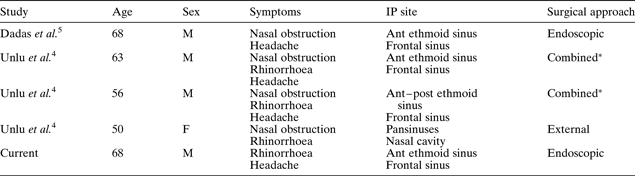
We report an extremely rare case of inverted papilloma with new bone formation, within the anterior ethmoid and frontal sinuses. This is only the fifth such case reported in the English literature (see Table I).Reference Unlu, Songu, Ovali and Nese4, Reference Dadas, Ercan and Başak5 This case highlights the existence of tumour-induced osteogenesis.
Reported cases of sinonasal inverted papilloma with osteogenesis
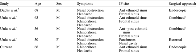
* Combined = endoscopic + external approach. IP = inverted papilloma; M = male; F = female; ant = anterior; post = posterior
In the initial pre-operative stage, we failed to correctly diagnose our patient's bony lesion, believing it to represent unilateral chronic sinusitis with calcification or osteoma. In hindsight, the pre-operative CT images should have been evaluated more carefully. Had we been more aware of tumour-induced osteogenesis, we might have suspected papilloma or another tumour, and determined the extent of the mass using MRI scanning. Then, complete excision of the mass could have been attempted during the initial surgery. Because of our patient's history of severe diabetes mellitus, together with the high density lesion delineated on CT, we considered the possibility of fungal sinus infection rather than inverted papilloma. Therefore, we performed an endoscopic sinus surgery approach and avoided any risk of complication during the first stage of surgery. However, the discovery of an easily bleeding, polypous mucosa should have prompted pathological examination of the mucosa to investigate the possibility of papilloma or other tumour, even though the mucosa appeared to show inflammatory changes. The immediate lesson learned from our experience was that the possibility of neoplastic disease should always be considered when investigating any unilateral sinus lesion.
In general, approaches to complete removal of inverted papilloma can be categorised as intranasal endoscopic, external and combined.Reference Wormald, Ooi, van Hasselt and Nair8 In the current case of stage III inverted papilloma (based on the Krouşe staging system), any of these three approaches could be indicated. If the inverted papilloma extended widely and laterally in the frontal sinus mucosa, an external frontal approach would be required. Alternatively, using an endoscopic sinus surgery approach and navigation system, we could endoscopically remove the tumourous mucosa and ethmoid and frontal sinus bony mass with a safety margin for radical healing at the second stage, because the residual frontal mucosa was intact.
Calcifications visualised on sinonasal CT scans as high density lesions are commonly characterised as: (1) entrapped bone structures enclosed by tumour, which originally were part of the existing middle or inferior turbinate or lateral sinonasal wall; or (2) primary neoplastic calcification created by the tumour itself. On CT, osteomas appear as very dense, homogeneous, well circumscribed masses attached to the adjacent bone.Reference Eller and Sillers9 Our case did not show these characteristics. Our patient's CT scans did however show bony density with a ground glass pattern, and a pattern of opacification in the centre of a soft tissue mass; these were similar to previously reported cases of new bone formation.Reference Unlu, Songu, Ovali and Nese4, Reference Dadas, Ercan and Başak5
Histopathological analysis revealed woven bone with osteoblasts, surrounded by inverted papilloma. Taken together with the CT appearance, these findings were compatible with new bone formation, rather than existing entrapped bone, tumourous calcification or osteoma. Although knowledge of the pathogenesis of new bone formation remains incomplete, the process may be similar to ectopic bone formation within nasal polyps.Reference Jacono, Sclafani, Van de Walter, McCormick and Frenz10
New bone formation within inverted papilloma is thought to be extremely rare (see Table I). Both of the two previous English language reports of such lesions were from Turkey.Reference Unlu, Songu, Ovali and Nese4, Reference Dadas, Ercan and Başak5 There may be other, similar, unreported cases in which surgeons removed an inverted papilloma but remained unaware that it contained new bone formation.
• A rare case of osteogenesis within sinonasal inverted papilloma is reported
• Histopathological analysis found that immature bony tissues (with osteoblasts rimmed around irregularly organised bone trabeculae) existed adjacent to areas of inverted papilloma
• Surgical treatment is necessary; endoscopic excision is recommended if available
• In cases of unilateral nasal or paranasal lesions with high density computed tomography delineation, the possibility of tumour-induced osteogenesis should be considered
In the future, additional investigation to clarify the pathogenesis of tumour-induced osteogenesis may provide helpful information for the treatment of inverted papilloma.