Introduction
The facial nerve has a complex anatomical structure, but the course of the facial nerve is well known and has been extensively studied using radio-anatomy, which contributes to the recognition of pathologies in this area. Examination of the facial canal and detection of dehiscences also contributes to differential diagnosis of ear pathologies.Reference Ho, Juliano, Eisenberg and Moonis1–Reference Gupta, Mends, Hagiwara, Fatterpekar and Roehm4
In the current study, patients with chronic mastoiditis were graded in terms of mastoid pneumatisation, and the relationship between mastoid pneumatisation and facial canal dimensions was statistically evaluated. Relationships between chronic mastoiditis and facial canal pathology were also assessed. The results of these measurements are intended to guide clinical and radiological use and contribute to the general medical literature on this subject.Reference Inal, Bayar Muluk, Dağ, Arıkan and Kara5–Reference Shambaug and Glasscock8
Examination of facial canal diameter, course and dehiscence facilitates identification of lesions in this area and contributes to the reduction of facial nerve damage during operations. In the current literature, similar measurements have been made in a limited number of studies. However, only a few of these studies used computed tomography (CT) to measure the anatomical structures of this region, and in some studies similar measurements are made in cadavers. In the current study, measurements were made using multidetector CT in a large population. In this respect, there is no similar study in the literature that examines chronic mastoiditis and facial canal dimensions using multidetector CT scans to make measurements of temporal bone.Reference Wysocki9–Reference Purcell, Johnson, Fischbein and Lalwani13
In this study, we evaluated the correlation between mastoid pneumatisation and facial canal dimensions at the labyrinthine, tympanic and mastoid segments.
Materials and methods
This retrospective study was conducted at Kirikkale University Faculty of Medicine, Turkey, in accordance with the principles of the Declaration of Helsinki. Multidetector CT images of temporal bone were obtained from the Kirikkale University Faculty of Medicine Radiology Department database. Ethics committee approval was obtained from the Clinical Research Ethics Committee of Adana City Training and Research Hospital, Turkey (approval number: 349; date: 5th December 2018). Study participants did not need to provide informed consent because the data was evaluated retrospectively.
Subjects
One hundred and sixty-nine multidetector CT scans of temporal bone were reviewed. The mean age of the patients was 44.6 ± 16.3 years (range, 17–91 years). Of these patients, 50.3 per cent (n = 85) were male and 49.7 per cent (n = 84) were female.
Patients who underwent multidetector CT scans of temporal bone because of vertigo or otological complaints or patients who had had multidetector CT scans of temporal bone due to any otological complaint (e.g. tinnitus) and whose result was evaluated as normal were included in the study. Patients were excluded if they had a history of previous trauma or surgery or had scans with movement or metal (surgical, prosthesis) artefacts causing low diagnostic quality.
Multidetector CT
All scans were obtained from routine temporal bone images taken with the patient in the supine position, and no contrast or sedation were used for the procedure. The images were acquired with a 64-slice CT scanner (Brilliance 64 MSCT scanner, Philips Medical Systems, Eindhoven, The Netherlands). All scans were obtained using the following parameters: tube voltage, 120 kV; effective mAs, 350; slice thickness, 0.67 mm; scan field of view, 180 mm; and image matrix, 768 × 768 mm were used. All images were transferred to a commercially available workstation. First images were taken in the axial plane and coronal and sagittal plane reconstructions of raw data were performed. Later investigations were carried out on these three-dimensional (3D) images.Reference Inal, Bayar Muluk, Ozveren, Çelebi, Şimşek and Burulday14
Measurements
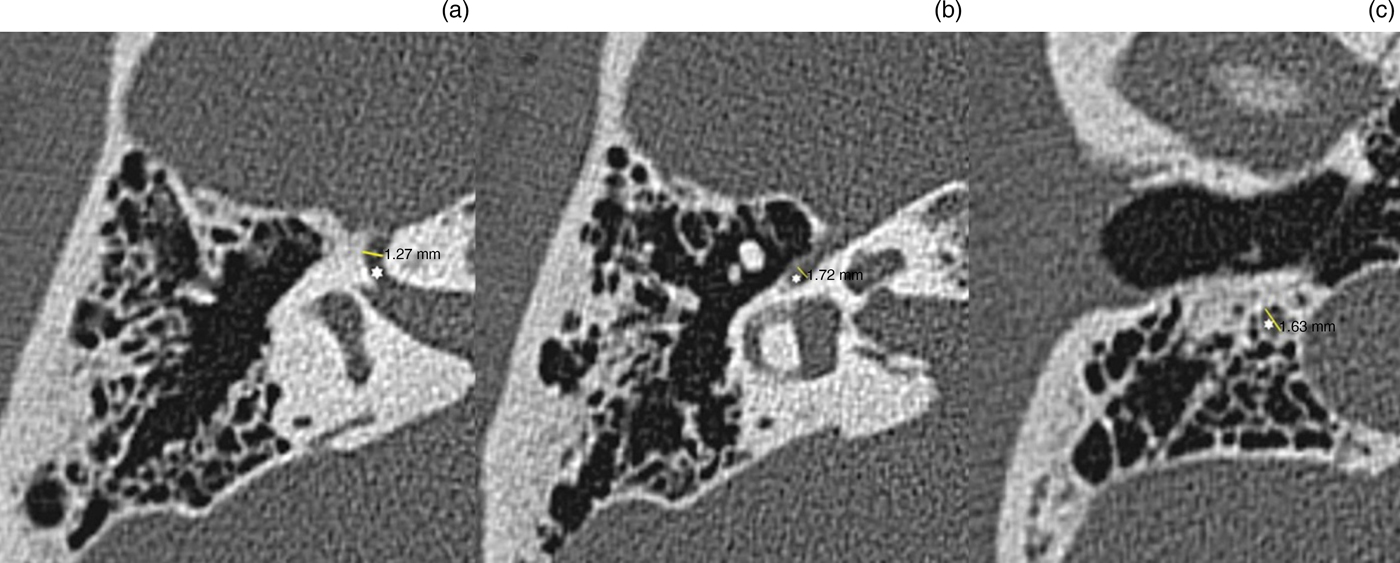
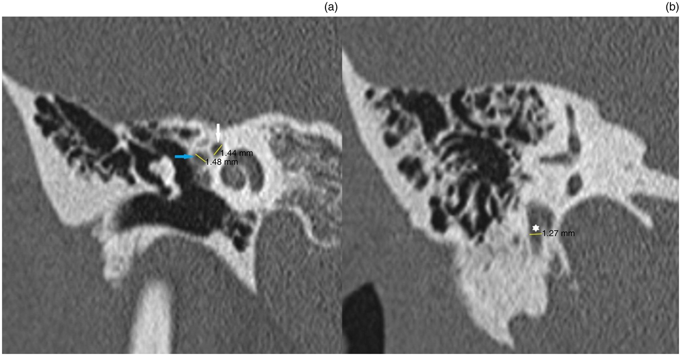
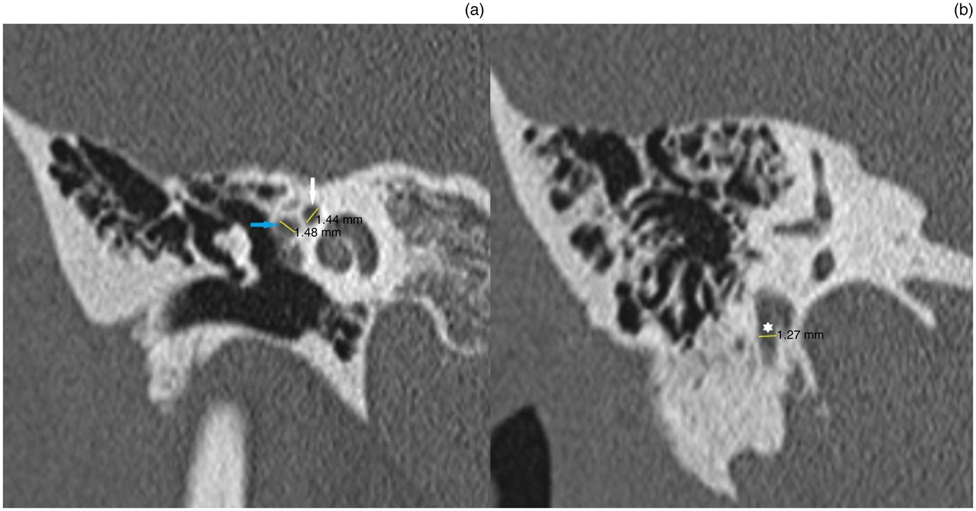
Facial canal dimensions were evaluated at the labyrinthine, tympanic and mastoid segments using axial plane (Figure 1) and coronal plane (Figure 2) multidetector CT scans of temporal bone. The measurements were performed at the maximum diameter.
Axial computed tomography images showing: (a) the facial nerve labyrinthine segment (white star) and measurement; (b) the facial nerve tympanic segment (white star) and measurement; and (c) the facial nerve mastoid segment (white star) and measurement.

Coronal computed tomography images showing: (a) the facial nerve labyrinthine segment (white arrow) and the facial nerve tympanic segment (blue arrow) and measurement; and (b) the facial nerve mastoid segment (white star) and measurement.
The mastoid pneumatisation was graded using the following scale: 0, no mastoid pneumatisation; 1, less than 25 per cent mastoid pneumatisation; 2, 25–50 per cent mastoid pneumatisation; 3, 51–75 per cent mastoid pneumatisation; and 4, more than 75 per cent mastoid pneumatisation.
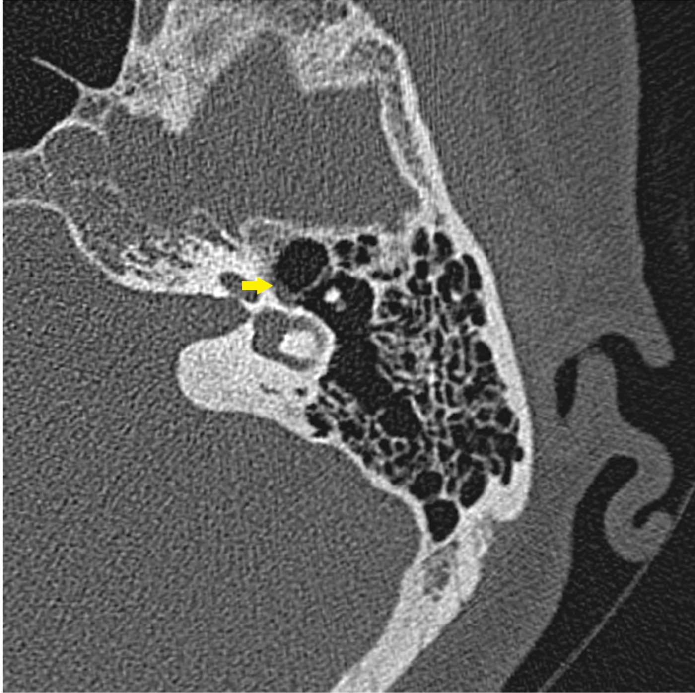
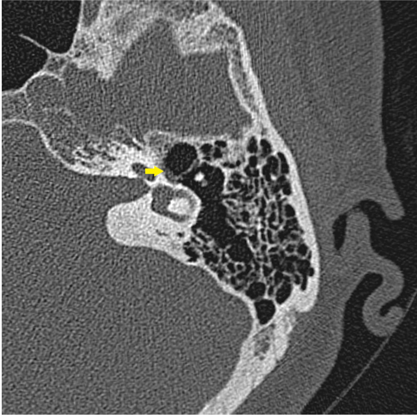
Facial canal dehiscence was measured (either as present or absent). If it was present, the length of the dehiscence was also evaluated (Figure 3).
Axial computed tomography image showing facial nerve dehiscence (yellow arrow).
Statistical analysis
All analyses were performed using SPSS® (version 16) statistical software. Independent sample t-tests, paired sample t-tests, chi-square tests, Pearson correlation tests and Spearman's correlation rho efficient tests were used. A p-value less than 0.05 was considered to be statistically significant.
Results
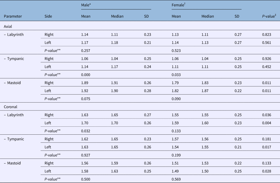
Facial canal measurements of axial and coronal plane sections are shown in Table 1. In both the axial and coronal sections, the difference between the labyrinthine, tympanic and mastoid segments of the facial canal in the males and females was analysed by independent sample t-tests.
Facial canal dimension measurements

*n = 85; †n = 84; ‡p-value shows the results of independent samples t-tests; **p-value shows the results of paired sample t-tests. SD = standard deviation
In the axial sections, the mean values of the facial canal dimensions in the mastoid segments of males (right: 1.89 mm, left: 1.92 mm) were significantly higher than those of females (right: 1.79 mm, left: 1.82 mm; p < 0.05) (Table 1). There were no significant differences between the labyrinthine and tympanic segments of males and females (p > 0.05) (Table 1).
In the coronal sections, the mean values of the facial canal dimensions in the labyrinthine segments of males (right: 1.63 mm, left: 1.70 mm) were significantly higher than those of females (right: 1.55 mm, left: 1.59 mm; p < 0.05). The mean values of the left tympanic segments in males (1.63 mm) were significantly higher than those of females (1.54 mm; p < 0.05). The mean values of the left mastoid segments in males (1.58 mm) were significantly higher than those of females (1.49 mm; p < 0.05) (Table 1).
Looking at males and females separately, the difference between the right and left sides of the labyrinthine, tympanic and mastoid segments in axial and coronal sections was analysed by paired samples t-tests. In both males (right: 1.06 mm, left: 1.14 mm) and females (right: 1.04 mm, left: 1.11 mm), the left axial tympanic facial canal dimensions were significantly higher than those of the right axial tympanic facial canal dimensions (p < 0.05) (Table 1). In males, the left coronal labyrinthine facial canal dimensions (1.70 mm) were significantly higher than the right ones (1.63 mm; p < 0.05) (Table 1).
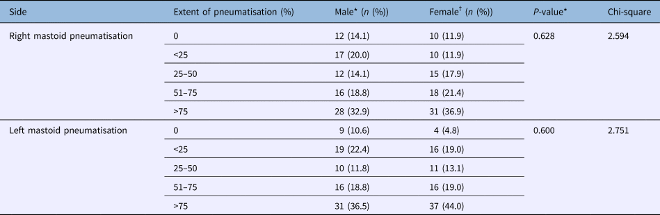
The mastoid pneumatisation values of males and females are shown in Table 2. The difference between the mastoid pneumatisation values of the males and females was analysed by the chi-square test. There were no significant differences in the right and left sides (p > 0.05). More than 75 per cent pneumatisation was the most detected level of pneumatisation in both genders and in both sides (Table 2).
Mastoid pneumatisation values for males and females

*n = 85; †n = 84
Facial canal dehiscence in males and females is shown in Table 3. On the right side, dehiscence was detected in 10 patients (5.9 per cent) comprising 5 males (5.9 per cent) and 5 females (6 per cent) (p > 0.05). The length of dehiscence was 2.03 ± 1.29 mm (range, 1.09–5.56 mm) on the right side. On the left side, dehiscence was detected in 11 patients (6.5 per cent) comprising 4 males (4.7 per cent) and 7 females (8.3 per cent) (p > 0.05). The length of dehiscence was 1.92 ± 0.44 mm (range, 0.86–2.51 mm) on the left side (Table 3).
Facial canal dehiscence in males and females

*n = 85; †n = 84
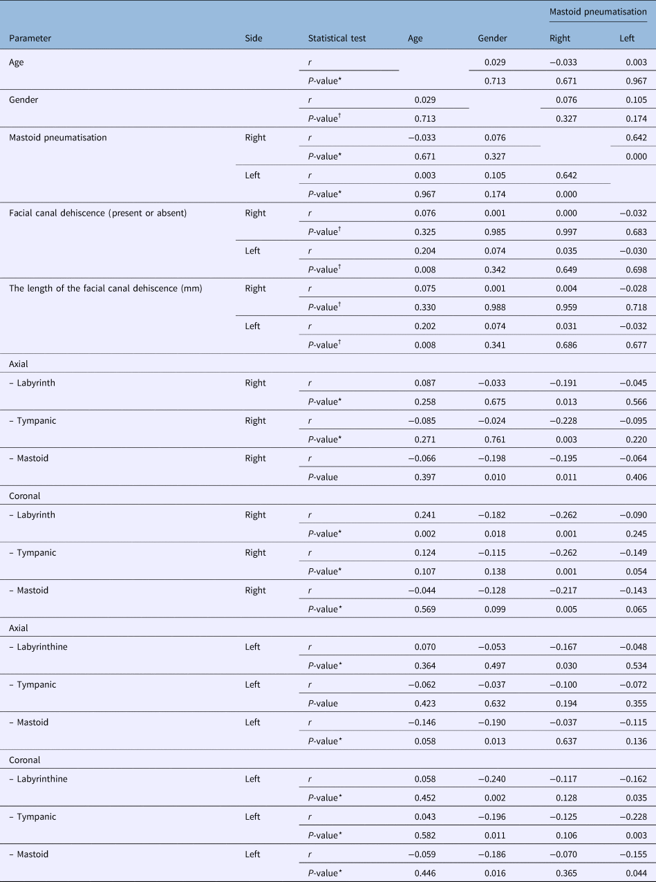
Correlation test results are shown in Table 4. There was a positive correlation between right and left side mastoid pneumatisation (p < 0.05) (Table 4). In females, right axial mastoid and coronal labyrinthine facial canal dimensions and left axial mastoid and coronal labyrinthine, tympanic and mastoid facial canal dimensions were smaller compared to males (p < 0.05) (Table 4).
Correlation tests

*p-value shows the results of the Pearson correlation test; †p-value shows the results of Spearman's correlation rho efficient test
In older subjects, left facial canal dehiscence was detected more, and the length of the dehiscence increased. Additionally, right coronal labyrinthine facial canal dimensions increased (p < 0.05) (Table 4). There were no significant correlations between mastoid pneumatisation and facial canal dehiscence (p > 0.05) (Table 4). On the right side, the axial and coronal labyrinthine, tympanic and mastoid facial canal dimensions decreased in more pneumatised mastoids (p < 0.05) (Table 4). On the left side, coronal labyrinthine, tympanic and mastoid facial canal dimensions decreased in more pneumatised mastoids (p < 0.05) (Table 3). As right mastoid pneumatisation increased, left axial labyrinthine facial canal dimensions decreased (p < 0.05) (Table 3).
Discussion
The term ‘pneumatisation’ generally refers to the presence of air cells within temporal bone. Temporal bones can be divided into extensively pneumatised, well pneumatised, poorly pneumatised, and non-pneumatised or sclerotic bones. The process of pneumatisation takes place from the perinatal period to puberty and is genetically determined, though greatly influenced by the environment that these bones are exposed to during development. The degree of pneumatisation varies significantly, and there are no two identical temporal bones, even from one person, which is what makes mastoid surgery such a difficult task.Reference Tos15 Mastoid cells are fully developed around 10 years of age and reach maturity between 15 to 20 years.Reference Ilea, Butnaru, Sfrângeu, Hedeşiu, Dudescu and Berce16–Reference Sadé and Ar18
Gupta et al. reported that 70 per cent of the temporal bones in the cadavers they studied were pneumatised, and only 10 per cent of the bones were completely sclerotic. The other 20 per cent of bones belonged to an intermediate group called diploic bones.Reference Gupta, Gupta and Singh19 The course of the facial nerve in the facial canal through the temporal bone is important. In trauma cases, tympanic and mastoid parts are affected more than the labyrinthine portion. In Bell's palsy, the labyrinthine portion may be the site of the pathogenesis.Reference Wadin and Wilbrand20 Ogawa and SandoReference Ogawa and Sando21 showed that the internal diameter of the main artery was smaller in the labyrinthine portion than in the tympanic and mastoid parts of the facial canal. The spatial relationship between the facial canal, facial nerve and vascularity of the nerve are important in the different parts of the facial canal.Reference Ogawa and Sando21
Murai et al.Reference Murai, Kariya, Tamura, Doi, Kozakura and Okano22 reported that in Bell's palsy, facial nerve oedema related to inflammation, ischemic changes and mechanical compression. Using CT scans of temporal bone, they measured the facial nerve cross-sectional area in unilateral Bell's palsy. The labyrinthine, horizontal and mastoid segments were measured. In the affected side, the cross-sectional area of the facial nerve was smaller in the labyrinthine and horizontal segments compared with the unaffected side. There were no differences in the mastoid segment. They concluded that because the labyrinthine segment is the narrowest part of the facial nerve canal, it is one of the risk factors for Bell's palsy. Ogawa and SandoReference Ogawa and Sando21 reported that there was less extra space for the nerve in the labyrinthine and tympanic segments compared with the mastoid segment. In a study by Lim et al.,Reference Lim, Lee, Hyun, Park, Kim and Lee23 patients with facial neuritis showed enhancement at the canalicular and anterior genu segments on contrast-enhanced 3D fluid-attenuated inversion recovery. Other studies also mentioned enhancement,Reference Kinoshita, Ishii, Okitsu, Okudera and Ogawa24–Reference Hong, Yi, Cha, Park, Kim and Lee26 including one studyReference Gebarski, Telian and Niparko25 in which 76 per cent of patients had facial neuritis at the geniculate ganglion and tympanic and mastoid segments related to arteriovenous plexus distribution along the facial nerve.
In the present study, we investigated facial canal dimensions and mastoid pneumatisation. Mastoid pneumatisation was not different in the right and left sides of males and females. More than 75 per cent pneumatisation was the most detected level of pneumatisation in both genders and in both sides. Sadé and Fuchs reported that there were no differences in the size of the mastoid between men and women.Reference Sadé and Fuchs27 Han et al. found that mastoid pneumatisation was not different between the right and left side.Reference Han, Song, Kim, Lee and Lee28
In our study, facial canal dimensions in the mastoid segments in males were significantly higher than those of the females, in the axial sections. In the coronal sections, facial canal dimensions in the labyrinthine segments (right and left), left tympanic and mastoid segments in males were higher than those of the females. Left coronal labyrinthine dimensions in males and left axial tympanic facial canal dimensions in both males and females were higher than the right sides.
On the right side, facial canal dehiscence was detected in 10 patients (5.9 per cent). The length of dehiscence was 2.03 ± 1.29 mm (range, 1.09–5.56 mm) on the right side. On the left side, dehiscence was detected in 11 patients (6.5 per cent). The length of dehiscence was 1.92 ± 0.44 mm (range, 0.86–2.51 mm) on the left side. In older subjects, left facial canal dehiscence was detected more, and the length of the dehiscence increased. There were no significant correlations between mastoid pneumatisation and facial canal dehiscence. In the study by Gupta et al.,Reference Gupta, Gupta and Singh19 the facial nerve dehiscence was more common in pneumatised specimens (16 per cent) when compared with poorly pneumatised specimens (7 per cent). Dehiscence was seen in the tympanic segment just above the oval window area in all specimens, with a maximum dehiscence of 2 mm. The length of dehiscence was estimated to be 1 mm in three specimens (10 per cent) and 2 mm in four specimens (13 per cent).Reference Gupta, Gupta and Singh19
In our study, right axial mastoid and coronal labyrinthine facial canal dimensions and left axial mastoid and left coronal labyrinthine, tympanic and mastoid facial canal dimensions were smaller in females compared with males. In older subjects, right coronal labyrinthine facial canal dimensions increased.
In the present study, there was a positive correlation between right and left mastoid pneumatisation. On the right side, axial and coronal labyrinthine, tympanic and mastoid facial canal dimensions decreased in more pneumatised mastoids. On the left side, coronal labyrinthine, tympanic and mastoid facial canal dimensions decreased in more pneumatised mastoids.
Gupta et al.Reference Gupta, Gupta and Singh19 investigated the facial nerve in relation to temporal bone pneumatisation in a cadaveric study, in which 30 cadaveric temporal bones were dissected. Temporal bones were divided into two groups: pneumatised and poorly pneumatised. The facial nerve and its branches were exposed from the lateral part of first genu to the outside of the stylomastoid foramen. The average length of the mastoid and tympanic segments of the facial nerve was found to be 12.6 mm and 14.7 mm, respectively, in pneumatised specimens, and 11.8 mm and 13.9 mm, respectively, among poorly pneumatised specimens. No significant difference was seen in the dimensions of facial nerve between the two groups (pneumatised and poorly pneumatised).Reference Gupta, Gupta and Singh19 In a radio-anatomical study, Dimopoulos et al.Reference Dimopoulos, Muren, Smedby and Wadin29 found that the dimensions of the tympanic and mastoid segments of the facial nerve were not influenced by mastoid pneumatisation. Our results are different from those of Gupta et al.Reference Gupta, Gupta and Singh19 and Dimopoulos et al.Reference Dimopoulos, Muren, Smedby and Wadin29 In our study, facial canal dimensions decreased in pneumatised mastoids.
• Facial canal dimensions decreased in pneumatised mastoids
• Facial canal dehiscence was detected in 5.9 and 6.5 per cent of the patients on the right and left sides, respectively
• There was no correlation between facial canal dehiscence and mastoid pneumatisation
The incidence of facial canal dehiscence was reported as 23 per cent by Gupta et al.,Reference Gupta, Gupta and Singh19 and 25 per cent by Beddard and SaundersReference Beddard and Saunders30 and MollicaReference Mollica31. The tympanic portion is the most frequent site of dehiscence.Reference Gupta, Gupta and Singh19 In their radio-anatomical study, Dimopoulos et al.Reference Dimopoulos, Muren, Smedby and Wadin29 found that 41 per cent of specimens exhibited dehiscence that ranged from 0.4 to 3.1 mm in size. However, in the study by Gupta et al.,Reference Gupta, Gupta and Singh19 none of the samples showed dehiscence of 3 mm.
Conclusion
Our study showed that facial canal dimensions decreased in pneumatised mastoids. Facial canal dimensions in females were smaller than in males. Facial canal dehiscence was detected in 5.9 per cent and 6.5 per cent of the patients at the right and left sides. No correlations were found between facial canal dehiscence and mastoid pneumatisation. During the surgery, facial canal dehiscence should be kept in mind to avoid complications.
Competing interests
None declared