Device closure of secundum atrial septal defect is now a routine procedure, and various devices are being used with a low incidence of complications.Reference Abaci, Unlu, Alsancak, Kaya and Sezenoz1,Reference Moore, Hegde and El-Said2 The Figulla-II Occlutech septal occluder (Occlutech, Jena, Germany), although not yet food and drug administration (FDA) approved, is Conformité Européene (European Conformity) marked and is being widely used with good early and mid-term results.Reference Haas, Soetemann and Ates3 Device malfunctions like cobra formation and bulging lateral edges of the left atrial disc have been reported.Reference Kilic, Ural and Sahin4 The device can usually be manipulated successfully to regain its shape in upto 70% cases; however, occasionally, it needs to be retrieved completely and reloaded.Reference Sadiq, Rehman and Mehmood5 The flat profile and globular hub of the Figulla-II Occlutech septal occluder makes retrieval difficult with currently available snares.Reference Jang and Kim6,Reference Jahrome, Stella and Leijdekkers7 We describe a case of atrial septal defect device closure using Figulla-II Occlutech septal occluder device where left atrial disc assumed an unusual shape after release leading to residual shunt.
Case report
A 17-year-old boy with a large secundum atrial septal defect with right ventricular volume overload and no pulmonary hypertension was evaluated for transcatheter closure. Transoesophageal echocardiography revealed a large defect (21 × 23 × 22 mm) with adequate all rims, including aortic rim. There was no septal floppiness or septal malalignment. The patient was taken to the catheterisation laboratory for transcatheter device closure under general anaesthesia.
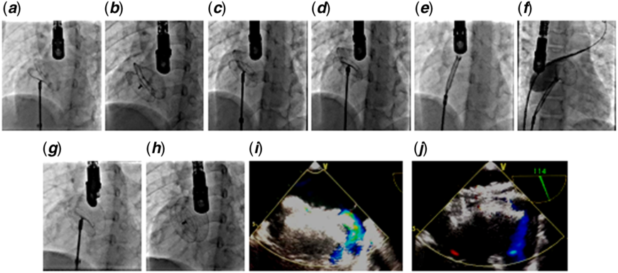
A 27-mm Figulla-II Occlutech septal occluder (Occlutech GmbH, Jena, Germany) was selected, loaded onto its delivery system, and released across the defect. On transoesophageal echocardiography, the device looked in a good position. Wiggle manoeuvre was carried out to ensure stability. Although overlooked at the time, the left atrium disc did assume a concave rather than a flat profile with an unusual hump (Fig 1a). Once released, the device malpositioned with abnormal left atrial disc orientation. Transoesophageal echocardiography showed a residual leak across superior vena cava (SVC) margin (Fig 1b and i). The device was stable with adequate positioning at all other rims. Keeping in view the malpositioned disc, it was decided to recapture the device and reposition after re-evaluation of the defect as well as the device. Considering the use of routine snares impractical under the circumstances, there arose a need for improvisation. The Figulla-II Occlutech septal occluder has a bioptome-type delivery system, which has the advantage that it allows a full circular movement of the device. We used the same delivery system to recapture the device successfully by grasping the right atrial disc ball (Fig 1c, d and e). The sheath was moved forward pulling the device back into the sheath.
Malpositioned device before and after release (a, b), device retrieval through same cable and sheath (c, d, e), redeployment with balloon assist technique (f), final position of the device (g, h), malposed and optimal positions of device on transoesophageal echocardiography (i, j).

There was no obvious deformity of the device on inspection after retrieval. Since we did not know the reason for device deformation, we wanted to use a proven technique, which ensures proper alignment of the left and right disc and hence obviates the risk of deformation again. Hence, balloon-assisted technique was used, and the same 27-mm Figulla-II Occlutech septal occluder was successfully redeployed (Fig 1f, g, h and j). The left atrial disc shape assumed its normal profile with no residual leak on transoesophageal echocardiography. The patient was discharged 24 hours later on Aspirin (5 mg/kg/day) with transthoracic echocardiogram showing an optimally placed device with no evidence of any complication related to the procedure.
Discussion
The various occluder devices used for atrial septal defect closure have some important structural differences with specific advantages and some potential disadvantages.8 The FDA approved Amplatzer atrial septal defect device is made up of nitinol wire clamped in two stainless steel hubs on each side of the disc connected at left atrial hub. Figulla-II Occlutech septal occluder in contrast has no left atrial hub, reducing the risk of thrombus formation on left atrial side and a single nitinol wire (40–200 microns) mesh with reduced wire concentration towards the centre giving the device additional flexibility. We feel that the unusual shape of the left atrial disc was due to this wire’s transient malfunction and loss of shape memory resulting in residual flow across SVC margin. Smaller amount of metal in the device, though beneficial for improving flexibility, might have lead to reduced shape memory and decreased resistance to compression. In the hindsight, we should not have released the device with that awkward left atrial disc profile and should have redeployed the device until the disc reconfigured. The other possibility could have been a second small defect but that could not be verified on transoesophageal echocardiography after device removal.
The retrieval and repositioning of same embolised Amplatzer devices is well reported in the literature.Reference Abaci, Unlu, Alsancak, Kaya and Sezenoz1 Unlike Amplatzer, Occlutech device has an integral ball and bioptome like locking delivery mechanism. Instead of a snare, we used the same delivery system to capture the right atrial hub. The jaws were able to close around the ball of the right atrial pin which was fortunately well aligned to the long axis of the sheath. A forward push was experienced by the device during this process, but as the device was still stable across the septum, we were able to slide the sheath on the captured pin and retrieved the device. We suggest that this technique should be equally effective under various other scenarios including situations where the body of the device is held securely by a secondary snare, where the device is already partly within the sheath or when the tip of the sheath is pressing the device against another cardiac structure such as left atrial or right ventricular wall.Reference Shebani, Rehman, Taliotis and Magee9 The use of same delivery system avoided the need to introduce a larger sheath for device retrieval.Reference Goel, Kapoor, Batra and Khanna10 Shebani et al used similar technique to electively retrieve an atrial septal defect device. They first used a snare to guide the right atrial pin into the tip of a Mullins sheath, but the right atrial pin was oriented obliquely and the ball could not be grasped. Mullins sheath was withdrawn slightly, exposing the right atrial pin. The jaws of the delivery cable were advanced, the Mullins sheath was used to steer the jaws, and the pin could be grasped allowing the device to be successfully removed.
In conclusion, the left atrial disc of the Figulla-II Occlutech septal occluder may assume an unusual shape due to transient wire malfunction or loss of shape memory. In such instance, device should be recaptured and redeployed till usual configuration is achieved. Secondly, Occlutech device gives a potential advantage of retrieval by using the same delivery cable due to its intrinsic design.
Financial support
This research received no grant from any funding agency, commercial, or not-for-profit sector.
Conflicts of interest
None.
Ethical standards
The authors assert that all procedures contributing to this work comply with the ethical standards of the relevant national guidelines on human experimentation (Pakistan Medical and Dental Council and Punjab Health Commission) and with the Helsinki Declaration of 1975, as revised in 2008 and have been approved by the institutional committees (Ittefaq Hospital Trust, Lahore, Pakistan). The authors assert that all procedures contributing to this work comply with the ethical standards of the relevant national guides on the care and have been approved by the institutional committee (Ittefaq Hospital Trust, Lahore, Pakistan).