Double-outlet left atrium is a very rare cardiac anomaly in which the left atrium drains into both ventricles, and the only outlet of the right atrium is an atrial septal defect or a patent foramen ovale. Only a few cases have been described in the literature thus far, always diagnosed postnatally and associated with various cardiac malformations.Reference Horiuchi, Saji, Osuka, Sato and Okada 1 – Reference Tripathi, Phadke, Nabar, Lanjewar and Kerkar 9 In this study, we report a fetus with double-outlet left atrium associated with dextrocardia and complex arrangement of the ventricles and the great vessels.
Case report
A pregnant woman was referred at 20 weeks of gestation for fetal echocardiography, as an abnormal cardiac four-chamber view had been detected on ultrasonic scanning. The routine scanning also showed cystic adenomatoid malformation of the right lung.
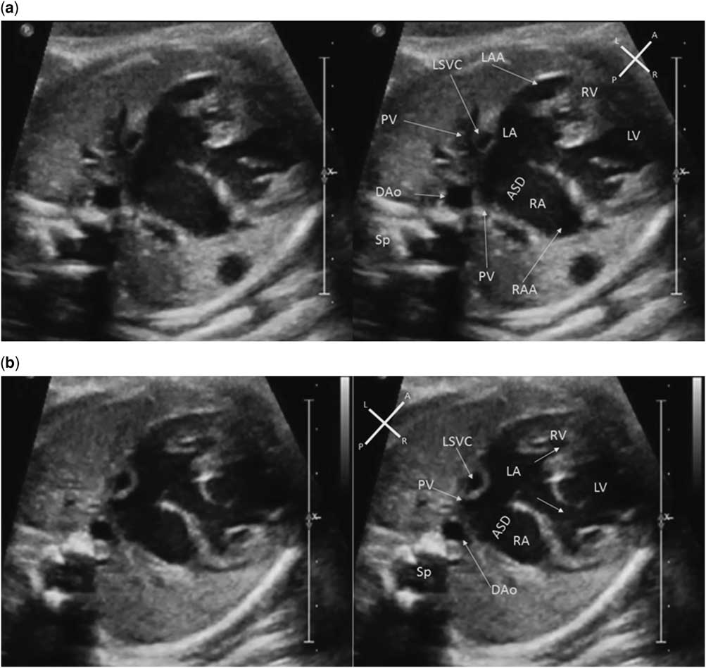
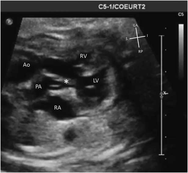
The fetal echocardiogram showed the heart to be right sided with the apex to the right. The situs was solitus with normal arrangement of the abdominal organs and atria. On four-chamber view, there was no connection between the right atrium and the ventricles. A large ostium secundum atrial septal defect without posterior rim was seen. The left atrium was connected to both ventricles through two atrioventricular valves. Atria were recognisable, thanks to their appendages. The left atrial appendage was narrow and tubular, and the right atrial appendage was wide and triangular. The morphological right ventricle was hypoplastic and left sided, and the morphological left ventricle was right sided and of normal size. A left superior caval vein draining into the coronary sinus was identified (Fig 1). The ventriculo-arterial connections were discordant with the aorta lying to the left and anterior relative to the pulmonary trunk. There was a large outlet ventricular septal defect with posterior deviation of the outlet septum creating subpulmonary stenosis (Fig 2). The aortic arch was left sided. Thus, the final diagnosis of the malformation was double-outlet left atrium with atrioventricular and ventriculo-arterial discordances {S,L,L} with dextrocardia, hypoplastic right ventricle, outlet ventricular septal defect with posterior malalignment of the outlet septum, and subpulmonary stenosis.
Four-chamber view of the heart. ( a ) In systole: dextrocardia is present. Atria are recognisable by their appendages. The morphological right ventricle (RV) is hypoplastic, and the tricuspid valve is recognisable because of its septal attachments and because of the more apical insertion of its septal leaflet comparatively with the anterior leaflet of the mitral valve. The mitral valve has no septal attachment. The morphological left ventricle (LV) is right sided and of normal size. The only outlet of the right atria (RA) is a large atrial septal defect without a posterior rim (ASD). There is a left superior caval vein (LSVC). ( b ) Same view in diastole showing the opening of the mitral and tricuspid valves (white arrows). Dao=descending aorta; LA=left atria; LAA=left atrial appendage; PV=pulmonary vein; RAA=right atrial appendage; Sp=spine.

Longitudinal view of the ventricles and the great vessels showing transposition of the great arteries, ventricular septal defect, and subpulmonary stenosis with posterior deviation of the outlet septum (*). Ao=aorta; LV=left ventricle; PA=pulmonary artery; RA=right atria; RV=right ventricle.

Fetal karyotype obtained with amniocentesis was normal. The parents decided to continue the pregnancy and the mother gave birth to a female infant weighting 2.8 kg at 37 weeks of gestation. The cardiovascular findings suspected prenatally were confirmed after birth by cross-sectional echocardiography. She needed a systemic–pulmonary anastomosis at 1 month of age. A bilateral superior cavopulmonary anastomosis was performed at 6 months of age. The infant has normal growth and is scheduled for a total cavopulmonary connection with resection of the cystic adenomatoid malformation of the right lung at 3 years of age.
Discussion
Double-outlet left atrium was initially considered as a form of atrioventricular septal defect;Reference Horiuchi, Saji, Osuka, Sato and Okada 1 , Reference Van Mierop 10 however, most cases reported since are due to a malalignment of atrial and ventricular septal with a straddling atrioventricular valve, with the other atrioventricular valve being either patentReference Gnanapragasam, Houston and Anderson 5 or atretic.Reference Bini, Pellegrino and Mazzucco 2 – Reference Suzuki, Hamada, Miura, Haneda, Horiuchi and Ogata 4 The atrioventricular connections can be concordant Reference Bini, Pellegrino and Mazzucco 2 – Reference Suzuki, Hamada, Miura, Haneda, Horiuchi and Ogata 4 , Reference Shetkar and Kothari 8 or more rarely discordant as in our case.Reference Otero Coto, Calabro, Marsico and Lopez Arranz 3 , Reference Gnanapragasam, Houston and Anderson 5 In a recent report describing a case very similar to ours, double-outlet left atrium was thought to be consecutive to a ventriculo-atrial malalignment, with an “extreme dextroposition of the atrial septum to the right”Reference Shetkar and Kothari 8 caused by excessive leftward movement of the ventricles and ventricular septum relative to the atria and the atrial septum during cardiac morphogenesis.Reference Van Praagh 11 The nature of the atrioventricular valves forming the outlet of the left atrium is debated: straddling atrioventricular valve or two distinct mitral and tricuspid valves. In our case, the left atrioventricular valve is inserted more apically than the right atrioventricular valve and has septal attachments, which is in favour of two morphologically distinct atrioventricular valves, a left-sided tricuspid valve, and a right-sided mitral valve. To the best of our knowledge, the description of this rare malformation is unique in the history of fetal diagnosis.
Surgical management depends on associated malformations. In our case, a biventricular repair was not possible because of right ventricular hypoplasia. In two previously reported cases, biventricular repair has been performed by re-positioning the atrial septum in order to deviate flow from the right atria to the right ventricle.Reference Horiuchi, Saji, Osuka, Sato and Okada 1 , Reference Suzuki, Hamada, Miura, Haneda, Horiuchi and Ogata 4
Acknowledgement
None.
Financial Support
This research received no specific grant from any funding agency or from commercial or not-for-profit sectors.
Conflicts of Interest
None.
Ethical Standards
The authors assert that all procedures contributing to this work comply with the ethical standards of the relevant French guidelines on human experimentation and with the Helsinki Declaration of 1975, as revised in 2008, and has been approved by the institutional committees of Hôpital privé de La Louvière.
Supplementary materials
To view supplementary material for this article, please visit https://doi.org/10.1017/S104795111600250X