Introduction
The skeletons of echinoderms mainly consisting of Mg-calcite biominerals showed hierarchical structures and excellent mechanical property.[Reference Seto, Ma, Davis, Meldrum, Gourrier, Kim, Schilde, Sztucki, Burghammer, Maltsev, Jager and Colfen1,Reference Cao, Li, Zhou, Li, Vecchio, Yang, Cui, Yang, Zhu, Guo and Zhang2] Echinoderm skeletons such as sea urchin (SU) spines[Reference Seto, Ma, Davis, Meldrum, Gourrier, Kim, Schilde, Sztucki, Burghammer, Maltsev, Jager and Colfen1,Reference Politi, Arad, Klein, Weiner and Addadi3] and teeth[Reference Killian, Metzler, Gong, Olson, Aizenberg, Politi, Wilt, Scholl, Young, Doran, Kunz, Tamura, Coppersmith and Gilbert4,Reference Ma, Aichmayer, Paris, Fratzl, Meibom, Metzler, Politi, Addadi, Gilbert and Weiner5] behave like single crystals. Recent studies revealed that these echinoderm skeletons were composed of well-oriented nanoparticles, called mesocrystals,[Reference Seto, Ma, Davis, Meldrum, Gourrier, Kim, Schilde, Sztucki, Burghammer, Maltsev, Jager and Colfen1,Reference Oaki and Imai6] like those existed in other biominerals[Reference Cai and Tang7,Reference Amini, Masic, Bertinetti, Teguh, Herrin, Zhu, Su and Miserez8] and metal alloys.[Reference Song and Colfen9–Reference Zhou, Li and Lu11] Mesocrystals form from the ordered array of amorphous precursor particles through a nonclassical nucleation pathway.[Reference Colfen and Antonietti12–Reference Raiteri and Gale15]
Natural calcium carbonate skeletons, like coral, cuttlebones, and SU spines, have been hydrothermally reacted to form β-tricalcium phosphate [β-TCP, Ca3(PO4)2] or hydroxyapatite [HA, Ca5(PO4)3OH] as bone grafts, considering their hierarchical open-cell structures suitable for tissue ingrowth. Compared to HA, β-TCP is biodegradable, which can be fully replaced by newly formed bone.[Reference Cao, Li, Zhou, Li, Vecchio, Yang, Cui, Yang, Zhu, Guo and Zhang2,Reference Ellies, Nelson and Featherstone16,Reference Yu, Liu, Jiang, Wang, Yang and Kang17] This is likely due to the relatively higher Ksp of β-TCP (10−28.9) than that of HA (10−58.4).[Reference Dorozhkin18] However, β-TCP is difficult to be fabricated in aqueous solutions at low temperature due to the preferential formation of HA under the same condition. Therefore, β-TCP powders were usually prepared by calcination of precursor materials or solid-state reactions at high temperature.[Reference Kivrak and Tas19,Reference Gibson, Rehman, Best and Bonfield20] Previous studies[Reference Cao, Li, Zhou, Li, Vecchio, Yang, Cui, Yang, Zhu, Guo and Zhang2,Reference Vecchio, Zhang, Massie, Wang and Kim21,Reference Roy and Linnehan22] showed that SU spines consisting of Mg‒calcite can be converted to Mg-substituted tricalcium phosphate (Mg-TCP) by a hydrothermal reaction. On the other hand, skeletons including coral, cuttlebone, and seashells consisting of aragonite or calcite without Mg2+ ion substitution were converted to HA.[Reference Vecchio, Zhang, Massie, Wang and Kim21–Reference Clarke, Walsh and Maggs23] Thus, Mg2+ ions played an important role in the hydrothermal reaction of Mg-calcite to Mg-TCP (not HA). However, the underlying mechanisms for the effects of Mg on the formation of Mg-TCP remain unclear.
In this study, natural calcite skeletons with various amounts of Mg are hydrothermally converted to Mg-TCP. Mg-calcite with different amounts of Mg2+ ions has been synthesized via the transition of amorphous calcium carbonate (ACC), which can be further hydrothermally converted to Mg-TCP. The effects of Mg substitution on the formation of Mg-TCP by hydrothermal reactions of Mg-calcite have been investigated.
Experimental section
Raw materials
Seashell (Veneroida Tridacnidae spp.), the spines and shells of SU (Heterocentrotus mammillatus), and the skeletons of starfish (SF, Protoreaster nodosus) were bought from a marine product store, which were cut and minced to obtain small granules. Samples were sieved using standard sieves (20 and 40 meshes) to obtain particle sizes in the range of 150–840 µm. These raw materials were rinsed and boiled in deionized water for 30 min and further soaked in a 10 wt% NaClO solution for 40 min with an ultrasonic concussion to remove the existing organic components. The resulting samples were rinsed with deionized water thrice followed by ethanol once, which were further dried in a vacuum oven at 80°C overnight. The seashell samples (mainly comprising of the aragonite phase without Mg) were polished to remove residual calcite and further calcined at 500°C for 24 h to obtain the calcite phase without Mg. All chemical reagents (analytical grade) used in the experiments in this study were bought from Sinopharm Chemical Reagent Co., Ltd. (Beijing, China).
Synthesis of Mg-calcite powders
Mg-calcite with different Mg contents were produced by wet-chemical precipitation. MgCl2 powders were dissolved into a 0.1 mol/L CaCl2 solution at the Ca/Mg molar rate of 1:1, 2:1, and 5:2 to obtain a homogeneous mixture. A 0.1 mol/L Na2CO3 solution with the same volume was then poured into the above mixture solution while stirring at 500 rpm for 10 s. The precipitates were collected by a vacuum filtration and further rinsed with deionized water thrice followed by ethanol once. The samples were further dried in a vacuum oven at 60 °C overnight to obtain Mg-calcite powders.
Hydrothermal reactions of CaCO3 scaffolds
One gram granules (150–840 µm) of seashell, SU spine, SU shell, and SF horn were separately mixed with 20 mL of 0.1 g/mL (NH4)2HPO4 solution in an autoclave container, which were then reacted at 200 °C for 24 h. The products were washed with deionized water thrice followed by ethanol once and further dried in a vacuum oven at 120°C overnight.
0.5 g of synthetic Mg-calcite powders were pressed to form solid tablets with a diameter of 13 mm under 50 MPa for 10 min. One gram of the Mg-calcite tablets with different Mg contents was separately mixed with 20 mL of 0.1 g/mL (NH4)2HPO4 solution at the temperature of 200°C for 24 h. The products were washed with deionized water thrice and ethanol once and further dried in a vacuum oven at 120 °C overnight.
Material composition analysis
Mg and Ca contents in the samples were analyzed using the inductively coupled plasma mass spectrometry (ICP-MS, Prodigy, Leeman Labs, Hudson, USA). The samples were dissolved in a 2 wt% nitric acid solution for ICP-MS tests, and three replicates were conducted. The mineral phases of samples were identified by XRD analysis using an X-ray diffractometer (Rigaku D/max 2400 diffractometer, Tokyo, Japan) operated at 40 kV with Cu Kα1 of 1.5418 Å. The 2θ range of 20–70° with a stepwise of 0.04° was used for all samples. Chemical bonding information for each sample was analyzed using a Fourier-transform infrared spectrometer (Tensor27, Bruker, Germany). Attenuated total reflection mode with the wavenumber in the range of 600–3000 cm−1 was conducted for each sample.
Microstructure analysis
Sample microstructures were observed with an LEO Supra 35 field-emission scanning electron microscope (Zeiss, Jena, Germany) at an accelerating voltage of 20 kV. The samples were coated with a gold film using the sputter coating instrument (Cressington, Watford, England) prior to SEM observation. The microstructures of the echinoderm skeletons before and after hydrothermal reactions were also characterized by 3D X-ray tomography (XRT) analysis using an Xradia VersaXRM-500 System (Carl Zeiss X-ray Microscopy Inc., Pleasanton, CA, USA).[Reference Zhang and Wang24] The X-ray source voltage was 80 kV, and the beam current was 87 µA. The powder samples were first ground by ball milling for 24 h and then dispersed in ethanol with ultrasonication at 100 Hz for 5 min. The suspension was dropwise added to a copper grid with a carbon film (Beijing Zhongjingkeyi Technology Co., Ltd, Beijing, China) for morphology and crystalline analysis by a Tecnai F30 transmission electron microscope (FEI, Hillsboro, OR, USA) at an accelerating voltage of 300 kV.
First-principle calculation analysis
The total energy of the lattice of β-TCP (space group R-3c, z = 21, Ca63P42O168) was performed by CASTEP of Materials Studio 8.0 software with generalized gradient approximation through the first-principle calculation analysis. Mg-TCP crystal models were constructed by substituting Ca atoms with Mg atoms from Ca(5) to Ca(4). An energy cutoff of 571.4 eV and k-point of 3*3*2 were used for the calculation of the total energy of β-TCP lattice with geometry optimization. The total energy convergence is smaller than 10−5 eV/atom. The relationship between the total energy of β-TCP lattice and Mg content [Mg/(Mg + Ca)] was analyzed.
Results and discussion
Hydrothermal conversion of Mg-calcite to Mg-TCP
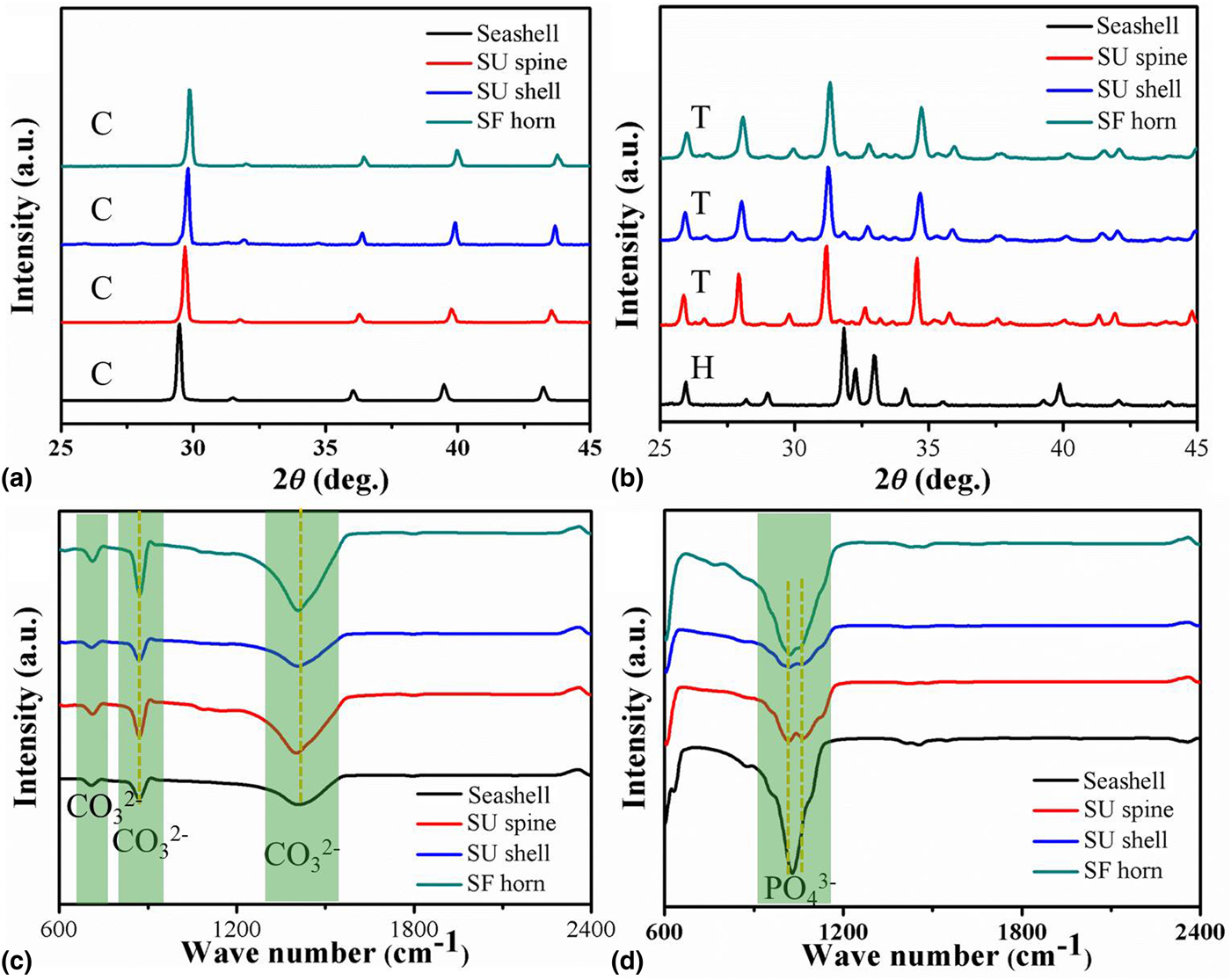
Biogenic CaCO3 skeletons have been widely used to produce calcium phosphate for bone defect repair. Previous studies showed that hydrothermal reactions of Mg-calcite, like SU spines, produced Mg-TCP. Aragonite skeletons (without Mg2+ ion) substitution, including coral, seashell and cuttlefish bone, produced HA.[Reference Lemos, Rocha and Quaresma25,Reference Marchegiani, Cibej and Vergni26] Our study revealed that seashell samples of calcite without Mg [Fig. 1(a)] were also converted to HA by a hydrothermal reaction [Fig. 1(b)]. Thus, Mg substitution is critical to the formation of Mg-TCP instead of HA by hydrothermal reactions. Mg contents [Mg/(Mg + Ca)] for β-TCP products from seashell, SU spine, SU shell, and SF horn were 0, 10.3 ± 0.3 mol.%, 14.9 ± 0.3 mol.%, and 16.4 ± 0.1 mol.%, respectively, based on ICP results (Supplementary Table S1). There was a slight decrease in the Mg content in the resulting β-TCP products compared to the original SU spine (11.5 mol.%), SU shell (15.3 mol.%), and SF horn (17.6 mol.%), likely due to the dissolution of Mg2+ ions into the solution. The seashell sample without Mg was hydrothermally converted to HA with characteristic peaks such as (2 1 1), (1 1 2), and (3 0 0) (JCPDF #09-0432), while all other Mg-calcite samples were converted to β-TCP with characteristic peaks such as (0 2 10), (2 1 4), and (2 2 0) [JCPDF #09-0169, Fig. 1(b)]. XRD patterns with 2θ range of 20–70° for seashell, SU spine, SU shell, and SF horn samples before and after hydrothermal reactions were shown in Supplementary Fig. S1. Only Mg-calcite skeletons can be hydrothermally converted to Mg-TCP, while pure calcite or aragonite skeletons were converted to HA, consistent with previous findings.[Reference Vecchio, Zhang, Massie, Wang and Kim21,Reference Roy and Linnehan22,Reference Lemos, Rocha and Quaresma25,Reference Marchegiani, Cibej and Vergni26]
XRD patterns (a) and (b) and FTIR results (c) and (d) of seashell, SU spine, SU shell, and SF horn samples before (a) and (c) and after (b) and (d) hydrothermal reactions at 200 °C for 24 h (C: calcite, T: β-TCP, H: HA).
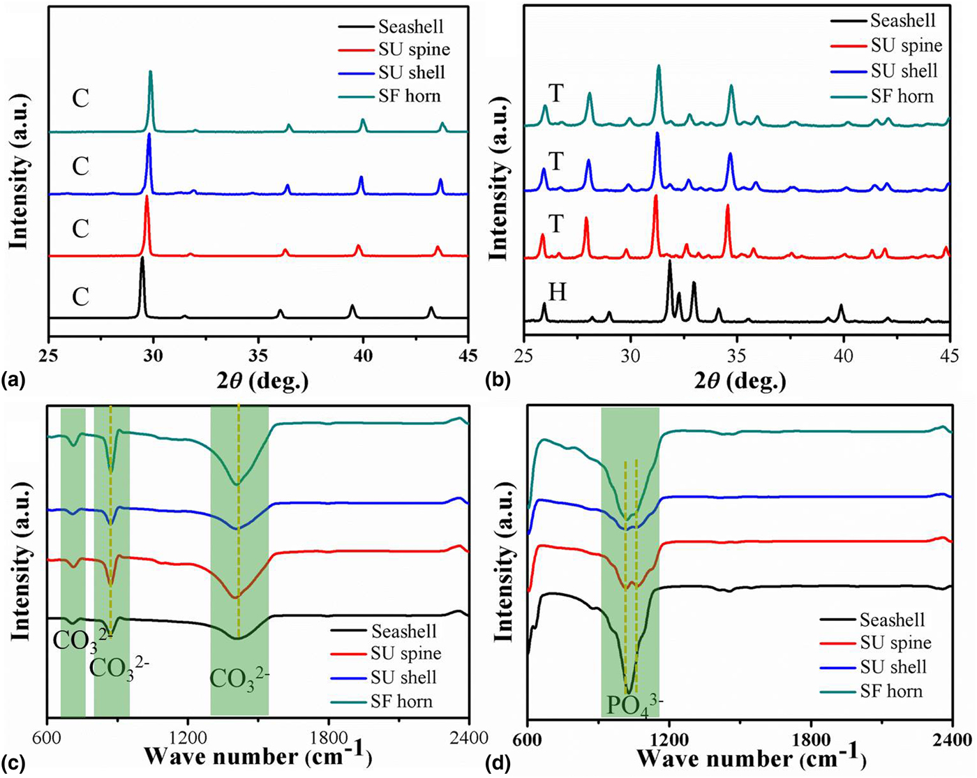
The 2θ range of the (0 2 10) peak of β-TCP produced from SU spine, SU shell, and SF horn was 31.23 ± 0.02°, 31.26 ± 0.01°, and 31.31 ± 0.02° [Fig. 1(b)], respectively, shifting to larger angles compared to that from the standard pattern (31.03° based on JCPDF #09-0169). These results indicated a larger amount of Mg content in Mg-TCP, greater lattice contraction, likely due to much smaller radium of Mg2+ ion (R = 0.72 Å) than that of Ca2+ ion (R = 1.00 Å). The presence of peaks of  ${\rm PO}_4^{3-}$ (about 604 cm−1 for γ4, 1018 and 1067 cm−1 for γ3) [Fig. 1(d)] in the FTIR spectrum confirmed the formation of Mg-TCP from Mg-calcite with peaks of
 ${\rm CO}_3^{2-}$ [about 711 cm−1 (γ4), 872 cm−1 (γ2), 1416 cm−1 (γ3)] [Fig. 1(c)] by hydrothermal reactions.
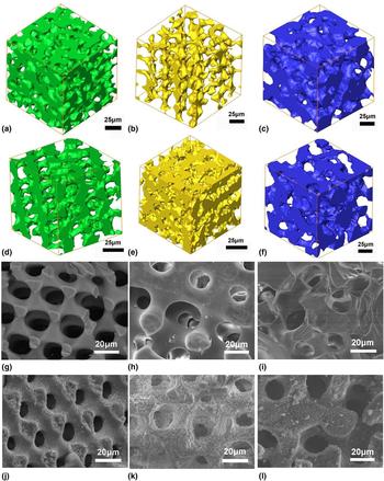
The calcium carbonate skeletons from SU spine and SF horn show three-dimensional open-cell structures, which give them superior mechanical strength at relatively lightweight.[Reference Maiti, Small, Lewicki, Weisgraber, Duoss, Chinn, Pearson, Spadaccini, Maxwell and Wilson27] The three-dimensional microstructures of SF horn, SU spine, and SU shell before and after hydrothermal reactions are shown by XRT results [Figs. 2(a)–2(f)]. The natural skeletons showed hierarchical open-cell structures and thin struts with smooth surfaces. The Mg-TCP scaffolds after hydrothermal reactions preserved the original open-cell structures, which are beneficial to cell and tissue ingrowth in vivo when used as bone grafts. In addition, the thin struts of the Mg-TCP scaffolds by hydrothermal conversion of SU spines can be easily degraded and replaced by newly formed bone in vivo.[Reference Cao, Li, Zhou, Li, Vecchio, Yang, Cui, Yang, Zhu, Guo and Zhang2] The morphology of pores and struts were further investigated using SEM images [Figs. 2(g)–2(l)]. The pore sizes of SF horn, SU shell, and SU spine were about 12.6–20.5, 6.9–14.4, and 15.2–40.0 µm (n = 15), whereas the strut thickness is 8.5–12.7, 2.3–8.7, and 8.0–32.5 µm (n = 15), respectively.
XRT images of SF horn (a) and (d), SU shell (b) and (e), and SU spine (c) and (f) before (a)–(c) and after (d)–(f) hydrothermal reactions at 200 °C for 24 h, and SEM images of SF horn (g) and (j), SU shell (h) and (k), and SU spine (i) and (l) before (g)–(i) and after (j)–(l) hydrothermal reactions at 200 °C for 24 h.
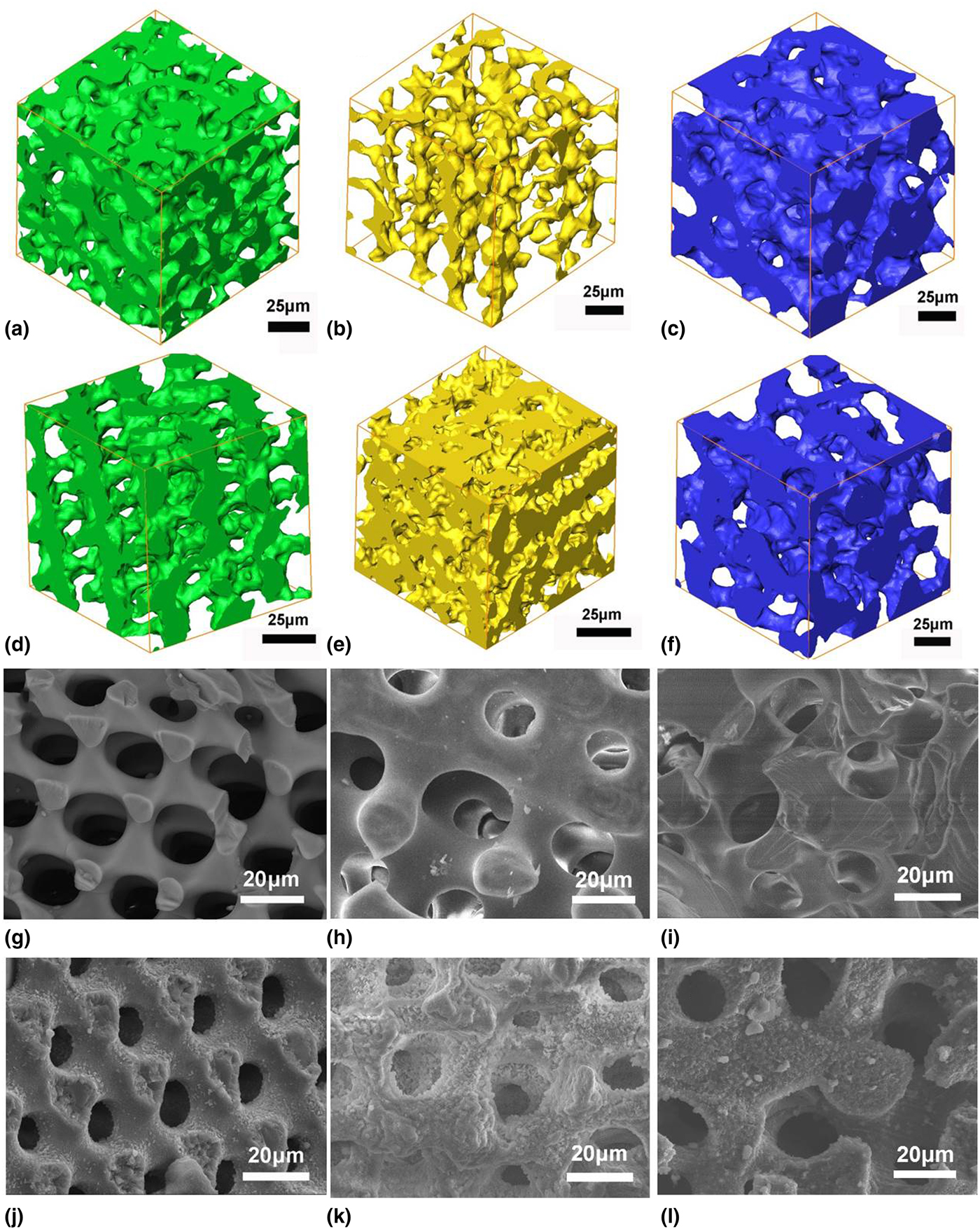
There was a slight dissolution of Mg-calcite from the strut surfaces of echinoderm skeletons, indicated by the rough surface morphology as well as a slight decrease in the Mg content in Mg-TCP products after hydrothermal reactions. However, the amount of dissolved Mg-calcite was neglectable, compared to that for the ion-exchange reaction. Therefore, natural echinoderm Mg-calcite skeletons can be hydrothermally converted to Mg-TCP.
Biomimetic synthesis
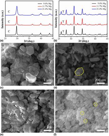
Inspired by biomineralization of natural Mg-calcite, Mg-ACC with different Mg contents was produced by wet-chemical precipitation from a mixture solution with different Mg/Ca molar ratios (0.4–1), which were then dehydrated to form Mg-calcite [Fig. 3(a)].[Reference Tomono, Nada, Zhu, Sakamoto, Nishimura and Kato28,Reference Ihli, Wong, Noel, Kim, Kulak, Christenson, Duer and Meldrum29] The molar percentage of Mg in calcite [Mg/(Mg + Ca)] was in the range of 9.6–21.4 mol.% based on ICP results (Supplementary Table S2), close to those from echinoderm skeletons. The synthetic Mg-calcite pressed into a solid tablet, which can be hydrothermally converted to Mg-TCP similar to echinoderm skeletons [Fig. 3(b)]. XRD patterns with the 2θ range of 20–70° for the products before and after hydrothermal reactions of Mg-calcite with different Mg contents (9.6, 11.7, and 21.4 mol.%) were shown in Supplementary Fig. S2. Moreover, SU spines particles (<45 µm) and synthetic Mg-calcite (11.7 mol.% Mg) by ball milling produced a large amount of HA besides Mg-TCP by hydrothermal reactions (Supplementary Fig. S3). Small particles by ball milling had relatively large surface areas, resulting in large amounts of Ca2+ ions dissolved to produce HA by the dissolution–reprecipitation reaction.
XRD patterns of the bulk powder samples before (a) and after (b) hydrothermal reactions of Mg-calcite tablets with different Mg contents (9.6, 11.7, and 21.4 mol.%). (C: calcite, T: β-TCP). SEM images of the interior (c) and outer surface (d) of the products fabricated by Mg-calcite tablets (9.6 mol.% Mg) through hydrothermal reactions at 200 °C for 24 h. SEM images of the interior (e) and outer surface (f) of the products fabricated by Mg-calcite tablets (11.7 mol.% Mg) through hydrothermal reactions at 200 °C for 24 h. The yellow boxes in (d) indicated the morphology and size of HA particles on the outer surface. The yellow circles in (f) indicated the particle size of Mg-TCP on the outer surface through the dissolution–reprecipitation reaction.
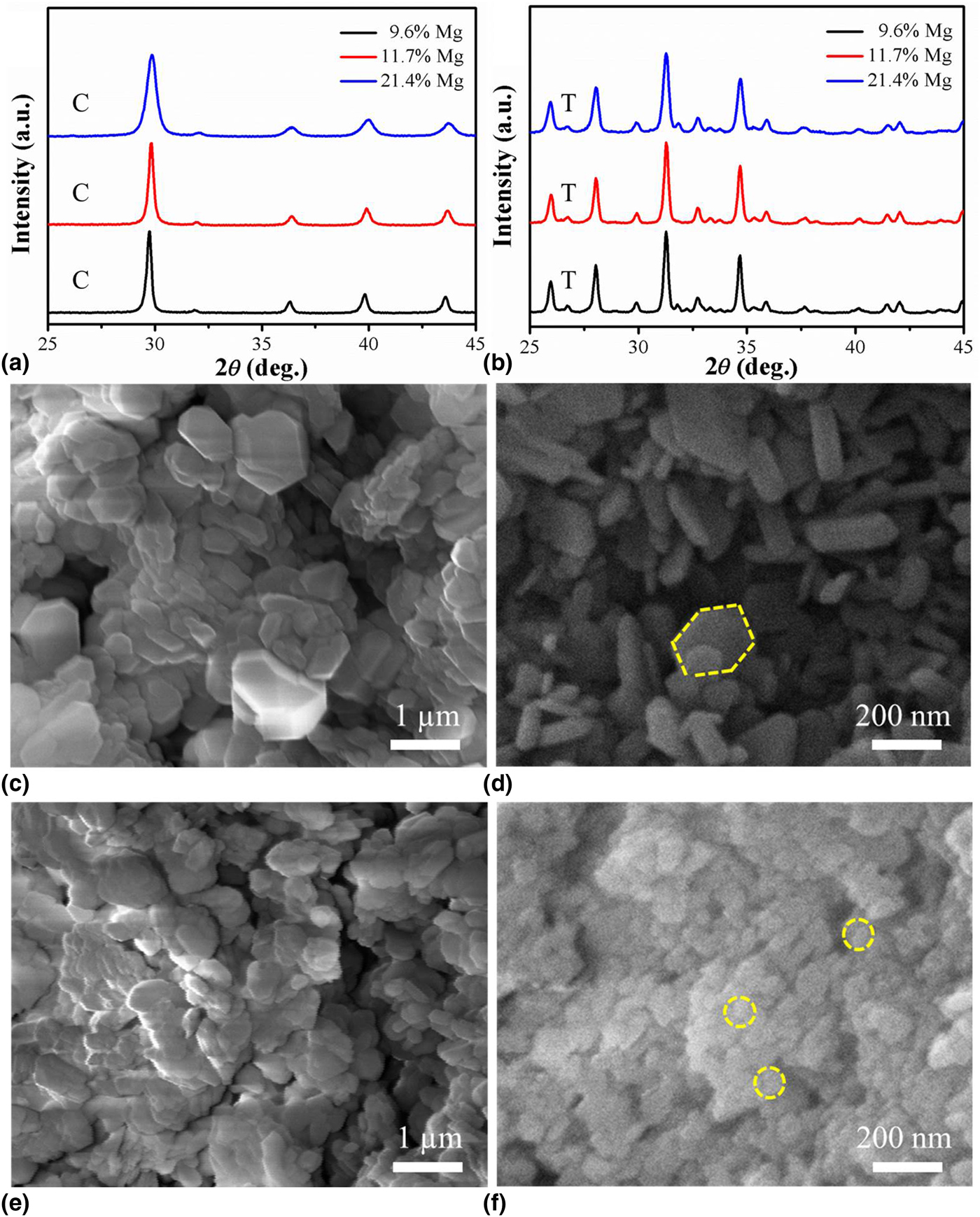
SEM images [Figs. 3(c) and 3(e)] showed that the interior was composed of polyhedral Mg-TCP particles ~0.3–1 µm (n = 15). The outer surface of the sample with 9.6 mol.% Mg was fully embedded with nano-size hexagonal HA prisms [Fig. 3(d)], likely formed through the dissolution–reprecipitation reaction. The XRD pattern of the tablet sample with 9.6 mol.% Mg [Supplementary Fig. S4(a)] indicated the presence of HA besides β-TCP. On the other hand, the outer surface of the Mg-TCP sample with 11.7 mol.% Mg was fully embedded with β-TCP nanoparticles ~40–70 nm (n = 15) [Fig. 3(f)]. The XRD pattern of the tablet sample with 11.7 mol.% Mg [Supplementary Fig. S4(b)] also showed the chemical composition of β-TCP. A previous study showed that the solubility of β-TCP is lower than HA with pH ~6.0 and at 25 °C, when Mg content increases to 10 mol.%.[Reference Ding, Pan, Xu and Tang30] Therefore, β-TCP nanoparticles formed on the outer surface of the Mg-TCP sample with 11.7 mol.% Mg in the hydrothermal system, instead of HA.
Reaction mechanisms
To reveal the mechanisms for the hydrothermal reactions, the crystalline information of HA and β-TCP are illustrated. The coordination number of Ca(1) and Ca(2) with O atoms in HA crystals (space group P63/m) is 9 and 7, respectively (Supplementary Fig. S5). It was extremely unstable for the substitution of Ca2+ ions by Mg2+ ions in the HA crystal lattice.[Reference Sun, Jayaraman, Chen, Persson and Ceder31] Matsunaga demonstrated that Ca2+ ions cannot be substituted by Mg2+ ions in the HA crystal lattice due to the relatively large formation energy ~0.8 eV/defect and 0.6 eV/defect for Ca(1) and Ca(2) at pH 7, respectively.[Reference Matsunaga32]
On the other hand, Ca2+ ions in the β-TCP crystal lattice (space group R-3c) with short Ca‒O bonds and small coordination numbers can be substituted by Mg2+ ions with the smaller ionic radius.[Reference Sun, Jayaraman, Chen, Persson and Ceder31] Substitution of Ca(5) sites (one of five types of Ca2+ ions ~9.52 mol.%, and sixfold coordination with oxygen atoms) by Mg2+ ions in the crystal lattice can occur in the first place, considering the bond length of Ca(5)‒O is 2.270 or 2.299 Å, shortest among all Ca‒O bonds (Supplementary Fig. S6). This finding is in good agreement with a previous study.[Reference Grigg, Mee, Mallinson, Fong, Gan, Dupree and Holland33] Ca(4) (threefold coordination with oxygen atoms, half occupied) with the bond length of Ca(4)‒O 2.371 Å can also be replaced by Mg2+ ions in the crystal lattice (maximum of 4.76 mol.%).[Reference Grigg, Mee, Mallinson, Fong, Gan, Dupree and Holland33] The formation of Mg-TCP from Mg-calcite led to crystal lattice contraction and increase in the formation energy due to the substitution of Ca2+ ions by Mg2+ ions, based on the first-principle calculation (Supplementary Fig. S7). There was a nearly linear relationship between the cell volume (V) [or formation energy variation (∆E)] and the Mg content [Eqs. (1) and (2)],
 $$V = -9.64x + 3687.58{\rm \;} \lpar {{\AA}^3} \rpar $$
 $$\Delta E = 17.84x-0.61{\rm \;} \lpar {{\rm eV}} \rpar \; $$
where x mol.% is the Mg content [Mg/(Mg + Ca), 0–9.52 mol.%]. First-principle calculation results showed that the cell volume of β-TCP crystal lattice decreased with the increase in the Mg content. After the substitution of Ca(5) by Mg2+ ions, the crystal lattice of Mg-TCP contracted with the decrease in the Ca(1, 2, 3, 4)‒O bond length. For example, the bond length of Ca(4)‒O decreased from 2.371 to 2.329 Å based on the first-principle calculation, although there was a significant increase in the formation energy [Supplementary Fig. S4(a)]. The relationship between the cell volume (V) [or formation energy variation (∆E)] and the Mg content was calculated using the first principle with the substitution of Ca(4) by Mg2+ ions [Eqs. (3) and (4)],
 $$V = -5.80x + 3642.81{\rm \;} \lpar {{\AA}^3} \rpar $$
 $$\Delta E = 37.30x-187.66{\rm \;} \lpar {{\rm eV}} \rpar $$
where x mol.% is Mg content [Mg/(Mg + Ca), 9.52–14.28%]. Compared with the substitution of Ca(5), the substitution of Ca(4) showed smaller bond length contraction and higher energy increase. Energy increase for the substitution of each Ca(4) atom is about twice of that for each Ca(5) atom, indicating that the substitution of Ca(5) sites by Mg2+ ions is preferred than Ca(4) sites. After the substitution of Ca(5) and Ca(4), there was a further contraction of the β-TCP crystal lattice, allowing the substitution of Ca2+ [Ca(1), Ca(2), or Ca(3)] by Mg2+ ions beyond 14.28 mol.% (Supplementary Table S3).
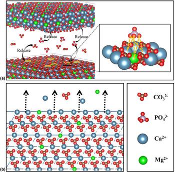
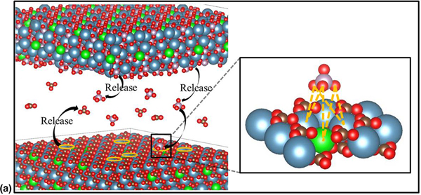
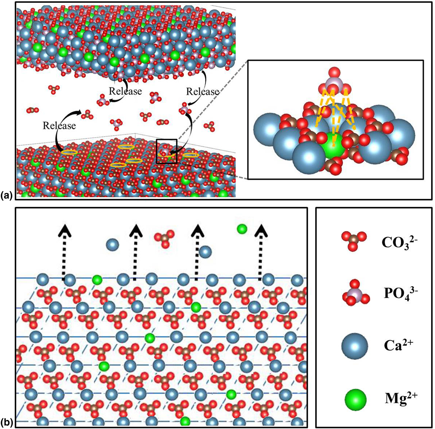
Figure 4 shows the mechanistic illustration for the hydrothermal conversion of Mg-calcite to Mg-TCP through ion-exchange reactions. The  ${\rm CO}_3^{2-}$ ions in the bulk Mg-calcite were substituted by
 ${\rm PO}_4^{3-}$ ions produced by
 ${\rm HPO}_4^{2-}$ and
 ${\rm CO}_3^{2-}$ in the solution, resulting in the formation of Mg-TCP, following Eq. (5). The echinoderm skeletons with thin struts were well preserved after hydrothermal reactions, likely due to the efficient transportation of the reactants to the porous structures (Fig. 2). Our previous study showed that chicken eggshells made up of Mg-calcite with ~2.0 mol.% Mg can be hydrothermally converted to Mg-TCP by ion-exchange reactions (data not shown).[Reference Cui, Song, Su, Yang, Yang, Li and Zhang34] In the hydrothermal system, there was also a dissolution–reprecipitation reaction as a side reaction on the surface, which produced HA following Eq. (6) (Mg content less than ~10 mol.%). The previous study reported that the solubility of Mg-TCP is lower than HA with pH about 6.0 and at 25 °C when Mg content increases to 10 mol.%,[Reference Ding, Pan, Xu and Tang30] consistent with our results.
 $$\eqalign{& 2{\rm Ca}\lpar {{\rm Mg}} \rpar {\rm C}{\rm O}_3 + 2\lpar {{\rm N}{\rm H}_4} \rpar _2{\rm HP}{\rm O}_4 \cr & \quad \to 2{\rm Ca}\lpar {{\rm Mg}} \rpar _3\lpar {{\rm P}{\rm O}_4} \rpar _2 + 2\lpar {{\rm N}{\rm H}_4} \rpar _2{\rm C}{\rm O}_3 + 2{\rm H}_2{\rm C}{\rm O}_3}$$
 $$\eqalign{& 10{\rm CaC}{\rm O}_3 + 6\lpar {{\rm N}{\rm H}_4} \rpar _2{\rm HP}{\rm O}_4 + 2{\rm H}_2{\rm O} \cr & \quad \to {\rm C}{\rm a}_{10}\lpar {{\rm P}{\rm O}_4} \rpar _6\lpar {{\rm OH}} \rpar _2 + 6\lpar {{\rm N}{\rm H}_4} \rpar _2{\rm C}{\rm O}_3 + 4{\rm H}_2{\rm C}{\rm O}_3}$$
(a) Schematic illustration of the ion-exchange reaction for the synthesis of Mg-TCP nanoparticles in the interior of Mg-calcite samples and (b) the dissolution–reprecipitation reaction for the synthesis of HA (or TCP) on the outer surface of Mg-calcite samples.
Conclusions
In this study, Mg-calcite from echinoderm skeletons was hydrothermally converted to Mg-TCP. Moreover, Mg-calcite powders were prepared by the transformation of ACC precursors, which can be further hydrothermally converted to Mg-TCP as solid tablets. Our results revealed that Mg-TCP formed through main ion-exchange reactions of Mg-calcite in the hydrothermal system. On the other hand, HA crystals formed on the outer surfaces of the samples through the dissolution–reprecipitation reaction. Therefore, Mg played significant roles in the hydrothermal synthesis of Mg-TCP from Mg-calcite.
Supplementary Material
The supplementary material for this article can be found at https://doi.org/10.1557/mrc.2019.110
Acknowledgments
This work was supported by the National Key Research and Development Program of China from the Ministry of Science and Technology (2016YFC1100502), Key the Research Program of Frontier Sciences from the Chinese Academy of Sciences (QYZDY-SSW-JSC031), Key Research and Development Program of Liaoning Province (201703031) and Shenyang Key R&D and Technology Transfer Program (Z17-7-023). We thank Professor Qingmiao Hu at the Institute of Metal Research, Chinese Academy of Sciences for help with the first-principle calculation analysis.