Background
Left ventricular-infundibular apical septal defects are distinct types of muscular ventricular septal defects that are characterised by the presence of a defect in the apical septum between the left ventricular apex and the infundibulo-apical recess of the right ventricle beneath the moderator band. Reference Kumar, Lock and Geva1,Reference Van Praagh, Mayer, Berman, Flanagan, Geva and Van Praagh2 Identifying the morphology and the hemodynamic implications of the defect is important in choosing the appropriate treatment modality.
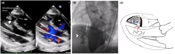
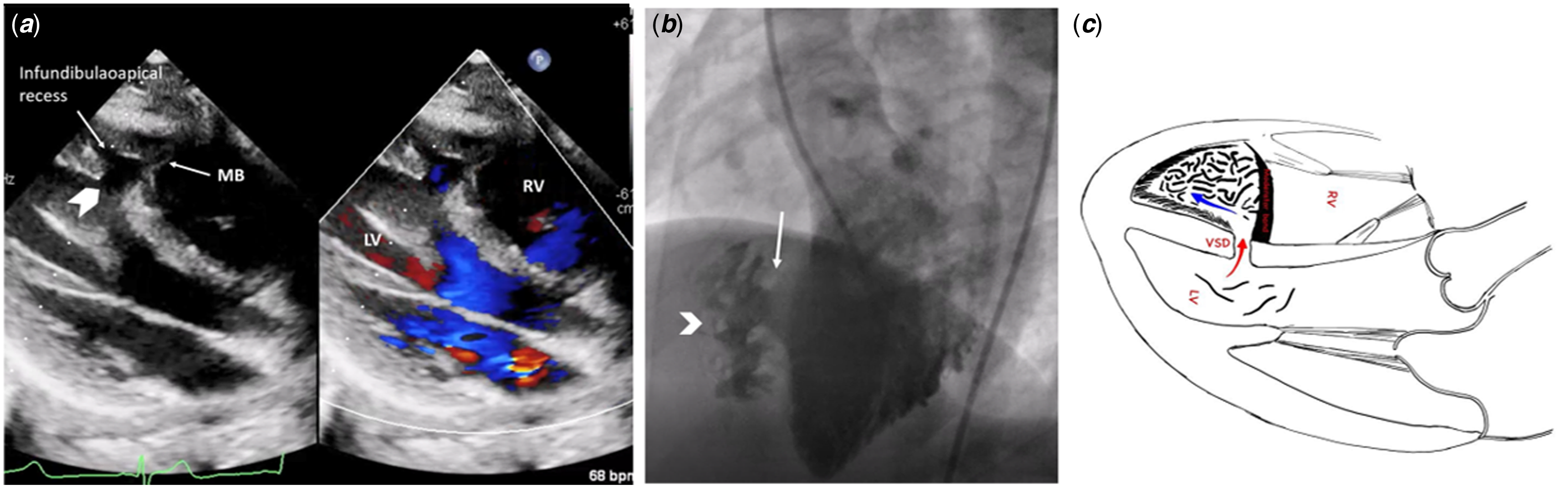
Case 1: A 17-year-old boy was diagnosed to have a large ventricular septal defect with bidirectional shunt and was referred for cardiac catheterisation to assess operability. As his oxygen saturation was 100%, a transthoracic echocardiogram was repeated which showed a large ventricular septal defect (9 mm) in the apical septum opening into the right ventricle beneath the moderator band (Fig 1a). Though there was bidirectional flow in the ventricular septal defect, there was no shunt into the right ventricle as the ventricular septal defect was excluded from the right ventricle body by the moderator band (Supplementary video 1). A cardiac catheterisation confirmed normal pulmonary artery pressure. Left ventricular angiogram showed a large ventricular septal defect filling a recess in the right ventricle which was closed by hypertrophied muscle bundles with intertrabecular recesses (Fig 1a, Supplementary video 2).
( a ) Transthoracic echo of case 1 in parasternal long-axis view showing the large apical ventricular septal defect opening into a recess in the right ventricle beneath the moderator band. ( b ) Left ventricular angiogram of case 1 in left anterior oblique view showing the large ventricular septal defect (arrow) restricted by hypertrophied muscular bands with intertrabecular recesses (Arrowhead). ( c ) Cartoon image of the same showing the ventricular septal defect restricted by the moderator band.
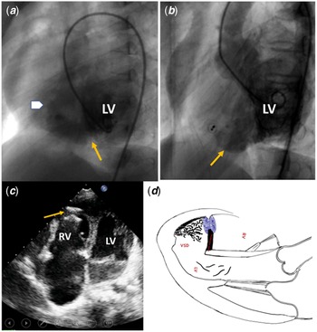
Case 2: A 4-year-old boy was diagnosed to have a large (15 mm) apical ventricular septal defect. Angiogram showed a large apical ventricular septal defect communicating with the infundibulum of the right ventricle through the infundibulo-apical recess (Fig 2a, Supplementary video 3). The ventricular septal defect was bounded superiorly by the moderator band which offered mild restriction to the shunt. A cardiac catheterisation confirmed normal pulmonary artery pressure (mean pulmonary artery pressure of 16 mm Hg) and Qp/Qs of 3:1. As there was no apical septum in the distal part of the ventricular septal defect, a size eight muscular ventricular septal defect occluder device was deployed at the exit of the ventricular septal defect at the right ventricle end (Fig 2c, Supplementary video 4), using the moderator band and other hypertrophied muscle bundles in right ventricle apex as rims for the device. The infundibulo-apical region of the right ventricle was thus incorporated into the left ventricle. On follow-up, he presented with cyanosis and effort intolerance. Ventricular angiogram done 10 years later showed no residual ventricular septal defect, the right ventricular apex was unobliterated, but with bald appearance of right ventricle (Fig 2b, Supplementary videos 5 and 6). There were small outpouchings in the right ventricle below the tricuspid valve and in the right ventricle outflow tract. Trans-oesophageal echocardiogram showed a patent oval foramen shunting right to left due to right ventricle diastolic dysfunction. The infundibulo-apical portion of the right ventricle gave the spurious appearance of a left ventricular diverticulum in left ventricular angiogram (Fig 2b). Cardiac MRI did not show any evidence of arrhythmogenic right ventricle dysplasia or features of other cardiomyopathies. There was mild left ventricular dysfunction, but there was no evidence of cardiomyopathy of the left ventricle in MRI.
( a ) Left ventricular angiogram of case 2 in left anterior oblique view showing the large apical ventricular septal defect (red arrow) into the infundibulo-apical recess and the exit of the recess (block arrow). ( b ) Left ventricular angiogram showing incorporation of the infundibulo-apical recess (arrow) as part of left ventricle. The septal occluder device is seen. ( c ) Transthoracic echocardiogram of case 2 in apical four-chamber view showing the large apical septal defect and closure of the defect at the right ventricular exit by a muscular ventricular septal defect occluder (arrow). ( d ) Cartoon of the same patient showing the ventricular septal defect in the parasternal long-axis view. The infundibular recess now functions as part of left ventricle .

Discussion
The echocardiographic, angiographic, and MRI images of the two illustrative cases are that of a rare variant of apical ventricular septal defect, the left ventricular-infundibular apical ventricular septal defect. These are a group of muscular ventricular septal defects that share a unique set of features: a large ventricular apical septal defect at the left ventricle end opening into the infundibulo-apical recess of the right ventricle which is bound superiorly by the moderator band and separated from the rest of the right ventricle body by thick muscle bundles which may have intertrabecular recesses. Reference Kumar, Lock and Geva1 Tsang et al. have attributed this particular character of the apical ventricular septal defect to incomplete compaction of the apical trabecular septum. Reference Tsang, Hsia, Yates and Anderson3 An important concept for understanding this entity is knowledge of the presence of two apices in the right ventricle: the inflow or sinus apex (posterior) to which the tensor part of the tricuspid valve is attached and the infundibular apex (anterior) which is related to the parietal band, the conal septum, septal band, and the moderator band. Reference Van Praagh, Mayer, Berman, Flanagan, Geva and Van Praagh2 The infundibulo-apical recess is located anterosuperior and leftward of the apex of the right ventricle inflow sinus. This recess extends to connect with the infundibulum of Right ventricular outflow tract, as demonstrated in the angiogram of case 2. Large apical ventricular septal defects that open beneath the moderator band into the right ventricle will open into this recess.
Spontaneous closure has been described in some of these defects Reference Kumar, Lock and Geva1 (as in our case 1), and small defects with insignificant shunt maybe followed up. Interventional closure has been described as a feasible and safe option, and the device can be placed at the true ventricular septal defect at the left ventricle septum. Reference Kumar, Lock and Geva1 Alternatively, the device can be deployed at the exit of the infundibulo-apical recess at the right ventricle end Reference Kumar, Lock and Geva1 (where the device is placed across the moderator band as in case 2), in which case the recess becomes part of the left ventricle. This may result in loss of right ventricle volume and right ventricle dysfunction later if the infundibulo-apical recess formed a substantial proportion of the right ventricle apex. Surgical closure can be done by right ventricle infundibulotomy Reference Van Praagh, Mayer, Berman, Flanagan, Geva and Van Praagh2–Reference Myhre, Duncan and Mee4 (incision placed over the infundibulo-apical recess, to the right of the anterior descending artery) and closing the ventricular septal defect with a patch. Care must be taken to close the true ventricular septal defect and avoid placing the patch at the right ventricle exit of the ventricular septal defect. Our second patient had loss of right ventricle volume due to exclusion of the right ventricle infundibular apex from the rest of the right ventricle. This may have led to the right ventricle diastolic dysfunction and subsequent right to left shunting across the patent oval foramen noted in him on follow-up. The right ventricle outpouchings noted in the follow-up angiogram and the bald appearance of the body of the right ventricle may point to an additional myopathic involvement (though MRI did not substantiate this), that has been described in some cases with large apicoinfundibular defects. The incorporation of the infundibulo-apical recess as part of left ventricle cavity acts as a large inert segment of the left ventricle, akin to an left ventricle apical aneurysm, and this could possibly be contributory to the mild left ventricle systolic dysfunction noted in our patient.
Conclusion
The prognosis of left ventricular-infundibular apical ventricular septal defect is determined by the size and morphology of the defect and the presence of associated anomalies. Early recognition and closure of the true septal defect has been shown to have good long-term prognosis. Associated defects like myocardial abnormalities can worsen the prognosis.
Supplementary material
For supplementary material accompanying this paper visit https://doi.org/10.1017/S1047951122000695
Acknowledgement
None.
Financial support
None.
Conflict of interest
None.