Halloysite is a 1:1 clay mineral that is chemically similar to kaolinite and has a hollow tubular structure in the sub-micrometre range (Tari et al., Reference Tari, Bobos, Gomes and Ferreira1999; Price et al., Reference Price, Gaber and Lvov2001; Joussein et al., Reference Joussein, Petit, Churchman, Theng, Righi and Delvaux2005). Kaolinite sheets are rolled into tubes due to the strain caused by lattice mismatch between adjacent silicon tetrahedral and aluminium octahedral sheets (Tari et al., Reference Tari, Bobos, Gomes and Ferreira1999; Joussein et al., Reference Joussein, Petit, Churchman, Theng, Righi and Delvaux2005). The halloysite particles have an external diameter of 50–70 nm, a lumen diameter of ~15 nm and a length of 1 ± 0.5 μm (Price et al., Reference Price, Gaber and Lvov2001). Large halloysite deposits, allowing for the supply of hundreds of tons of pure product, are located in Australia, the USA, China, New Zealand and Turkey, although minor amounts of halloysite nanotubes may occur at any kaolinite deposit (Yuan et al., Reference Yuan, Southon, Liu, Green, Hook, Antill and Kepert2008; Lvov et al., Reference Lvov, Wang, Zhang and Fakhrullin2016). Significant advantages of halloysite are its availability and relatively small price, together with its biocompatibility (Shchukin & Mohwald, Reference Shchukin and Mohwald2007; Lvov et al., Reference Lvov, Shchukin, Mohwald and Price2008; Fix et al., Reference Fix, Andreeva, Lvov, Shchukin and Mohwald2009; Veerabadran et al., Reference Veerabadran, Mongayt, Torchilin, Price and Lvov2009). Clay nanotube-based hybrid materials with antibacterial properties have been produced and investigated widely (Suner et al., Reference Suner, Sahiner, Akcali and Sahiner2020). Halloysite is a good template of the formation of nanoparticles and is an exceptionally convenient platform for building nanoarchitectonic materials (Stavitskaya et al., Reference Stavitskaya, Novikov, Kotelev, Kopitsyn, Rozhina and Ishmukhametov2018; Vinokurov et al., Reference Vinokurov, Stavitskaya, Chudakov, Glotov, Ivanov and Gushchin2018; Lvov et al., Reference Lvov, Panchal, Fu, Fakhrullin, Kryuchkova and Batasheva2019).
Nanostructures containing silver, copper, zinc and iron oxides and their combinations are well known to kill antibiotic-resistant bacteria (Cervini-Silva et al., Reference Cervini-Silva, Camacho, Palacios, del Angel, Pentrak and Pentrakova2016; Karthikeyan et al., Reference Karthikeyan, Mitu, Pandian, Anbarasu and Rajavel2017). Antibacterial applications of metal nanoparticle-based materials are important due to the increasing bacteria resistance to conventional antibiotics (Wei et al., Reference Wei, Lin, Zainal, Williams, Zhu and Kruzic2002; Moran et al., Reference Moran, Israela and Meital2007; Ramstedt et al., Reference Ramstedt, Cheng, Azzaroni, Mossialos, Mathieu and Huck2007; Kumar et al., Reference Kumar, Vemula, Ajayan and John2008; Jana et al., Reference Jana, Kondakova, Shevchenko, Sheval, Gonchar, Timoshenko and Vasiliev2017). The use of halloysite may help us to overcome the aggregation of nanoparticles, to prolong their activity and to reduce free nanoparticle release into the environment.
Silver ions have very strong bactericidal properties with a wide antibacterial spectrum, including antibiotic-resistant bacteria (Liong et al., Reference Liong, France, Bradley and Zink2009; Lv et al., Reference Lv, Su, He, Huang, Hu and Li2010; Zhao et al., Reference Zhao, Wang, Huo, Cui, Zhang and Ni2011; Shu et al., Reference Shu, Zhang, Yang and Yang2017; Mallakpour & Hatami, Reference Mallakpour and Hatami2018). Recently, Stavitskaya et al. (Reference Stavitskaya, Batasheva, Vinokurov, Fakhrullina, Sangarov, Lvov and Fakhrullin2019) reviewed various methods of the formation of silver particles or rods on halloysite. In addition, several reports demonstrated the formation of silver nanoparticles (AgNPs) on clay nanotubes and their antibacterial efficiency (Zhang et al., Reference Zhang, Zhang, Chen, Du, Zhang and Zhang2012; Zhang et al., Reference Zhang, Chen, Zhang, Zhang and Liu2013; Ouyang et al., Reference Ouyang, Guo, Fu, Yang, Hu and Tang2016; Song et al., Reference Song, Zhao, Qiu, Zhu, Wu, Ju and Dong2019). However, it remains challenging to achieve simple and efficient AgNP synthesis using halloysite.
In this work, a new synthesis strategy was applied to obtain AgNP templates on clay nanotubes via reduction–stabilization with tannic acid (TA). Tannic acid was used as both a reduction and stabilization agent to reduce the chemicals used in the reaction. Tannic acid is a natural, non-toxic and biodegradable polyphenolic compound extracted from plant sources (Luo et al., Reference Luo, Lai, Zhang, Liu, Liu and Liu2016). It is one of the typical hydrolysable tannins present widely in wood (Hagerman et al., Reference Hagerman, Riedl, Jones, Sovik, Ritchard, Hartzfeld and Riechel1998). Due to a number of phenolic hydroxyl groups in its structure, this natural extracted compound can not only chelate but also reduce silver ions to metallic AgNPs (Bulut O'acar, Reference Bulut O'acar2009; Dadosh, Reference Dadosh2009; Huang et al., Reference Huang, Wu, Liao and Shi2010; Bastús et al., Reference Bastús, Merkoçi, Piella and Puntes2014; Cao et al., Reference Cao, Zheng, Ji, Liu, Xie and Yang2014; Luo et al., Reference Luo, Zhang, Lai, Liu and Liu2015; Sahiner et al., Reference Sahiner, Sagbas and Aktas2015). Moreover, the utilization of TA does not require toxic chemical reductants (hydride, sodium borohydride) or stabilizers during synthesis, making it compatible with the green and sustainable development principles. Tannic acid has significant potential in medical applications due to its antioxidant and antimicrobial properties and its capacity for surface modification (Sagbas et al., Reference Sagbas, Aktas and Sahiner2015).
As a model organism for antibacterial investigation, we used Serratia marcescens. Serratia marcescens is a Gram-negative microorganism belonging to the Enterobacteriaceae family. Currently, this microorganism is understood to be a causative agent of nosocomial infections, causing conjunctivitis, keratitis, inflammatory diseases of the respiratory system, the gastrointestinal tract and the urinary tract and, in especially severe cases, bloodstream infections and meningitis (Cristina et al., Reference Cristina, Sartini and Spagnolo2019). It also causes outbreaks of infectious diseases in neonatal institutions, as well as in patients with severe immunodeficiency or in seriously sick patients, especially in intensive care units. Strains of S. marcescens are often multi-resistant, showing resistance to several classes of antibiotics. In the World Health Organization (WHO) list of priority pathogens for research and development in the discovery of new antibiotics, representatives of the Enterobacteriaceae family, to which S. marcescens belongs, are listed within the first priority category (critically high priority level) (Hong et al., Reference Hong, Xiao, Lazarus, Good, Maitta and Jacobs2016). According to the European Centre for Disease Prevention and Control (ECDC) in 2016, Serratia spp. were the sixth most frequently isolated microorganisms in patients with pneumonia hospitalized in European intensive care units (5.2 per 100 microorganisms), and they were in tenth and ninth places, respectively, for patients with bloodstream infections (3.3 per 100 microorganisms) and urinary tract infections (1.6 per 100 microorganisms).
In vivo investigations were performed using Caenorhabditis elegans, a free-living soil nematode. To analyse the possible negative effects on living invertebrate organisms, all samples were used as food for the nematodes. Nemotodes, both free living and parasitic, have been studied extensively due to their biological diversity and effects on human well-being and agriculture. Nematodes have been successfully employed as versatile model organisms in previous investigations by the present authors (Fakhrullina et al., Reference Fakhrullina, Akhatova, Lvov and Fakhrullin2015, Reference Fakhrullina, Akhatova, Kibardina, Fokin and Fakhrullin2017, Reference Fakhrullina, Khakimova, Akhatova, Lazzara, Parisi and Fakhrullin2019; Akhatova et al., Reference Akhatova, Fakhrullina, Khakimova and Fakhrullin2018). Use of the C. elegans nematode, as a typical and perhaps the most studied model organism, is extremely popular in biomedicine and nanoscience.
Here, we report halloysite-templated AgNPs stabilized with TA as being very active in killing S. marcescens and as being non-toxic to C. elegans at the same time. For the first time, AgNPs were synthesized on both pristine and modified clay nanotubes using TA as a reducing and stabilization agent. Tannic acid adsorption studies were also performed.
Materials
Halloysite nanoclay (Hall; Al2Si2O5(OH)4⋅2H2O), silver nitrate (AgNO3) ≥99.0%, (3-aminopropyl)trimethoxysilane (APTES; H2N(CH2)3Si(OCH3)3) 97% and TA (C76H52O46) were purchased from Sigma Aldrich. Distilled water and ethanol (96%) were used as solvents and reaction media.
Experimental
Synthesis of tannic acid-stabilized silver–halloysite nanomaterials
Halloysite nanotube modification with APTES
A total of 1 g of halloysite was dispersed in hydrogen peroxide at a mass to volume ratio of 1:20. The mixture was sonicated for 30 min and then left overnight on a magnetic stirrer. It was then centrifuged with water and dried at 110°C.
The following procedure was modified from Yang et al. (Reference Yang, Du, Zhang, Zhu, Wang and Zou2015). The purified halloysite was dispersed in 100 mL of ethanol in an ultrasonic bath for 30 min. Then, 0.2 mL of APTES per 1 g of halloysite was added and the mixture was kept in a capped flask in the ultrasonic bath for 10 min. The mixture was stirred overnight at 60°C, then it was centrifuged, washed with ethanol three times and dried at 80°C for 8 h. As a result, Hall_NH2 was obtained.
Tannic acid adsorption on the surface of nanotubes
Tannic acid was used as a reducing and stabilizing agent to obtain AgNPs on halloysite nanotubes. Pristine halloysite (Hall) and APTES-modified nanotubes (Hall_NH2) were used as templates for AgNP formation. A total of 0.1 g of Hall or Hall_NH2 and 0.002 g of TA were dispersed separately with ultrasound in 50 mL of water or ethanol. Then, the dispersion of nanotubes was placed on the stirrer and a TA solution was added to obtain a TA:Hall ratio of 2:100 by mass. The mixture was stirred for 24 h, centrifuged and washed twice with water (w) or ethanol (eth) solvent. The resultant hybrid materials were Hall_TA(w), Hall_TA(eth), Hall_NH2_TA(w) and Hall_NH2_TA(eth).
In situ synthesis of silver nanoparticles on carriers
Totals of 0.1 g of the Hall_TA(w), Hall_TA(eth), Hall_NH2_TA(w) and Hall_NH2_TA(eth) samples were dispersed in 10 mL of solvent (ethanol or water) for 30 min. A total of 0.004 g of silver nitrate solution in 5 mL of the same solvent was added dropwise to the halloysite dispersion. The colour of the mixture turned to yellow, and it was stirred for 24 h at room temperature. The resulting materials were centrifuged, washed three times with the solvent and dried at 60°C. Four samples of hybrid antibacterial materials were obtained: Ag/Hall_TA(w), Ag/Hall_TA(eth), Ag/Hall_NH2_TA(w) and Ag/Hall_NH2_TA(eth).
Sample characterization
Transmission electron microscopy (TEM; JEM-2100, JEOL, Japan) was used to study the morphology of the samples. The samples were dispersed in water placed on copper grids and analysed under accelerating voltages of 200 kV. The images obtained were processed using the ImageJ program developed by the National Institutes of Health (USA). Particle-size distributions were obtained from a minimum of 200 nanoparticles.
Thermogravimetric analysis (TGA) was performed with a Mettler Toledo TGA/DSC1 analyser. The sample was placed in a 70 μL Al2O3 crucible and heated in air atmosphere (air supply rate 10 mL min–1), in the range 30–800°C at a heating rate of 10°C min–1. The data obtained were processed with the service program STARe. The accuracy for temperature measurement was 0.3°C and that of mass loss was 0.1 μg.
Elemental composition was determined with an ARL™ QUANT'X X-ray fluorescence energy-dispersive spectrometer (XRF-EDS). The sample was weighed in a cuvette and analysis was performed at atmospheric pressure. The concentration of the components was obtained after determination of the valence state of the metals (oxide or individual) and selection of the background model.
Antibacterial activity investigation
Serratia marcescens ATCC 9986® strain (American-type culture collection) was used as a test culture. A microorganism culture was grown in Nutrient Broth medium (Oxoid Ltd) with a volume fraction of glycerol of 1%.
The antibacterial activity of Ag-loaded materials was determined using semi-liquid agar (1% pepton, 0.5% NaCl and 0.3% agar) in Petri dishes. A night culture of S. marcescens at a concentration of 0.4 by optical density (OD) at 595 nm was added to semi-liquid agar, distributed evenly and poured into Petri dishes. Then, 100 μL of the test sample suspension at a concentration of 10 mg mL–1 was added to the centre of the plate and cultured for 24 h at 28°C. Native halloysite nanotubes were used as a control.
The antibacterial activity of TA-modified and Ag-loaded samples was investigated by counting colony-forming units (CFUs) on dense nutrient media. The samples studied at a concentration of 1 mg mL–1 were cultured with S. marcescens for 18–20 h at 30°C in a nutrient medium. Then, S. marcescens was seeded using a micro-method on glucose–peptone agar. The colonies grown were counted after 20 h; native halloysite tubes were used as a control.
Determination of the dynamics of growth and inhibition of samples at a concentration of 1 mg mL–1 against planktonic strains of S. marcescens was carried out on a Мultisсan FC microplate photometer (ThermoScientific) for 48 h, followed by the construction of the growth curve.
In vivo studies of tannic acid-stabilized silver–halloysite nanomaterials
The free-living soil nematode C. elegans N2 Bristol was chosen as a model for studying the distribution of particles in nematodes. As a food, we used the usual food substrate of soil nematodes: the bacterium Escherichia coli OP50. In a six-well plate with age-synchronized adult nematodes, 200 μL of a suspension of bacteria and studied materials in concentrations of 1 mg mL–1 were added and incubated for 24 h. An Olympus BX41 upright microscope operating in dark-field mode (using a CytoViva condenser) was used to investigate the distribution of the nanoparticles in the nematodes. The development of the nematodes was monitored with a Nikon SMZ-745T stereomicroscope.
Results and discussion
Tannic acid is a well-known natural compound that acts as a stabilization and reducing agent at the same time. In addition, it is well adsorbed on mineral surfaces. Both the TA and unmodified halloysite have negative charge. Modification of halloysite with APTES results in a positively charged surface. To investigate the changes during the modification of halloysite and TA adsorption from various solvents, ζ-potential measurements were conducted after each stage of modification. The ζ-potential of pristine halloysite (Hall) changed from –44 mV to + 13 mV after APTES modification, suggesting successful NH2 grafting. The TA adsorption on the surface of Hall_NH2 from water resulted in a ζ-potential of clay nanotubes of –26 mV. The TA adsorbed on the tube surface from ethanol changed the ζ-potential to –12 mV. Hall-based materials showed the same values of –26 mV after TA adsorption.
To investigate the amount of TA adsorbed on the surface of pristine and modified halloysite, we tested Hall_TA(w), Hall_TA(eth), Hall_NH2_TA(w) and Hall_NH2_TA(eth) using TGA. Figure 1 shows the TGA profiles of halloysite and APTES-modified halloysite before and after TA adsorption from water or ethanol. The TGA profiles of halloysite and halloysite modified with silane as well as TA have been described in previous works (Horváth et al., Reference Horváth, Kristóf, Kurdi, Makó and Khunová2011; Keeling, Reference Keeling, Pasbakhsh and Churchman2015; Yang et al., Reference Yang, Du, Zhang, Zhu, Wang and Zou2015; Drits et al., Reference Drits, Sakharov and Hillier2018). Due to the simultaneous dehydration of the interlayer spacing of halloysite and the decomposition of TA, the amount of TA adsorbed was calculated by subtracting the weight loss percentage of the carrier from that of the hybrid material at 750°C. The adsorption of TA depends on the solvent as well as on the carrier (Fig. 1a,b). In both cases, more tannin was adsorbed from the water solution compared to ethanol. The tannin content in the Hall_TA samples was ~0.5 wt.% for the ethanol system and ~1.5 wt.% for the water system. More tannin is adsorbed on Hall_NH2 in water medium (~2.5%) than in ethanol medium (~1%). The TGA data suggest that the modified halloysite samples adsorb TA better than pristine nanotubes, which is due to the positively charged surface of amino-grafted halloysite that adsorbs negatively charged TA. The polarity of the solvent affects the surface charge, as was determined from the decomposition curves.
TGA profiles of (a) Hall_TA produced form water (w) or ethanol (eth) and (b) Hall_NH2_TA produced from water (w) or ethanol (eth).

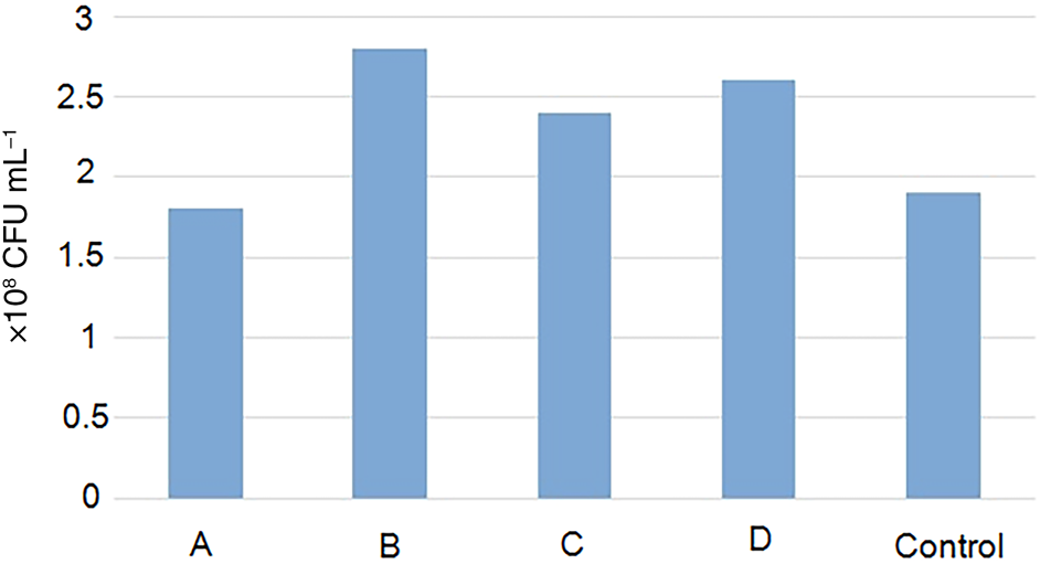
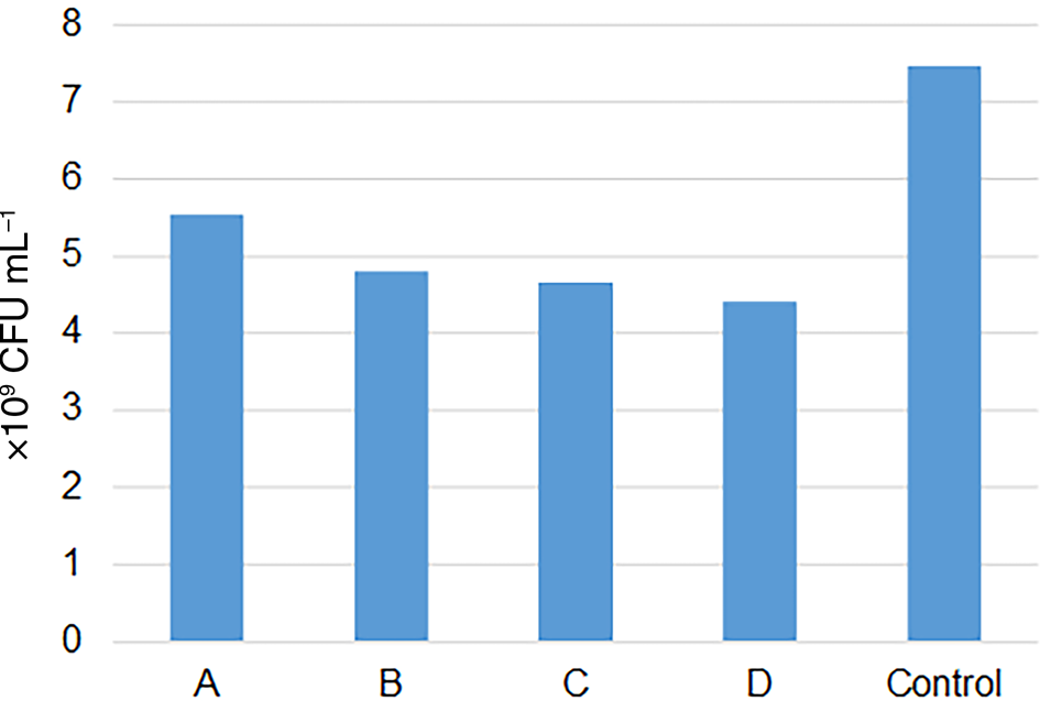
To confirm the effect of silver in antibacterial materials, we performed antibacterial tests of TA-adsorbed samples (Fig. 2). Only Hall_TA(eth) had mild antibacterial activity. Other samples showed intensification of bacteria growth instead of inhibition.
Concentration of S. marcescens after 24 h in: A = S. marcescens with Hall_TA(eth); B = S. marcescens with Hall_TA(w); C = S. marcescens with Hall_NH2_TA(eth); D = S. marcescens with Hall_NH2_TA(w); and Control = S. marcescens with pristine halloysite.
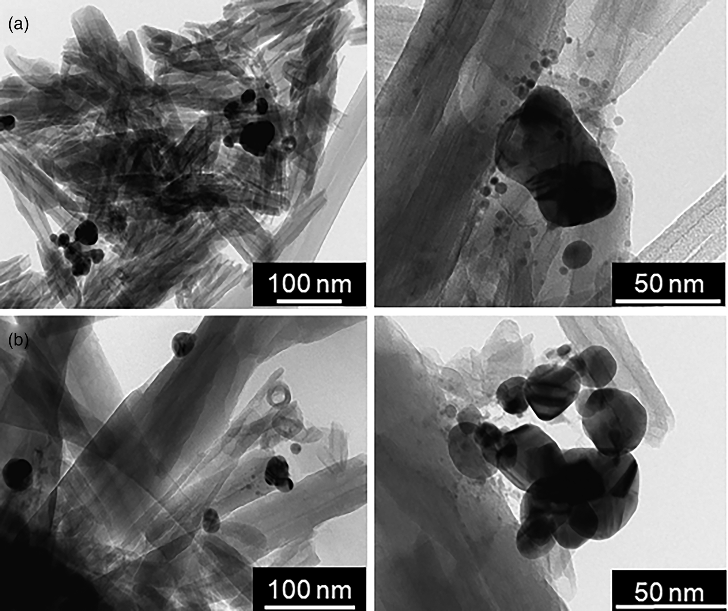
To investigate the mechanism of AgNP formation on TA, a modified halloysite solution of AgNO3 in the corresponding solvent was added to the hybrid material dropwise, and the resulting dispersion was left for 24 h at room temperature. The particles formed in ethanol are of better quality and the samples are more homogeneous. The morphology of water-based samples demonstrates the formation of numerous aggregates and the agglomeration of silver particles into larger structures (Fig. 3). This might be due to the high polarity of the solvent and its greater surface tension causing particle interactions.
TEM images of (a) Ag/Hall_NH2_TA(w) and (b) Ag/Hall_TA(w) particles.

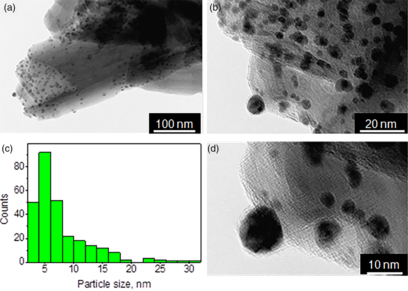
The TEM images demonstrating the morphologies of the Ag/Hall_TA(eth) and Ag/Hall_NH2_TA(eth) samples are shown in Figs 4 and 5. The formation of nanoparticles with an average size of ~7 nm is characteristic of the sample produced by the adsorption of TA from ethanol on pristine halloysite with further silver ion reduction on the surface in the same solvent (Fig. 4). Due to the relatively low adsorption values not all nanotubes were covered with TA and, as a result, with AgNPs. The high-resolution TEM image (Fig. 4d) shows the formation of the TA layer on the surface of the nanoparticles.
(a, b, d) Morphology and (c) particle-size distribution of Ag/Hall_TA(eth) particles.

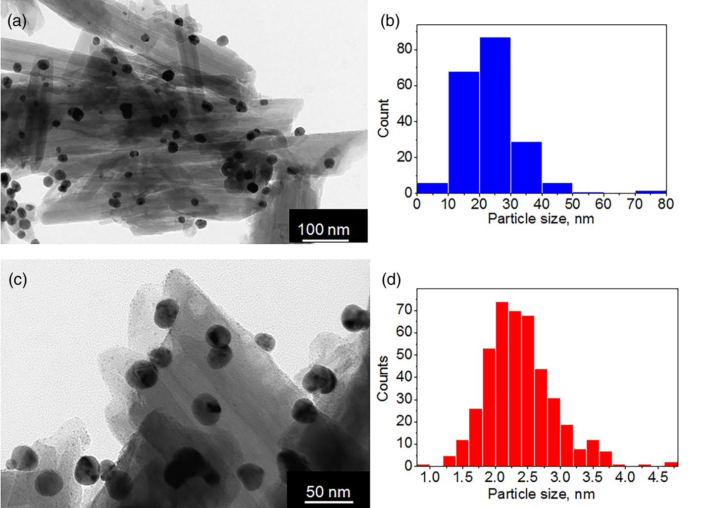
(a, c) Morphology and (b, d) particle-size distribution of Ag/Hall_NH2_TA(eth).

Figure 5 shows TEM images and the particle-size distribution of Ag/Hall_NH2_TA(eth). For the amino-modified halloysite, two types of nanoparticles were formed (Fig. 5b,d). Ultra-small silver particles with sizes of ~2.5 nm were adsorbed homogeneously on the surface of halloysite, probably showing APTES positions. The formation of 2.5 nm nanoparticles on modified halloysite has not been described in previous works and may have potential utility in various fields. Due to the high concentration of TA in the samples, especially in Ag/Hall_NH2_TA(w), larger nanoparticles with sizes of ~20–40 nm were also formed in both Hall_NH2 samples. These particles have an almost spherical shape and are strongly attached to the halloysite surface. The stabilization layer of TA covers both small and large particles.
In contrast to the proposition that a higher concentration of TA is capable of Ag reduction in water-based samples, elemental analysis showed that Ag/Hall_TA(w) contains 0.5 wt.% Ag and Ag/Hall_NH2_TA(w) contains 0.7 wt.% Ag. Materials produced from ethanol showed greater silver concentrations of 1 and 1.4 wt.% Ag in Ag/Hall_TA(eth) and Ag/Hall_NH2_TA(eth), respectively.
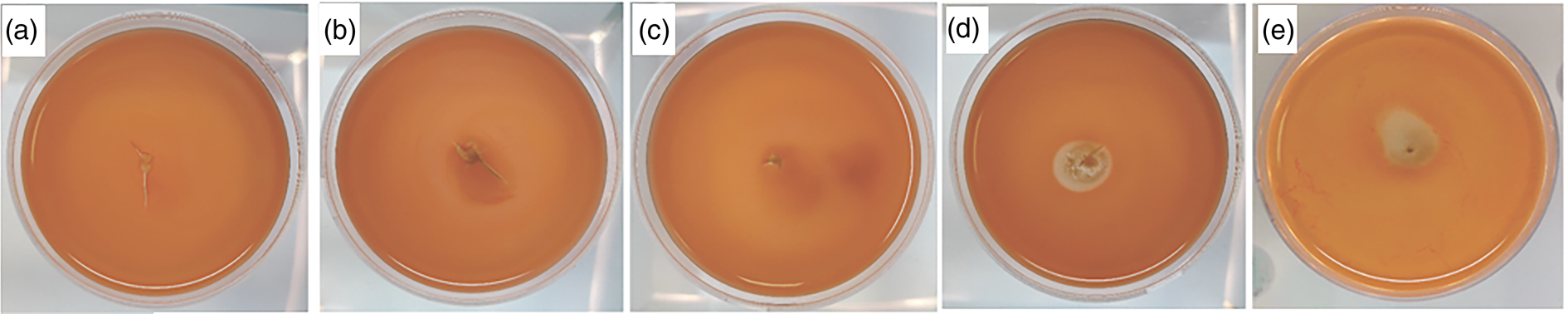
Most studies on the antibacterial activity of silver-based halloysite nanomaterials apply a single method of investigation of antibacterial activity. In the present work, we show that a complex study of materials may better demonstrate the mechanisms of antibacterial action. Figure 6b–e shows the contact of silver-based material dispersions in buffer solutions with target bacteria after 24 h of growth. In the cases of Ag/Hall_NH2_TA(w) and Ag/Hall_NH2_TA(eth), there are no growth-inhibition zones, which is similar to the control sample. The presence of a growth-inhibition zone in semi-liquid agar at the studied concentration was observed only in samples Ag/Hall_TA(eth) and Ag/Hall_TA(w), showing good antibacterial effect.
Antibacterial activity of the samples against S. marcescens. (a) Control S. marcescens with pristine halloysite; (b) S. marcescens with Ag/Hall_NH2_TA(w); (c) S. marcescens with Ag/Hall_NH2_TA(eth); (d) S. marcescens Ag/Hall_TA(w); (e) S. marcescens with Ag/Hall_TA(eth). The diameter of the cups is 60 mm.

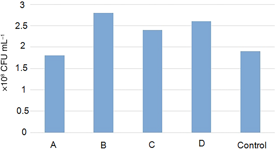
In general, the contact method is often applied for antibacterial activity investigation, including of silver–halloysite nanomaterials. It may be concluded that Ag/Hall_NH2_TA(w) and Ag/Hall_NH2_TA(eth) are not efficient as antibiotics. The inhibition zones of Ag/Hall_TA(eth) and Ag/Hall_TA(w) being almost the same size may lead to the conclusion of their similar activity. As a result, the investigation of nanomaterials with low or no ion release should be conducted using complex methods. By counting CFUs, it was observed that all of the samples in liquid nutrient media inhibited the growth of S. marcescens bacteria after 18–20 h (Fig. 7). Ag/Hall_NH2_TA(w) inhibits growth by 26.3%, Ag/Hall_NH2_TA(eth) by 35.7%, Ag/Hall_TA(w) by 37.5% and Ag/Hall_TA(eth) by 41.0% relative to the control sample.
Concentration of S. marcescens after 24 h in: A = S. marcescens with Ag/Hall_NH2_TA(w); B = S. marcescens with Ag/Hall_NH2_TA(eth); C = S. marcescens Ag/Hall_TA(w); D = S. marcescens with Ag/Hall_TA(eth); and Control = S. marcescens with pristine halloysite.
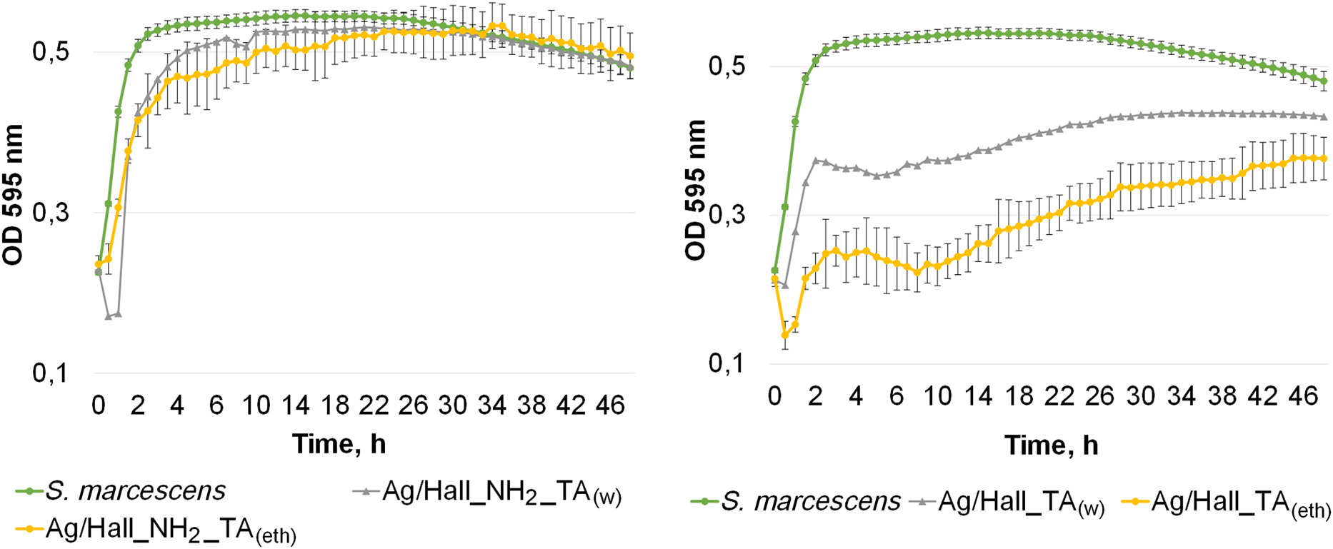
The growth curve uses the absorbance data from a microplate photometer and shows that very high antibacterial activity was observed in all samples during the first 0.5–1.0 h (Fig. 8). However, an increase in OD is observed in the samples produced from amino-modified halloysite, which indicates the growth of S. marcescens bacteria. Throughout the experiments, the indicators in these samples did not statistically and significantly differ from those of the control sample. The Ag/Hall_NH2_TA samples demonstrated pronounced antibacterial activity. The antibacterial effect in these samples was observed throughout the study from the first hours of setting up the experiment. The concentration of bacteria in Ag/Hall_TA(w) decreased on average by 1.25 times and in sample Ag/Hall_TA(eth) by 1.6 times relative to the control. A slight decrease in the activity of these samples is observed after 24 h of incubation.
The growth curves of S. marcescens. The antibacterial effects of the various samples against S. marcescens at a concentration of 1.0 mg mL–1 are shown.

We conclude that the best antibacterial effect is observed in the samples obtained by the reduction of silver on pristine halloysite using both water and ethanol due to the two-fold greater silver concentration in the ethanol-based sample. The bacterial colony counts demonstrated that the effect of the least active water-based Ag/Hall_NH2_TA is also viable. Although Ag/Hall_NH2_TA samples contain greater silver concentrations than the pristine halloysite-based materials, they showed much lower activity. This result may be evidence of capped nanoparticle formation and different antibacterial mechanisms.
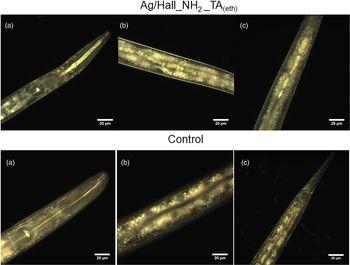
Figure 9 represents the distribution of TA-stabilized silver–halloysite nanomaterials in a nematode body. Image-based analysis has shown that the nanomaterials are located only in the intestinal tracts of the nematodes. The investigation showed that none of the samples was harmful to the nematodes or their reproductive systems.
Dark-field images showing the distribution of Ag/Hall_NH2_TA(eth) in the bodies of C. elegans compared to control C. elegans: (a) the pharynx; (b) the middle section of the intestine; and (C) the back of the intestine.

Conclusions
A new simple process of the in situ synthesis of AgNPs was performed using halloysite nanotubes as a template. Nanoparticle formation was achieved through the simultaneous reduction and stabilization of silver by TA adsorbed on pristine or amino-modified halloysite nanotubes. The TA is better adsorbed on the surface of modified and pristine halloysite from a water solution. The formation of nanoparticles of various sizes was observed on various carriers, with 5–7 nm particles formed on pristine halloysite and 20–30 nm particles formed on amino-grafted nanotubes, as well as ultra-small 2.5 nm particle formation. All samples showed antibacterial activity according to a complex study of S. marcescens. The best and most long-lasting activity was attributed to the sample produced from pristine halloysite with AgNPs formed by TA reduction in ethanol medium. The nanoparticles formed in situ using APTES-modified halloysite from ethanol are highly monodispersed and of very good quality, though they showed quite low antimicrobial activity under the studied conditions. In vivo studies showed that none of the samples has negative impacts on C. elegans. The nanomaterials accumulated only in the intestinal tract of the nematodes, demonstrating their possible application for the targeted delivery of antimicrobial nanocomposites.
Financial support
This work was supported by the Russian Science Foundation under grant 18-73-00342. This work is also performed according to the Russian Government Program of Competitive Growth of Kazan Federal University.