Implications
Piglets are prone to diarrhoea after they have been weaned. Protein restriction and succedent realimentation can decrease diarrhoea without reducing the ultimate growth performance of weaned piglets. This study reveals the effects of protein restriction and succedent realimentation on ileal microbiota and suggests that protein restriction and succedent realimentation, as a nutrition strategy, does not impair ileal morphology. Rather, it maintains ecological homeostasis in the ileum and decreases diarrhoea. The application of this nutrition strategy in animal husbandry will benefit the livestock industry.
Introduction
Post-weaning diarrhoea (PWD), a leading cause of death in piglets, is a common problem in animal husbandry (McLamb et al., Reference McLamb, Gibson, Overman, Stahl and Moeser2013). It has become increasingly clear that nutritional factors are associated with PWD and play an important role in the gastrointestinal health of animals. Some studies have highlighted that a low-protein diet (CP level <18%), as a vital nutritional strategy, may reduce protein fermentation indices (e.g. the concentration of ammonia in the intestine of piglets) and PWD (Heo et al., Reference Heo, Kim, Yoo and Pluske2015). Intestinal microbial metabolites from undigested dietary protein fermentation include ammonia, indole and various phenols. These metabolites are toxic and can provoke PWD (Villodre Tudela et al., Reference Villodre Tudela, Boudry, Stumpff, Aschenback, Vahjen, Zentek and Pieper2015). However, it has also been reported that piglets who were fed a low-protein diet had reduced growth performance compared with piglets on a high-protein diet (Yue and Qiao, Reference Yue and Qiao2008). Thus, the concept of compensatory growth can be applied to this situation. Compensatory growth is a phenomenon in which animals exhibit a faster growth rate after their nutritional levels return to normal due to prior malnutrition or artificial restriction (Chaosap et al., Reference Chaosap, Parr and Wiseman2011; Madsen and Bee Reference Madsen and Bee2015). Although the compensatory growth induced by protein restriction may reduce diarrhoea levels without degrading the ultimate growth performance, the effects of protein restriction and succedent realimentation on gut microbiota in weaned piglets are still unclear.
In addition to the large intestine, a number of microbes also colonise in the ileum, and bacterial fermentation also takes place in the small intestine, predominantly in the ileum (Piccolo et al., Reference Piccolo, Mercer, Bhattacharyya, Bowlin, Saraf, Pack, Chintapalli, Shankar, Adams, Badger and Yeruva2017). The dietary CP level is closely related to the balance of ileal microbiota, which further influences ileal morphology, digestive and absorptive functions, barrier functions and host metabolism (Pieper et al., Reference Pieper, Tudela, Taciak, Bindelle, Pérez and Zentek2016). A previous study has shown that a reduction in dietary protein (from 16% to 13%) could improve the ileal bacterial community structure and barrier functions in adult pigs, whereas the over-reduction of dietary protein (from 16% to 10%) damaged ileal morphology and disturbed the bacterial community structure (Fan et al., Reference Fan, Liu, Song, Chen and Ma2017). Surprisingly, little is known about the impact of protein realimentation on the ileal microbiota in pigs. Thus, it is necessary to explore the effects of protein restriction and realimentation on the ileal microbiota and its metabolites. We therefore hypothesise that the ileal microbial composition and metabolites change according to the dietary protein restriction and realimentation and influence ileal morphology and barrier functions. In the present study, we investigated the effects of protein restriction and realimentation on ileal morphology, ileal microbial composition and metabolites in weaned piglets.
Material and methods
Animal trial
Thirty-six 28-day-old weaned piglets (initial body weight 6.47 ± 0.04 kg) from a pig farm in the Jiangsu province of China were randomly divided into a control group and a treatment group with six pens per group and three piglets housed in each pen. During the protein restriction period (days 0 to 14), the dietary CP level was normal (18.83%) in the control group and low (13.05%) in the treatment group. Six piglets from each group were euthanised on day 14, and the rest of the piglets continued with the protein realimentation experiment. During the protein realimentation period (days 14 to 28), the dietary CP level was normal (18.83%) in both groups. Six piglets from each group were euthanised for sampling on day 28. The ingredient and nutrient composition of both diets are shown in Supplementary Table S1. All of the piglets had free access to food and water during the entire trial. The daily food intake of the piglets in each pen was accurately recorded; the piglets were weighed every other day, and the index of growth performance was calculated. After the piglets were euthanised, the abdominal cavity of each piglet was opened. The ileum was then removed and saved for experiments with its mucosa, digesta and tissue.
Assessment of diarrhoea and calculation of diarrhoea index
The faecal consistency was checked between 1800 h and 2000 h every night and determined according to the following scoring criteria: 1 = hard, dry and cloddy; 2 = firm; 3 = soft, with shape; 4 = soft and liquid; 5 = watery and dark; 6 = watery and yellow; and 7 = foamy and yellow. A faecal consistency score >3 represented piglets with diarrhoea (Sugiharto et al., Reference Sugiharto, Poulsen, Canibe and Lauridsen2015). The diarrhoea index (DI) in each pen was calculated as:
 $${\rm{DI }} = X/({\rm{14}}\,\cdots\, Y)\,\cdots\, {\rm{1}}00\% ,$$
where X is the number of piglets with diarrhoea (faecal score >3) per pen and Y is the number of piglets per pen.
Microbial DNA isolation and high-throughput sequencing
The DNA from 0.3 g of ileal digesta was extracted, according to the method used by Zoetendal et al. (Reference Zoetendal, Akkermans and Vos1998). Following this extraction, the PCR of the V3–V4 regions of the bacterial 16S ribosomal RNA (rRNA) gene was amplified with the forward primer 319F (ACTCCTACGGGAGGCAGCAG) and reverse primer 806R (GGACTACHVGGGTWTCTAAT). The PCR augmentation program was performed with an initial denaturation temperature of 95°C for 2 min, followed by 25 cycles of 95°C for 30 s, 55°C for 30 s, 72°C for 30 s and a final extension of 72°C for 7 min (Zhu et al., Reference Zhu, Niu, Shi, Wang and Zhu2017). The amplicons were purified using the AxyPrep DNA Gel Extraction Kit (Axygen Biosciences, Union City, CA, USA) according to the manufacturer’s instructions. The amplicons were then quantified using QuantiFluor™-ST (Promega, Madison, WI, USA). The purified amplicons were then pooled in an equimolar and paired-end sequenced on an Illumina MiSeq platform according to the standard protocol (Caporaso et al., Reference Caporaso, Lauber, Walters, Berg-Lyons, Huntley, Fierer, Owens, Betley, Fraser, Bauer, Gormley, Gilbert, Smith and Knight2012).
The raw sequencing data were uploaded to the GenBank database of the National Center for Biotechnology Information with the accession number SRP154413. Then the raw FASTQ files generated from 16S rRNA MiSeq sequencing were de-multiplexed and quality-filtered using QIIME software (version 1.70; Flagstaff, AZ, USA) with standard criteria (Mao et al., Reference Mao, Zhang, Liu and Zhu2015). Operational taxonomic units (OTUs) were clustered with a 97% similarity cut-off using UPARSE (version 7.1; http://drive5.com/uparse/), and chimeric sequences were identified and removed using UCHIME (Edgar, Reference Edgar2010). Mothur v.1.30.1 software was used to assess microbial community diversity, including the number of observed OTUs, Chao richness estimators and Simpson and Shannon indices. The between-specimen diversity (β-diversity) was visualised through a principal coordinate analysis, which was assessed by calculating distance and dissimilarity metrics.
Determination of microbial metabolites
The concentration of lactic acid in the ileal content was measured using a commercial kit (Nanjing Jiancheng Bioengineering Institute, China) according to the manufacturer’s instructions. The concentrations of short-chain fatty acids (SCFAs) in the ileal content were determined by gas chromatography (Shimadzu, GC-14A with a hydrogen flame ionisation detector, Japan), following the method of a previous study (Shi et al., Reference Shi, Zhu, Niu, Wang, Wang and Zhu2018). The temperatures of the injector, column and detector were 110 °C, 150 °C and 180°C, respectively. The concentrations of ammonia in the ileal digesta were analysed using the colorimetric method (Nyachoti et al., Reference Nyachoti, Omogbenigun, Rademacher and Blank2006).
Ileal morphology
After being rinsed with normal saline, the ileum tissues were quickly placed in a 4% paraformaldehyde solution. The samples were made into paraffin sections and stained with haematoxylin and eosin. The slices were observed under computer-aided video microscopy (DXM1200C; Nikon) with ×4 objective, and villus height and crypt depth were measured using an image analysis system (NIS-Elements BR software, version 2.20; Nikon). Representative photographs of ileal morphology were collected using the capture button in the NIS-Elements BR software.
Immunohistochemistry
The paraffin sections were de-waxed in xylene and washed in absolute alcohol, then in 85% ethanol, then in 75% ethanol and finally in distilled water. Antigen retrieval was performed in a citrate buffer (pH 6.0) in a microwave oven. The slides were blocked with 3% bovine serum albumin (Servicebio, China) at room temperature for 30 min and then incubated with antibodies against zonula occludens-1 (ZO-1) and Claudin-1 in a humidified chamber at 4°C overnight. The stained sections were then washed three times with phosphate buffer solution (pH 7.4) for 5 min, incubated with an anti-rabbit (or mouse) secondary antibody for 50 min and treated with 3,3’-diaminobenzidine (DAB) chromogenic solution and hematoxylin (Servicebio, China). The cell nucleus with hematoxylin staining was blue, and the positive expression of the target protein with DAB was a brownish yellow colour. Images were obtained by light microscopy (XSP-C204, CIC) and representative photographs were processed with CaseViewer software.
RNA extraction and quantitative real-time PCR
The ileal mucosa was ground with liquid nitrogen, and the total RNA of the ileal mucosa was extracted using TRIzol (Invitrogen, China). The samples were reversely transcribed to complementary DNA (cDNA) with a PrimeScript RT Reagent Kit (TAKARA Biotechnology, Dalian, China) according to the manufacturer’s protocol. qPCR amplification was implemented using StepOne-Plus (Applied Biosystems, CA, USA) with StepOne software (version 2.2.2; Applied Biosystems). A 20 μL reaction system of PCR consisted of 10 μL SYBR Premix Ex Taq (TliRNaseH Plus), 0.4 μL of each primer, 0.4 μL of ROX reference dye (50×), 6.8 μL of double-distilled H2O and 2 μL of cDNA. The program of PCR amplification was performed with an initial denaturation step of 95°C for 30 s, followed by 40 cycles of 95°C for 5 s and 60°C for 34 s. The last stage was at 95°C for 15 s, 60°C for 1 min and 95°C for 15 s. The primers used are listed in Supplementary Table S2. The ileal mucosal genes measured in this study included the following barrier function-related genes: ZO-1, Occludin, Claudin-1, Claudin-2, mucin-2 ( MUC-2 ), transforming growth factor-β ( TGF-β ), and interleukin-1B ( IL-1B ). The following digestion- and absorption-related genes were also measured: amino-peptidase A ( APA ), amino-peptidase N ( APN ), peptide transporter-1 ( PEPT-1 ) and sodium-glucose transporter-1 ( SGLT-1 ). The relative mRNA expression of these genes was calculated relative to the expression of β-actin using the 2−ΔΔCt method (Herfel et al., Reference Herfel, Jacobi, Lin, Fellner, Walker, Jouni and Odle2011).
Statistical analysis
Data were analysed with SPSS 25.0 and reported as mean ± SEM. The Student’s t-test was applied to compare differences between the two groups. The normality of variable distribution was tested with the Shapiro–Wilk test. The variables that had a non-normal distribution were analysed using the non-parametric methods. A P-value <0.05 was regarded as statistically significant.
Results
Growth performance and diarrhoea index
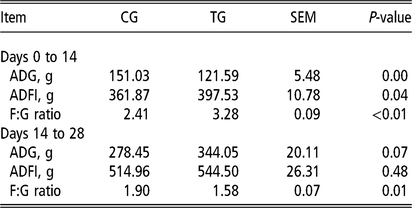
In the present study, we observed that a sharp decline in dietary CP, from 18.83% to 13.05%, in a weaned piglet trial reduced average daily gain (ADG; P < 0.01; Table 1) but augmented average daily feed intake (ADFI; P < 0.05) and the feed–gain ratio (F:G ratio; P < 0.01) during the 14-day protein restriction. After a succedent 14-day realimentation, no significant differences were observed in ADG (P = 0.07) and ADFI in the two groups, but the treatment group had a lower F:G ratio compared with the control group (P = 0.01). As shown in Figure 1a, the body weight of piglets was significantly decreased in the treatment group compared with the control group on day 14 (P < 0.05), while this difference between the two groups was not significant on day 28. Our results also found that the treatment group had a decreased DI during the protein restriction stage (P < 0.05; Figure 1b) and succedent realimentation stage (P < 0.05) compared with the control group.
Effect of protein restriction and realimentation on the (a) average body weight, (b) diarrhoea index, and (c, d) beta diversity, namely PCoA of ileal bacterial community in the control and treatment groups of weaned piglets. The values are expressed as means ± SEM, with six piglets per group. CG = control group; TG =, treatment group; PCoA = principal coordinates analysis. *, a significant difference (P < 0.05).

Effect of protein restriction and realimentation on the growth performance of piglets

CG = control group; TG = treatment group; ADG = average daily gain; ADFI = average daily feed intake; F:G ratio = feed–gain ratio.
Bacterial alpha diversity, beta diversity and bacterial community composition in the ileal content
To assess the impact of protein restriction and realimentation on bacterial communities, the sequences of the 16S rRNA gene were amplified. Estimators of ileal bacterial community diversity (Shannon and Simpson) and richness (Chao and OTU) are shown in Supplementary Table S3. None of these measures were significantly affected by the protein restriction or realimentation stages. As shown in Figure 1c and d, ileal digesta samples were apparently clustered separately in the two groups on day 14, but there was no statistical significance between the two groups on day 28. The bacterial community composition was assessed at different taxonomic levels. At the phylum level, the dominant groups were Firmicutes, Proteobacteria, Actinobacteria and Cyanobacteria in the ileum; they accounted for >99% of the total ileal bacterial community (Supplementary Figure S1a). One of the most dominant phyla was Firmicutes, accounting for 75.38% to 92.95% of the total sequences (Supplementary Table S4). There was no statistical difference in the abundance of Firmicutes and Proteobacteria between the two groups on day 14. The abundance of Actinobacteria was significantly increased in the treatment group compared with the control group on day 14 (P < 0.01). However, the abundance of Firmicutes was significantly increased and that of Proteobacteria was significantly decreased in the treatment group on day 28 (P < 0.05) compared with the control group.
At the family level, Lactobacillaceae was the most predominant family (27.22% to 71.23%; Supplementary Table S5; Supplementary Figure S1b). The abundance of Lactobacillaceae dropped dramatically in the treatment group compared with the control group on day 14 (P < 0.01). At the same time, an increased abundance of Clostridiaceae 1 and Halomonadaceae was observed in the treatment group on day 14 (P < 0.01). Furthermore, the abundance of Lactobacillaceae and Clostridiaceae 1 increased and that of Pseudomonadaceae, Enterobacteriaceae, Moraxellaceae and Halomonadaceae declined in the treatment group on day 28 (P < 0.05) compared with the control group.
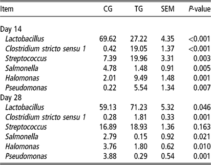
At the genus level, the most dominating genus was Lactobacillus (27.22% to 71.23%; Table 2; Supplementary Figure S1c). Compared with the control group, the abundance of Lactobacillus and Salmonella decreased, and that of Clostridium sensu stricto 1, Streptococcus, Halomonas and Pseudomonas sharply increased in the treatment group on day 14 (P < 0.01). However, the abundance of Lactobacillus and Clostridium sensu stricto 1 increased and that of Salmonella, Halomonas and Pseudomonas decreased in the treatment group compared with the control group on day 28 (P < 0.05).
Altered relative abundance of microbiota at the genus level in the ileal content of piglets

CG = control group; TG = treatment group.
Microbial metabolites in the ileal content
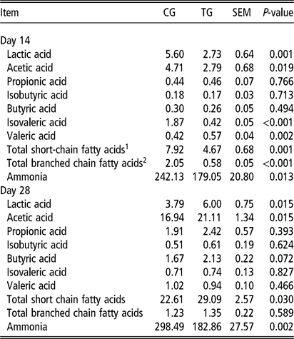
To assess microbial metabolism in the ileal content, the concentrations of lactic acid, SCFAs and ammonia were measured (Table 3). The concentrations of lactic acid, acetic acid, isovaleric acid, total SCFAs and total BCFAs (isobutyric acid and isovaleric acid) in the ileal content of piglets in the treatment group significantly declined on day 14 (P < 0.05) compared with the control group. However, the concentrations of propionic acid, butyric acid and isobutyric acid in the ileal content were not significantly different between the two groups. On the contrary, the concentrations of lactic acid, acetic acid and total SCFAs in the ileal content in the treatment group increased significantly compared with the control group on day 28 (P < 0.05). We did not observe any significant difference in the concentrations of propionic acid, butyric acid, valeric acid, isobutyric acid, isovaleric acid and total BCFAs in the ileal content between the two groups on day 28. Specifically, the ammonia concentration in the ileal content in the treatment group decreased on both days 14 and 28 (P < 0.05) compared with the control group.
Effect of protein restriction and realimentation on the concentrations of lactic acid (mmol/L), SCFAs (μmol/g) and ammonia (μg/g) in the ileal content of piglets

CG = control group; TG = treatment group.
1 Acetic acid, propionic acid, isobutyric acid, butyric acid, isovaleric acid and valeric acid.
2 Isobutyric acid and isovaleric acid.
Intestinal morphology
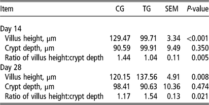
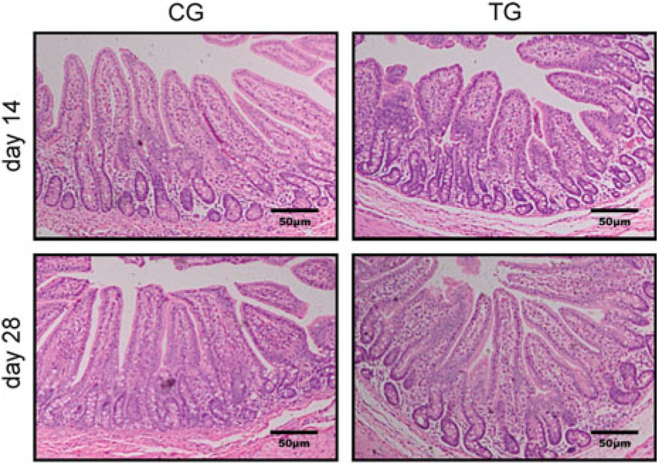
Compared with the control group, the treatment group showed a significantly decreased villus height and ratio of villus height:crypt depth of the ileum on day 14 (P < 0.01; Table 4; Figure 2). The crypt depth of the ileum showed no significant statistical difference between the two groups on day 14. However, the villus height and ratio of villus height:crypt depth were higher in the treatment group than in the control group on day 28 (P < 0.05). Similarly, the crypt depth of the ileum did not show a significant statistical difference between the two groups on day 28.
Effect of protein restriction and realimentation on ileal morphology of piglets

CG, control group; TG, treatment group.
Mucosal morphology by haematoxylin and eosin staining in the ileum of weaned piglets. CG = control group; TG = treatment group.

Intestinal barrier
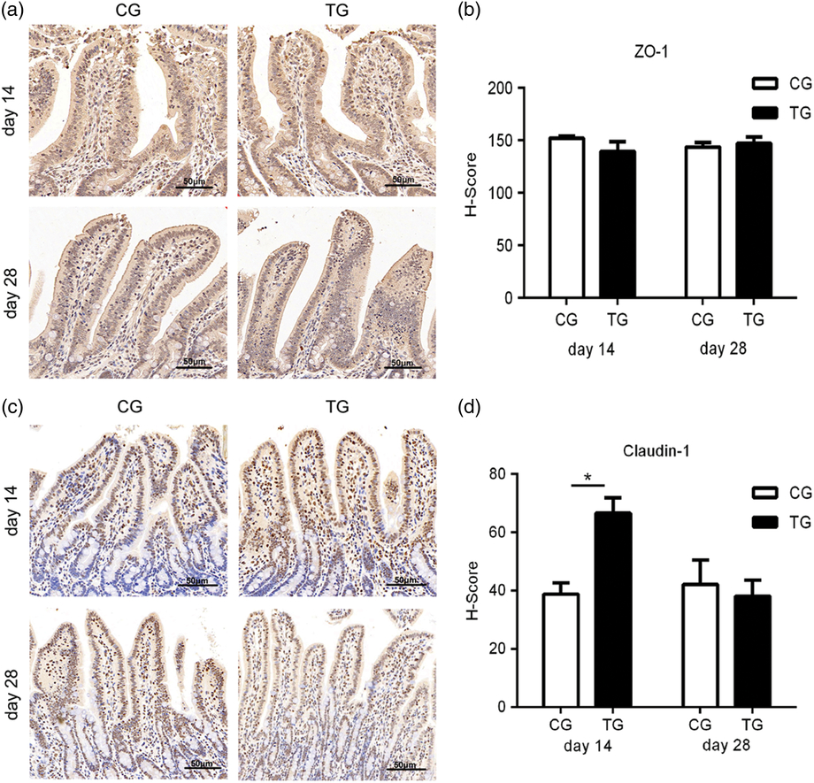
As shown in Figure 3a, the treatment group had lower ZO-1 and MUC-2 mRNA levels in the ileum compared with the control group on day 14 (P < 0.05). However, Occludin, Claudin-1 and Claudin-2 mRNA levels did not show any difference between the two groups. Meanwhile, no statistically significant difference was observed in the relative mRNA expression of ZO-1, Occludin, Claudin-1, Claudin-2 and MUC-2 between the two groups on day 28 (Figure 3b). The treatment group also showed a significant increase in the relative mRNA expression of TGF-β in the ileum compared with the control group on day 14 (P < 0.01), whereas there was no significant statistical difference in the relative mRNA expression of TGF-β between the two groups on day 28. Protein restriction and succedent realimentation did not influence the relative mRNA expression of IL-1β between the two groups on day 14 or 28. In addition, the physical barrier depended on the movement of tight junction proteins from the cytoplasma to the membrane, so we used immunohistochemical staining to detect the tight junction proteins. As shown in Figure 4, the histochemistry score (H-score) of ZO-1 in the ileum did not show a significant statistical difference between the two groups on days 14 and 28, whereas the treatment group had a higher H-score of Claudin-1 compared with the control group on day 14 (P < 0.05).
Relative mRNA expression of (a, b) ileal barrier-related genes of weaned piglets, including ZO-1, Occludin, Claudin-1, Claudin-2, MUC-2, TGF-β and IL-1β, and (c, d) ileal digestion- and absorption-related genes, including APA, APN, PEPT-1 and SGLT-1. The values are expressed as means ± SEM, with six piglets per group. CG = control group; TG = treatment group. ZO-1 = zonula occludens-1; MUC-2 = mucin-2; TGF-β = transforming growth factor-β; IL-1β = interleukin-1β; APA = amino-peptidase A; APN = amino-peptidase N; PEPT-1 = peptide transporter-1; SGLT-1 = sodium-glucose transporter-1. *, a significant difference (P < 0.05), and **, a very significant difference (P < 0.01).

Detection of tight junction proteins by immunohistochemical staining in the ileal mucosa of weaned piglets. (a, c) Detection of ZO-1 and Claudin-1 protein by immunohistochemical staining. (b, d) Histochemistry score (H-score) of ZO-1 and Claudin-1 proteins. H-score = ∑(PI × I) = (percentage of cells of weak intensity × 1) + (percentage of cells of moderate intensity × 2) + (percentage of cells of strong intensity × 3), where I is tinting intensity. The positive expression of target protein with 3,3’-diaminobenzidine was brownish yellow. The values are expressed as means ± SEM, with six piglets per group. CG = control group; TG = treatment group; ZO-1 = zonula occludens-1. *, a significant difference (p < 0.05). Scale bars, 50 μm.

Digestion- and absorption-related genes in ileum
As shown in Figure 3c and d, the piglets in the treatment group had higher relative mRNA expression levels of APN, PEPT-1 and SGLT-1 in the ileum on day 14 (P < 0.05) and had higher PEPT-1 and SGLT-1 relative mRNA expression levels in the ileum on day 28 (P < 0.01) than those in the control group. The relative mRNA expression of APA in the ileum did not show significant statistical difference between the two groups on day 14 and 28.
Discussion
Stein and Kil (Reference Stein and Kil2006) have shown that protein restriction followed by realimentation reduced diarrhoea levels in weaned piglets without degrading their ultimate growth performance. Our results indicated that piglets fed with a low CP diet for 2 weeks post-weaning had a reduced growth performance and diarrhoea rate. When the low CP diet was replaced with a normal protein diet for the proceeding 2 weeks, the piglets experienced compensatory growth and still had low diarrhoea rates. Furthermore, we also explored the effects of protein restriction and realimentation on ileal morphology, ileal microbial composition and metabolites.
We evaluated the effect of protein restriction and succedent realimentation on ileal morphology. Clearly, protein restriction decreased the villus height and ratio of villus height:crypt depth of the ileum on day 14, indicating that protein restriction had damaged the morphology of ileal mucosa. It is extremely likely that this was caused by the low CP levels in the treatment group’s diet, which was 5.78% lower in CP than that of the control group’s diet. Several studies have indicated that insufficient dietary CP (e.g. falls of ≥4%) will reduce growth performance, restrain growth and development and impair the structural integrity of intestines in piglets (Gu and Li, Reference Gu and Li2001). The damage of ileal morphology can reduce the ileum’s capacity to digest food and absorb nutrients. However, ileal expressions of genes related to digestion and absorption (APN, PEPT-1 and SGLT-1) were both upregulated in the treatment group on day 14. The upregulated expressions of those genes are conducive to food digestion and nutrient absorption. Nevertheless, completely distinct responses were observed in ileal morphology after succedent protein realimentation, showing that the ileum mucosa was repaired. Ileal PEPT-1 and SGLT-1 expressions still increased significantly in the treatment group on day 28. These changes were beneficial to the absorption of small peptides and glucose in the intestine. In another study, 3-week-old piglets were fed with 5% severely-deficient dietary CP for 7 weeks and then fed with 21% dietary CP for another 8 weeks. It was found that mucosal morphology was damaged in piglets who were fed with a diet low in protein, but their mucosal structure basically returned to normal after 8 weeks of protein recovery (Pond et al., Reference Pond, Ellis, Mersmann, Heath, Krook, Burrin, Dudley and Sheng1996). Consequently, our nutrition strategy is also feasible.
Protein restriction and succedent realimentation also affected the ileal barriers. The intestinal barriers comprise the physical barrier, chemical barrier, biological barrier and immune barrier (Diao et al., Reference Diao, Jiao, Yu, He, Yu, Zheng, Huang, Luo, Luo, Mao and Chen2017). The tight junction, adherens junction and desmosome keep the intestinal physical barrier intact (Chasiotis et al., Reference Chasiotis, Kolosov, Bui and Kelly2012). Among them, ZO-1, Occludin, Claudin-1 and Claudin-2 are important proteins that play an essential role in maintaining intestinal barrier functions (Tossou et al., Reference Tossou, Liu, Bai, Chen, Cai, Duraipandiyan, Liu, Adebowale, Aldhabi and Long2016). In addition, the mucus layer, consisting mainly of mucins, separates the intestinal epithelium from the lumen and creates a chemical barrier. MUC-2 is considered the most common mucin. In the present study, although the piglets in the treatment group showed no pathological phenomenon except lower levels of diarrhoea on day 14, protein restriction decreased the relative mRNA expression of ZO-1 and MUC-2 on day 14; this is consistent with a damaged ileal morphology. This could be related to the high abundance of Clostridium sensu stricto 1 in the treatment group on day 14. It was reported that Clostridia (mainly Clostridium sensu stricto 1) can impair intestinal barrier functions through altered tight junctions and mucus composition (Pieper et al., Reference Pieper, Tudela, Taciak, Bindelle, Pérez and Zentek2016). Moreover, SCFAs could promote ZO-1, Occludin and MUC-2 expression and improve epithelial barrier functions (Hamer et al., Reference Hamer, Jonkers, Venema, Vanhoutvin, Troost and Brummer2008). In our study, the concentrations of total SCFAs in the ileal content of piglets in the treatment group declined significantly on day 14. However, succedent protein realimentation increased the concentrations of total SCFAs, improving the intestinal barrier functions. No difference was observed in the relative mRNA expression of ZO-1, Occludin and MUC-2 between the two groups on day 28. The dramatic drop in the abundance of Clostridium sensu stricto 1 (relative to day 14) and increased concentration of lactic acid in the treatment group on day 28 were beneficial to the compensatory growth of piglets. For the immune barrier, we found that the treatment group had significantly increased relative mRNA expression of TGF-β compared with the control group on day 14. TGF-β is anti-inflammatory. With the recovery of the protein, there was no significant statistical difference between the two groups. Thus, the relative mRNA expression of ZO-1 and MUC-2 was lower in the treatment group than in the control group on day 14, leading to a vulnerable intestine. However, the ileal barrier function improved in piglets after the timely protein realimentation, which could be a viable nutrition strategy in the future.
It is necessary to investigate ileal microbes, since the gut microbiota is closely related to the physiological functions of animals. Diet usually has a great influence on the intestinal bacterial community. In the present study, the diversity and richness of the ileal bacterial community were almost unaffected by different dietary CP levels in the two groups. The bacterial relative abundance, which was obtained by sequencing the V3–V4 regions of bacterial 16S rRNA genes, was assessed at different taxonomic levels. The ileal microbial communities were dominated by Firmicutes (75.38% to 92.95% of the total microbial content), Proteobacteria (5.01% to 21.00%) and Actinobacteria (0.39% to 2.52%). Lactobacillus, Streptococcus and Clostridium sensu stricto 1 all belong to the Firmicutes and were the dominant bacterial genera in the ileal content. Consistent with this result, previous studies showed that Firmicutes are the uppermost phylum in the intestinal microbes of monogastric animals (Ji et al., Reference Ji, Kong, Li, Zhu, Guo and Yin2017). The abundance of Lactobacillus in the treatment group was significantly lower than that of the control group on day 14. This is consistent with Zhou et al.’s (Reference Zhou, Fang, Sun, Su and Zhu2016) study, suggesting that the proportion of Lactobacillus in the cecum decreased in the lower-protein group compared with the normal-protein group. Previous studies reported that commensal bacteria (i.e., Lactobacillus) can selectively ferment soybean meal (Zhou et al., Reference Zhou, Kong, Yang and Yin2012). Consistent with this line of thought, Zhu et al. (Reference Zhu, Shi, Niu, Wang and Zhu2018) have shown that the differences between soybean meal and corn in the low-protein group and the normal-protein group led to differences in the abundance of Lactobacillus in the jejunum of compensatory growth rats induced by protein restriction. In our study, the content of soybean meal in the low-protein diet was less than that of the normal protein diet. This is why the Lactobacillus was low in the treatment group on day 14. Unlike Lactobacillus, Streptococcus ferments mainly carbohydrates. The abundance of Streptococcus in the treatment group was more than that of the control group on day 14 due to the fact that the low-protein diet had a higher content of corn than the normal-protein diet. Moreover, the abundance of Halomonadaceae, Halomonas and Pseudomonas, which all belong to the Proteobacteria, increased in the treatment group on day 14 and decreased on day 28 compared with the control group. Studies have shown that Halomonas can secrete protease in the intestine (Vijayaraghavan and Vincent, Reference Vijayaraghavan and Vincent2012). The low abundance of Halomonas in the control group may be inhibited by Lactobacillus (proteolytic bacteria), but the relatively high abundance of Halomonas in the treatment group is beneficial to the digestion and absorption of protein in the ileum on day 14. Pseudomonas is an old genus. In most cases, bacteria in Pseudomonas are considered to be opportunistic pathogenic bacteria (Palleroni, Reference Palleroni, Starr, Stolp, Trüper, Balows and Schlegel1981). In our study, the decreasing abundance of Pseudomonas in the treatment group on day 28 is beneficial for maintaining the ileal health of piglets.
The abundance of Salmonella, which belongs to the Proteobacteria phylum, was significantly less in the treatment group than in the control group on days 14 and 28. Enterobacteriaceae was lower in the treatment group than in the control group on day 28. As reported, Salmonella can lead to diarrhoea and can quickly become a sanitation issue in pig production (Eisenmenger, Reference Eisenmenger2009). In our study, the decrease in diarrhoea in the treatment group during both trial stages may be related to the reduced abundance of Salmonella in the treatment group compared with the control group. In addition to microbes, microbial metabolites also affect diarrhoea rates. In the present study, protein restriction significantly reduced the concentrations of ammonia and BCFAs in the ileal content. Furthermore, the concentration of ammonia in the treatment group was still lower than that of the control group during protein realimentation, suggesting a faint bacterial protein fermentation. The content of fermentable substrates in pig diets has been reported to affect the microbes and microbial metabolites in the gastrointestinal tract (Aumiller et al., Reference Aumiller, Mosenthin and Weiss2014). The deamination of amino acids produces ammonia, and the remaining carbon skeleton can further yield SCFAs and BCFAs. BCFAs are the index of the microbial metabolism of amino acid. Too much ammonia can have a negative impact on the host and causes diarrhoea in pigs. The results of the study of Caco-2 monolayers by Hughes et al. (Reference Hughes, Kurth, McGilligan, McGlynn and Rowland2008) also showed that ammonia increased the permeability of macromolecules. Overall, our data showed a decrease in the concentrations of ammonia and BCFAs in the treatment group, indicating that reduced protein fermentation by ileal microorganism decreased diarrhoea levels during the entire trial period. In the long run, the application of this nutrition strategy in animal husbandry will benefit the livestock industry.
Conclusion
In summary, reducing dietary CP levels from 18.83% to 13.05% and then returning to 18.83% induced compensatory growth and reduced diarrhoea levels in piglets. Thus, protein restriction and succedent realimentation could change ileal microbial community composition and microbial metabolites, especially the abundance of Lactobacillus, Clostridium sensu stricto 1 and Salmonella, and maintain ecological homeostasis in the ileum. At the same time, this nutrition strategy for weaned piglets did not impair ileal morphology or barrier functions.
Acknowledgements
The authors thank the National Center for International Research on Animal Gut Nutrition for financial support. This study was supported by the Fundamental Research Funds for the Center Universities, China (KY201722).
J. Wang 0000-0001-9496-6276
Ethics statement
All experimental proposals and procedures were approved by the Animal Experiment Committee of Nanjing Agricultural University, following the Experimental Animal Care and Use Guidelines of China (Chinese Science and Technology Committee, 1988).
Declaration of interest
The authors declare that they have no conflicts of interest.
Software and data repository resources
All data are available upon request.
Supplementary material
To view supplementary material for this article, please visit 10.1017/S1751731119000776