The liver is necessary for survival because it is essential for the coordination of metabolism in the body, including glucose homeostasis, xenobiotic metabolism and detoxification. The liver is also a major site for steroid hormone synthesis and degradation and synthesis of plasma proteins. It continuously provides energy to the whole body by managing the systemic supply of nutrients. In addition, the liver is a mediator of systemic and local innate immunity and an important site of immune regulation (Refs Reference Nemeth, Baird and O'Farrelly1, Reference Dienes and Drebber2).
The liver is a complex organ comprised of parenchymal cells, sinusoidal cells and perisinusoidal cells. Parenchymal cells include hepatocytes, which account for 60% of the adult liver cell population and ~78% of the total liver mass, and bile duct epithelia (cholangiocytes). Sinusoidal cells are composed of hepatic sinusoidal endothelial cells, which account for 20% of total liver cells, and Kupffer cells (hepatic macrophages), which account for 80–90% of the total population of fixed macrophages in the body. Perisinusoidal cells consist of hepatic stellate cells (also known as Ito cells, vitamin-A-storing cells, fat-storing cells or lipocytes), which represent 5–8% of all liver cells, and pit cells, which are the natural killer cells of the liver. The functions of the various classes of liver cells are integrated in the extracellular matrix through direct cell-to-cell communication, paracrine secretion, intracellular signalling, interaction with the extracellular matrix, and generalised response to endocrine and metabolic fluxes (Refs Reference Misdraji, Sleisenger and Feldman3, Reference Carithers, Mcclain, Sleisenger and Feldman4). Although histologically the liver appears to be a uniform mass of tissue, it is morphologically, histochemically and functionally heterogeneous. Cells located in the periportal zone differ from those in the perivenous zone in oxygen content, hormones, and cell-to-cell and cell-to-biomatrix interactions. There are also zonal differences in key enzymes, receptors and subcellular structures. Therefore, cells in different regions have different functional capacities for metabolism (Refs Reference Jungermann and Kietzmann5, Reference Gebhardt6). This heterogeneity results from the unique blood supply to the different regions of the liver. The liver receives approximately 70% of its blood supply and 40% of its oxygen from the portal vein, with the remainder supplied by the hepatic artery (Ref. Reference Malarkey7).
The liver is the initial site of contact for many types of orally ingested therapeutic drugs, alcohol and other xenobiotics from intestinal absorption, thus making this organ particularly susceptible to chemical-induced injury. The spectrum of chemical-induced liver diseases is wide, including dose-dependent hepatotoxicity, other cytopathic toxicities and acute steatosis. Acute and chronic hepatitis, granulomatous hepatitis, cholestasis with bile duct injury, cholestasis with or without hepatitis, steatohepatitis, vascular disorders and tumours are among other known forms of chemical-induced liver diseases. The severity of chemical-induced liver injury varies from minor nonspecific changes in hepatic structure and function to acute liver failure, cirrhosis and liver cancer (Ref. Reference Carithers, Mcclain, Sleisenger and Feldman4). The predominant clinical presentation is acute hepatitis or cholestasis, although almost any clinical pathological pattern of acute or chronic liver disease can occur (Ref. Reference Gunawan and Kaplowitz8). Chemical-induced hepatotoxicity can recapitulate clinicopathological features of several kinds of acute and chronic liver diseases, and thus clinically it presents a diagnostic challenge to health care professionals. However, certain individual drugs tend to have a characteristic clinical signature (Ref. Reference Gunawan and Kaplowitz8).
In this paper, we will summarise our current knowledge of the molecular mechanisms of the pathogenesis of chemical-induced liver injury. More detailed information on the mechanisms of progression of liver disease can be found in other reviews such as those of Henderson and Iredale (Ref. Reference Henderson and Iredale9) and Guicciardi and Gores (Ref. Reference Guicciardi and Gores10).
Exposure of the liver to xenobiotic chemicals
Xenobiotics are chemicals that are not normally produced or expected to be present in the body. Humans are exposed to many xenobiotics, and several of these xenobiotics are known to have hepatotoxicant potential. Toxic exposure to these chemicals results most often from absorption through the gastrointestinal tract after oral ingestion. Inhalation and absorption through the skin are other common routes of exposure for xenobiotics. There are several hundred industrial chemicals that have been identified as hepatotoxicants; examples include N-nitrosodimethylamine, hydrazine and carbon disulfide (see the Centers for Disease Control and Prevention website for a full list: http://www.cdc.gov/niosh/npg/). Hepatotoxins can come directly from food, such as hepatotoxic mushrooms and fruits, or they can be generated during food processing, such as polycyclic aromatic hydrocarbons (Ref. Reference Hochstein, Arnesen and Goshorn11). Fungal mycotoxins (such as aflatoxin B1) can be found in contaminated grains and nuts (Ref. Reference Kensler12). Alcohol consumption can result in serious alcoholic liver diseases, which is a major health care problem in both Europe and the USA (Refs Reference Carithers, Mcclain, Sleisenger and Feldman4, Reference Stubbs and Morgan13). Liver injury also results from environmental exposure to inorganic elements. For example, beryllium is known to produce midzonal liver necrosis as a result of phagocytosis of insoluble beryllium phosphate by Kupffer cells (Ref. Reference Zimmerman14).
Adverse effects to the liver are one of the most frequently cited reasons for discontinuing the development of drug candidates in the pharmaceutical sector. In addition, hepatotoxicity is recognised during postmarketing phases as one of the main causes for withdrawing medications from the market (Ref. Reference Goldkind and Laine15). Drug-induced liver necrosis is also one of the leading causes of acute liver failure. Acetaminophen-induced liver injury is most common among all cases of drug-induced liver injuries (Refs Reference Reuben, Koch and Lee16, Reference Chun17). The use of vitamins, dietary supplements, herbals and nonproprietary remedies is a popular component of complementary and alternative medicine (Ref. Reference Rhee, Garg and Hershey18). However, many of these dietary supplements, such as Chaparral and Ma Huang, are known potent hepatotoxins and their use has resulted in cases of acute liver failure (Ref. Reference Seeff19).
Hepatic biotransformation of xenobiotics
Lipophilic xenobiotics are converted to more hydrophilic forms by biotransformation (xenobiotic metabolism) (Ref. Reference Lehman-McKeeman, Casarett and Doull20). This increase in hydrophilicity of xenobiotics makes their excretion more efficient. Enzymes responsible for xenobiotic metabolism can be found in subcellular compartments such as the endoplasmic reticulum (ER), cytosol, lysosomes, mitochondria, plasma membrane and blood (Ref. Reference Parkinson, Ogilvie, Casarett and Doull21). The liver is the principal site of xenobiotic metabolism immediately after their absorption from the gastrointestinal tract. The liver also has the highest supply of biotransformation enzymes of all organs in the body. Therefore, it has a key role in xenobiotic detoxification and protection against chemical toxicity.
Biotransformation can be categorised into hydrolysis, reduction, oxidation and conjugation reactions. Depending on the chemical composition of xenobiotics, different types of xenobiotic-metabolising enzymes are involved in these reactions. Enzymatic hydrolysis either produces two molecules or leads to the opening of a ring in the molecule. Reduction is a reaction in which a particular atom, such as oxygen, gains electrons. The reduction of a xenobiotic proceeds either enzymatically or nonenzymatically by interacting with reducing agents such as glutathione (GSH), FADH, FMNH and NAD[P]H. In contrast to reduction reactions, oxidation is a reaction that transfers electrons away from a particular atom (Refs Reference Parkinson, Ogilvie, Casarett and Doull21, Reference Testa and Kramer22). The most important enzyme system for oxidative reactions is cytochrome P450 (CYP). CYP enzymes are mixed-function monooxygenases that catalyse redox and isomerisation reactions in the presence of molecular oxygen (O2). More than 90% of drugs are substrates for the different CYP family of enzymes. The biotransformation of xenobiotics often begins with redox reactions particularly catalysed by CYP enzymes. In addition to biotransformation of xenobiotics, CYPs also have important roles in the synthesis of endogenous compounds such as steroid hormones, bile acids, fat-soluble vitamins and eicosanoids. The highest levels of CYP enzymes involved in xenobiotic biotransformation are found in the liver (Refs Reference Parkinson, Ogilvie, Casarett and Doull21, Reference Testa and Kramer23).
Conjugation refers to the transfer of an endogenous molecule or moiety, usually in the form of a cofactor, to functional chemical groups that either are present on the xenobiotic or are introduced or exposed during oxidation, reduction or hydrolysis reactions. The endogenous molecule or moiety usually has high polarity (hydrophilic). These conjugation reactions usually result in considerable increases in xenobiotic hydrophilicity (with the exception of methylation and acetylation). Conjugation reactions include glucuronidation, sulfation, amino acid conjugation, acetylation, methylation and GSH conjugation (mercapturic acid synthesis). They are catalysed by specific enzymes such as glucuronosyl (or glucuronyl) transferases, sulfotransferases, GSH S-transferases and acetyl and amino acid N-transferases. Some conjugation reactions proceed nonenzymatically. Most conjugation enzymes are located in the cytosol, whereas the family of UDP–glucuronosyltransferases is found in the ER (Refs Reference Parkinson, Ogilvie, Casarett and Doull21, Reference Testa and Kramer24).
Hydroxylation, reduction and oxidation are traditionally called Phase I reactions, which expose or introduce a functional group (−OH, −NH2, −SH or −COOH), usually resulting in increased hydrophilicity of xenobiotics. The functional groups exposed or introduced during Phase I biotransformation are often sites for Phase II conjugation reactions, which usually render the compound even more hydrophilic. The combined effect of Phase I and II reactions makes it easier for xenobiotics to be excreted into urine or bile.
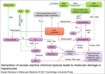
Transporters such as the ABCC (ATP-binding cassette transporter family, subfamily C) efflux proteins and the multidrug resistance proteins (MDRs) have an important role in hepatic xenobiotic elimination by mediating the excretion of parent molecules, metabolites and conjugates into bile or sinusoidal blood in an energy-dependent manner. Transporters can be found in both the canalicular (apical) and sinusoidal membranes of hepatocytes (Refs Reference Trauner and Boyer25, Reference Alrefai and Gill26, Reference Kosters and Karpen27). Transmembrane transporters are important determinants of the overall xenobiotic metabolism and clearance by the liver. The function of these transporters has been described by some investigators as Phase III of the xenobiotic metabolism process. Moreover, coevolution of conjugative enzymes and transporters is believed to have occurred so that the formation of polar conjugates (e.g. glucuronides and GSH derivatives) is coupled to their active excretion from the liver. The net result of these coordinated functions is an efficient removal of xenobiotics and their metabolites from the body (Refs Reference Nies, Schwab and Keppler28, Reference Pang, Maeng and Fan29, Reference Zamek-Gliszczynski30) (Fig. 1). For any given compound, one, two or all three of these steps can participate in its elimination.
Generation of excess reactive chemical species leads to molecular damage in hepatocytes. Reactive species (pink boxes) are generated during biotransformation of toxicophores, which can then be detoxified by drug-metabolising enzymes and antioxidant systems (green boxes). Excess reactive chemical species react with biomolecules to form molecular adducts, peroxides or broken molecular fragments (purple box), resulting in dysfunction or change in the structure of biomolecules. Green arrows indicate detoxification pathways; red arrows show toxification pathways. CAT, catalase; GSH, glutathione; GPX, glutathione peroxidase; GST, glutathione S-transferase; NOS, nitric oxide synthase; NQO1, NAD(P)H quinone oxidoreductase 1; Prx, peroxiredoxin; SOD, superoxide dismutase; SULT, sulfotransferase; UGT, glucuronosyltransferase. Parts of this figure are adapted from portions of figures from Refs Reference Gregus, Casarett and Doull33, Reference Testa and Kramer34.

Harmful reactive chemical species can be produced during biotransformation of xenobiotics and under physiological and pathological conditions
The conversion of xenobiotics from their lipophilic forms to more stable hydrophilic forms favours their excretion from the body and usually results in loss of pharmacological or biological activity. However, biotransformation of certain xenobiotics can also produce short-lived, unstable, highly reactive chemical species that can interact with functional biomolecules and potentially lead to adverse effects. The functional groups or structural motifs in xenobiotics that can produce harmful reactive intermediates have been defined as ‘toxicophores’ (Ref. Reference Williams and Naisbitt31). Toxicophores in drugs and their bioactivation pathways have been empirically catalogued (Ref. Reference Kalgutkar32). Reactive chemical species produced during biotransformation reactions include electrophiles, nucleophiles, free radicals and redox-active reactants (Fig. 1).
Electrophiles are produced by oxidation or reduction reactions. Usually, the parent molecule is changed by the addition of an atom of oxygen or nitrogen, which extracts electrons. This results in a partially or fully positively charged centre in electrophilic molecules. For example, aromatic compounds can be oxidised to epoxides, quinines, quinone imines and methides. Nitroarenes can be reduced to reactive N-oxygenated species and nitrenium ions. Thiolcarbonyl compounds can be oxidised to sulfenes. Electrophiles such as acyl halides, aldehydes, ketones and arene oxides are reactive because their electron-deficient centre allows them to react by sharing electron pairs with electron-rich atoms in nucleophiles. By contrast, nucleophiles such as thiols, amines and hydrazines are species that are strongly attracted to positively charged regions in other chemicals. Nucleophiles can be oxidised to free radicals and electrophiles, whereas electrophiles can be reduced to nucleophiles (Fig. 1). In general, the formation of toxic reactive nucleophiles is rare. The majority of all reactive chemical species are electrophilic (Refs Reference Gregus, Casarett and Doull33, Reference Testa and Kramer34).
A free radical is a molecule or molecular fragment that contains one or more unpaired electrons. Radicals are formed when a chemical accepts or loses an electron. Homolytic fission of a covalent bond also generates radicals. Free radical intermediates are known to occur during the metabolism of a wide variety of xenobiotics, such as polyhalogenated alkanes, polyphenols and quinines (Fig. 1). Free radical metabolites can, in principle, be generated during the metabolism of almost any aromatic xenobiotic (Refs Reference Mason and Chignell35, Reference Mason and Pryor36). Reactive oxygen and nitrogen free radical species can also be generated in response to exposure to toxic concentrations of metals such as iron, copper and arsenic (Ref. Reference Valko, Morris and Cronin37). The one-electron reduction of nitroaromatic compounds, quinones and other chemicals can be catalysed enzymatically by several of reductases and dehydrogenases. Xenobiotics can also be oxidised to free radicals by peroxidases (Ref. Reference Aust38). Free radicals usually extract a hydrogen atom from other molecules and can also add to the double bonds of a biomolecule, usually resulting in new free radicals (Ref. Reference Gregus, Casarett and Doull33).
Reactive oxygen species (ROS) are another important type of free radical. This term is used to describe a collection of oxygen-derived free radical and nonradical species such as superoxide anion (O2−•), hydroxyl (HO•), peroxyl (RO2•), alkoxyl (RO•) radicals, and hydrogen peroxide (H2O2) (Ref. Reference Halliwell and Cross39). Superoxide anion is a starting material in the formation of hydrogen peroxide, which can then be converted to hydroxyl radical in the presence of iron. Superoxide is also used in the production of peroxynitrite (ONOO−), which rapidly reacts with CO2 to ultimately form reactive free radicals such as nitrogen dioxide (•NO2) and carbonate anion radical (•CO3−) (Refs Reference Ferrer-Sueta and Radi40, Reference Gutteridge41) (Fig. 1). Superoxide anions are among the least reactive ROS whereas hydroxyl radicals react rapidly with virtually all molecules found in vivo. At low physiological levels, ROS act as ‘redox messengers’ in intracellular signalling and regulation of gene expression. However, when generated in excess amounts, ROS can be harmful to cellular components and tissues by inducing oxidative modifications to cellular macromolecules (Ref. Reference Circu and Aw42). ROS are normal byproducts of metabolism during xenobiotic exposure. Superoxide anion and hydrogen peroxide are released when the cytochrome P450 catalytic cycle is interrupted (uncoupled) following the introduction of the first and the second electron, respectively (Ref. Reference Parkinson, Ogilvie, Casarett and Doull21). Monooxygenases (e.g. cytochrome P450s) in ER contribute greatly to the increased levels of cellular hydrogen peroxide and superoxide anion, especially in the absence of substrates. More than 60% of electron transfer haemoproteins and about 20–30% of membrane-bound flavoproteins are localised in the ER (Ref. Reference Zangar, Davydov and Verma43). The mitochondrion is another major intracellular source of ROS, where ROS are constantly produced as a byproduct of oxidative respiration under normal conditions. Moreover, mitochondrial ROS production can increase significantly in response to drugs or metabolites that interfere with the respiratory chain (Refs Reference Kovacic44, Reference Murphy45). The peroxisome also contributes to cellular hydrogen peroxide production under normal physiological conditions (Ref. Reference Fritz46).
Detoxification of harmful reactive chemical species
During normal biotransformation of endobiotics and nutrients, reactive chemical species such as ROS can also be produced. Depending on the magnitude of their formation, reactive chemical species can be quenched or disposed of by endogenous cytoprotective mechanisms, including Phase II conjugation enzymes and their cofactors, efflux transporters, antioxidant enzymes and small molecules such as vitamin C (Refs Reference Gregus, Casarett and Doull33, Reference Davies47) (Fig. 1). The tight coupling of bioactivation and detoxification reactions serves to alleviate or eliminate the reactivity of intermediate chemical species (Ref. Reference Williams48).
Conjugative reactions have evolved from their normal physiological functions to fulfil a variety of cytoprotective functions against potentially harmful xenobiotics (Ref. Reference McCarver and Hines49) (Fig. 1). Nucleophiles are generally detoxified by conjugation with a soluble moiety or cofactor at the nucleophilic functional group. Hydroxylated compounds are conjugated by sulfation or glucuronidation, whereas thiols are methylated or glucuronidated. Amines and hydrazines are acetylated. These reactions prevent peroxidase-catalysed conversion of the nucleophiles to free radicals and biotransformation of phenols, aminophenols, catechols and hydroquinones to electrophilic quinones and quinone imines (Ref. Reference Gregus, Casarett and Doull33). Although some GSH conjugates, such as the GSH thiyl radical and GSH disulfide (Ref. Reference van Bladeren50), are highly reactive, a general mechanism for the detoxification of electrophilic toxicants is conjugation with the thiol group of the nucleophile GSH. This reaction may occur spontaneously or can be facilitated by GSH S-transferases (Refs Reference Whalen and Boyer51, Reference Dragovic52) (Fig. 1). The resulting conjugates are subsequently excreted by efflux transporters into the bile or urine (Refs Reference Nies, Schwab and Keppler28, Reference Pang, Maeng and Fan29, Reference Zamek-Gliszczynski30).
To combat oxidative stress and maintain a proper reducing environment, the liver is well equipped with antioxidant mechanisms, including micronutrients such as α-tocopherol (vitamin E) and ascorbic acid (vitamin C). Also, thiol-rich proteins, such as metallothionein, and other metal-sequestering proteins, such as ferritin, are important in preventing the potentially damaging effects of certain metals, such as cadmium and copper. Enzymes such as epoxide hydrolases, catalase, superoxide dismutase (SOD) and GSH peroxidases also have a role in the metabolism and neutralisation of reactive intermediates. For example, superoxide anion, an important ROS, can be converted into hydrogen peroxide by SODs in the cytosol and the mitochondria. Subsequently, hydrogen peroxide is reduced to water by catalase in the peroxisomes, by selenocysteine-containing GSH peroxidases in the cytosol and mitochondria, and by peroxiredoxins in the cytosol, mitochondria and ER. Peroxynitrite, a nitrogen-reactive species, is reduced to nitrite (ONO−) by the selenocysteine-containing GSH peroxidase and peroxiredoxins. In addition, peroxynitrite reacts with oxyhaemoglobin, haem-containing peroxidases or albumin (Ref. Reference Ferrer-Sueta and Radi40). The detoxification of superoxide anions, peroxynitrites and peroxides prevents the formation of free radicals produced from the rapid reaction of peroxynitrite with carbon dioxide (CO2) (Ref. Reference Ferrer-Sueta and Radi40) and also prevents the formation of extremely reactive hydroxyl radicals, which cannot be quenched enzymatically or by the antioxidants GSH, vitamin E and vitamin C.
The most important antioxidant in the mammalian liver is GSH. Hepatic levels of GSH are usually high and can be readily modulated by regulating its biosynthetic pathway (Refs Reference Forman, Zhang and Rinna53, Reference Lu54). GSH and redox status are compartmentalised within the hepatocyte, with the highest concentration of GSH found in the cytosol. The relative redox states of the different subcellular compartments from most reducing to most oxidising are mitochondria > nuclei > cytoplasm > ER > extracellular space (Refs Reference Hansen, Go and Jones55, Reference Jones56, Reference Circu and Aw57). GSH is a critical cofactor for several antioxidant pathways, including thiol–disulfide exchange reactions, GSH peroxidase and GSH S-transferases. GSH protects cells from oxidative injury by reducing hydrogen peroxide and scavenging reactive oxygen and nitrogen radicals (Refs Reference Yuan and Kaplowitz58, Reference Han59). The oxidised form of glutathione (GSSG) is reduced back to GSH by glutathione reductase. This reductive reaction needs NADPH, which is generated in the mitochondria in an ATP-dependent manner. Therefore, the ability of the liver to withstand oxidative stress depends on its ability to generate the necessary energy to carry out these vital reactions (Ref. Reference Kaplowitz60). Thus, GSH has an important role in the detoxification of both electrophiles and free radicals.
In addition to regulating redox status, GSH is also an effector of cell signalling. GSH-induced shifts in redox status are known to mediate the dynamic regulation of protein function by the formation of reversible intermolecular, intramolecular and mixed disulfide bonds between protein cysteines and GSH (S-glutathiolation). GSH also stabilises extracellular proteins and protects proteins against irreversible oxidation of critical cysteine residues. The DNA binding of certain transcription factors such as nuclear factor-κB, p53 and activator protein-1 is redox dependent, and redox signalling resulting from oxidative stress can be transduced into altered expression of a wide variety of genes implicated in cell proliferation, differentiation and apoptosis. These processes have been implicated in the pathogenesis of many liver diseases (Refs Reference Yuan and Kaplowitz58, Reference Han59, Reference Biswas, Chida and Rahman61).
Other redox systems, such as thioredoxin, thioredoxin reductase, peroxiredoxin, glutaredoxin and pyridine nucleotide redox couples, regulate the activities of multiple transcription factors, work as growth factors, serve as enzyme cofactors, and also participate in cell signalling and modulation of cell function. In addition, the thioredoxin and glutaredoxin systems have an important role in cellular defences against oxidative stress and in maintaining redox homeostasis by regulating thiol–disulfide exchange. NADPH-dependent thioredoxin reductase, which reduces a broad range of substrates including the oxidised form of thioredoxin, can also directly reduce lipid peroxides, hydrogen peroxide, and dehydroascorbic and lipoic acids (Refs Reference Circu and Aw42, Reference Kalinina, Chernov and Saprin62).
Modification of biomolecules and shifting of the reducing environment of cells by reactive chemical species
When the balance between bioactivation and detoxification reactions is altered, and the amount of reactive intermediates becomes abnormally elevated, reactive intermediates can overwhelm or bypass the defence systems, resulting in oxidative stress and damage to proteins, nucleic acids and lipids (Fig. 1).
Reactive intermediates can covalently bind to proteins and form protein adducts. Sulfhydryl groups and primary or secondary amino groups in serine and threonine residues react readily with electrophilic species such as alkyl and benzylic carbonium ions, iminium ions, aldehydes, epoxides, enones, quinone imines and Michael acceptors. Lysine and histidine residues in proteins are similarly targeted by these electrophiles (Refs Reference Srivastava63, Reference Zhou64). The identity of many proteins targeted by reactive intermediates of xenobiotics is well documented. Halothane and acetaminophen have been demonstrated to form adducts with specific proteins (Ref. Reference Cohen65). The formation of 3-(cystein-S-yl)-acetaminophen adducts between N-acetyl-p-benzoquinone imine and hepatocellular proteins has been used as a serum biomarker of acetaminophen toxicity in patients with acute liver injury (Refs Reference Davern66, Reference James67). More detailed information on the identity of proteins modified covalently by xenobiotics can be found in the web-accessible resource known as the reactive metabolite target protein database (Ref. Reference Hanzlik68). In addition, proteins can be modified by phosphorylation, acylation, methylation, ubiquitylation and sumoylation under electrophilic or oxidative stress conditions (Ref. Reference Liebler69). Adduct formation can lead to several adverse consequences, such as interference with protein function, stimulation of fibrogenesis and induction of immune responses. In alcohol consumers, covalent modification of proteins and other cellular constituents by acetaldehyde has a role in the sequence of events leading to alcoholic liver disease (Ref. Reference Thiele, Klassen and Tuma70). The role of protein covalent binding of reactive intermediates in the mechanism of toxicant action has been the subject of extensive investigations for several decades (Ref. Reference Rubino71), yet we do not have a clearly defined role of covalent binding in hepatotoxicity. It is thought that drug–protein adducts contribute to toxicity either through impaired function of the modified proteins or through immune-mediated mechanisms.
DNA can also be modified by reactive chemical species. Oxygen atoms of purine and pyrimidine bases and endocyclic nitrogens of purine bases in DNA are also targets of electrophiles (Ref. Reference Srivastava63). This is the case for metabolites of a spectrum of carcinogenic polycyclic hydrocarbons (Refs Reference Benigni72, Reference Kho73). Diffusible hydroxyl radicals can react either with the sugar units by hydrogen abstraction or with the base moieties by addition (Ref. Reference Dizdaroglu74). Metals such as iron and copper also cause various modifications of DNA bases by mediating the formation of free radicals. Lipid peroxides, which result from the reaction between radicals and polyunsaturated fatty acid residues of phospholipids, can further react with metals and finally produce mutagenic and carcinogenic malondialdehyde, 4-hydroxynonenal (HNE) and other exocyclic DNA adducts (etheno and propano adducts) (Refs Reference Valko, Morris and Cronin37, Reference Burcham75). When DNA damage is poorly repaired, deleterious genetic changes can occur.
Lipid peroxidation is usually the consequence of self-propagating chain reactions of free radicals. Peroxyl and hydroxyl radicals react with unsaturated and mainly polyunsaturated fatty acids in membranes, with major roles in initiating lipid peroxidation (Ref. Reference Dix and Aikens76). Lipid peroxidation produces various electrophiles, such as 4-hydroxynonenal (HNE), a major endproduct of peroxidation of membrane N-6-polyunsaturated fatty acids. The reaction of HNE with important signalling proteins might have a role in the progression of liver diseases (Refs Reference Poli, Biasi and Leonarduzzi77, Reference Smathers78). The peroxidative breakdown of membrane lipids results in impaired function of mitochondria, ER, lysosomes, plasma membranes and Golgi, and thus has a role in acute liver necrosis and the amplification and progression of liver injury (Ref. Reference Poli, Albano and Dianzani79).
Proper redox homeostasis is crucial for cell survival. The redox status in the cell is maintained by various redox couples, among which GSSG–2GSH is the major one that maintains redox regulation through protein glutathionylation (Refs Reference Jones56, Reference Ghezzi, Bonetto and Fratelli80, Reference Gallogly and Mieyal81, Reference Schafer and Buettner82). Normally, the GSH concentration is high in hepatocytes, which is indicative of the reducing environment in the cell. This reductive environment serves as a protective mechanism against oxidative stress. It is also essential for maintaining the integrity of the structure and function of biomolecules (Refs Reference Farout and Friguet83, Reference Klatt and Lamas84). Disturbance in the equilibrium of redox couples by reactive chemical species results in imbalances in redox status. Electrophiles extract electrons from thiols and reduce the amount of GSH. Similarly, free radicals, including reactive oxygen or nitrogen species, abstract hydrogens from the reducing milieu of the cell. These reactions proceed at the expense of reducing equivalents, thus resulting in oxidative stress. Under oxidative stress, redox-dependent proteins are modified, redox signalling pathways are disrupted, and peroxides and free radicals are produced, leading to damage of cell components, including proteins, lipids and DNA (Refs Reference Farber85, Reference Turrens86). For example, ethanol-induced oxidative stress results in aldehyde–protein adducts in the liver (Ref. Reference Smathers87). Also, liver microsomal malondialdehyde–protein adducts are produced as a result of oxidative stress resulting from chronic dietary iron overload (Ref. Reference Valerio and Petersen88). In addition, HNE–protein adducts are considered to be good biomarkers of lipid oxidation during liver injury (Refs Reference Poli, Biasi and Leonarduzzi77, Reference Smathers87). Redox-dependent post-translational modifications most frequently occur in protein thiols. These are mediated by environmental and endogenously generated reactive species that can trigger cell signalling events leading to toxic effects (Ref. Reference Rudolph and Freeman89). Chloroform, carbon tetrachloride and GSH depletion induce secondary genotoxicity in liver cells through oxidative stress (Ref. Reference Beddowes, Faux and Chipman90). In some cases, mitogen-activated protein kinases (MAPKs), extracellular signal-regulated kinase 1/2 (ERK1/2), c-Jun N-terminal kinase (JNK) and the nuclear factor kappa B (NF-κB) pathways are involved in hepatocyte oxidative injury (Refs Reference Czaja91, Reference Allen and Tresini92), as seen in alcoholic liver injury. Activating protein 1 (AP-1) is also involved in the deterioration of cell and organ function by toxic xenobiotics (Refs Reference Zima and Kalousova93, Reference Wu and Cederbaum94).
Adaptation to and repair of damaged biomolecules
In addition to having mechanisms and substances that prevent toxic reactions between reactive chemical species and biomolecules, cells have evolved protective mechanisms that promote repair, removal and replacement of damaged cellular constituents (Fig. 2) (Refs Reference Gregus, Casarett and Doull33, Reference Davies47). Damaged proteins, chemically altered DNA and peroxidised lipids can be repaired by different mechanisms. The mitochondrial matrix contains repair systems for oxidised proteins, including thioredoxin, thioredoxin reductase, glutaredoxin, GSH and GSH reductase. These systems reduce disulfide bridges or sulfenic acids catalytically. Similarly, methionine sulfoxide reductase reverses methionine sulfoxide back to methionine residues in proteins (Ref. Reference Ugarte, Petropoulos and Friguet95). Molecular chaperones such as heat-shock proteins can rescue denatured proteins by ATP-dependent disaggregation and refolding, or assist in their degradation by proteasomes by engaging the chaperone-dependent ubiquitin ligase CHIP (C-terminus of heat-shock protein cognate 70-interacting protein) (Ref. Reference Lee and Tsai96). There are distinct proteolytic systems for different physiological tasks under changing environmental and pathophysiological conditions. Damaged proteins can be eliminated by proteolysis in lysosomes (Ref. Reference Zhang97). Proteins damaged by oxidative stress are also removed by the proteasomes in the cytosol and nucleus. Under oxidative stress conditions, there is high proteasome content in the nucleus. This protects the nucleus from formation and accumulation of nondegradable proteins (Ref. Reference Breusing and Grune98). Lipid peroxides are repaired by a complex process that operates in concert with a series of reductants along with GSH peroxidase and GSH reductase. Different types of damage to DNA are corrected by specialised mechanisms, each using a different set of repair proteins (Refs Reference Christmann99, Reference Camenisch and Naegeli100, Reference Ciccia and Elledge101). Despite its high reactivity with electrophiles and free radicals, nuclear DNA is remarkably stable, in part because it is packaged in chromatin and because of the several repair mechanisms that correct alterations in DNA. Mitochondrial DNA is more prone to damage because of the lack of histones or the type of repair mechanisms present in the nucleus.
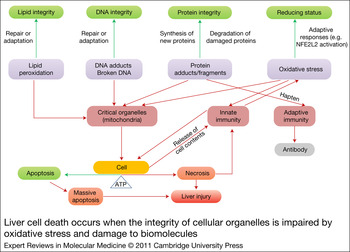
Liver cell death occurs when the integrity of cellular organelles is impaired by oxidative stress and damage to biomolecules. Damage to biomolecules and oxidative stress can be reversed or tolerated by activation of compensatory responses. Excessive damage or modification of biomolecules by oxidative stress can overwhelm the capacity of repair and adaptive processes, leading to uncorrected changes to critical organelles, among which mitochondria is the most important. This can lead to activation of immune responses, which could further aggravate cellular damage. The interplay between xenobiotic-induced molecular changes, organelle dysfunction and activation of immunity results in cell death and liver injury. Cellular ATP availability is an important determinant of the type of cell death that occurs. Programmed cell death or apoptosis requires ATP, whereas oncotic death (necrosis) predominates when ATP stores are depleted. However, massive apoptosis also leads to apoptotic necrosis. Green boxes indicate normal homeostasis; boxes of other colours show events leading to liver cell injury. Green arrows indicate pathways leading to cell recovery; red arrows show pathways leading to cell damage or death.

Although it has been proposed that biological adaptation to repeated exposure to certain xenobiotics can in some instances result in long-term toxicity (Ref. Reference Barouki102), adaptive responses to acute exposure are generally known to preserve or restore biological homeostasis in the face of chemical insult. These responses involve sensing the initial damage or dysfunction and initiating compensatory changes in gene expression. Adaptive responses are beneficial because they enhance the capacity of different cellular structures to respond more robustly to chemical-induced stress. They are also reversible and critical for preserving cell viability. Disruption of this equilibrium at levels of the molecules, organelle and other cellular organisations leads to an adverse effect or toxicity. Induction of xenobiotic-metabolising enzymes mediated by the xenosensors aryl hydrocarbon receptor (AhR), constitutive androstane receptor (CAR, NR1I3), pregnane X receptor (PXR, NR1I2) and peroxisome proliferator-activated receptor (PPAR) provides examples of adaptive responses to chemical stressors (Refs Reference Xie103, Reference Ramadoss, Marcus and Perdew104). In response to oxidative stress, KEAP1-NFE2L2 [kelch-like ECH-associated protein 1-nuclear factor (erythroid-derived 2)-like 2] is activated and induces GSH synthesis and alters gene expression for multiple xenobiotic metabolising enzymes and transporters (Refs Reference Copple105, Reference Shen and Kong106). In those instances when oxidative stress is severe, or when the function of protective enzymes is deeply compromised, cells may ‘sacrifice themselves’ by apoptosis, which protects the surrounding healthy tissue from further damage. Only under the most severe oxidative stress conditions and when adaptation mechanisms fail will cells then undergo necrotic death, which exposes the surrounding nondamaged cells to immune responses (Refs Reference Gregus, Casarett and Doull33, Reference Williams and Iatropoulos107).
Oxidative stress and dysfunction of biomolecules result in dysregulation of cell activities, leading to cell death
When damage to cellular biomolecules remains unrepaired or the cellular adaptive responses are overwhelmed by unquenched reactive chemical species, the cell structural components and essential biochemical processes become impaired. These biochemical disorders result in functional and structural damage to certain organelles. Cell death can ensue and, depending on the severity of the damage, clinical manifestations can become evident (Fig. 2). Any intracellular compartment, such as mitochondria, cytoskeleton, lysosome, ER, nucleus or plasma membrane, can be affected by toxicants, initiating different types of cell death. The different pathways mediating modes of cell death could crosstalk through a molecular ‘switch’, such as p53. Inhibition of one particular mode of cell death might activate an alternative pathway to cell death (Refs Reference Green and Kroemer108, Reference Orrenius, Nicotera and Zhivotovsky109).
Mitochondrial dysfunction is a major mechanism of chemical-induced liver injury. The mitochondrion is a key organelle involved in pyruvate oxidation, fat oxidation and ATP production. It is also a site where external and intracellular stress-response pathways converge to provoke cell death. The opening of permeability transition pores under stress conditions results in sudden increases in permeability of the inner mitochondrial membrane. Depending on the magnitude of this event, mitochondrial membrane permeabilisation can lead to apoptosis by release of cytochrome c, which is a known activator of caspases in the cytosol. Under more serious circumstances, mitochondrial membranes can rupture, leading to ATP depletion and subsequent liver cell necrosis. Abnormal mitochondrial function can also lead to fat accumulation. In the most severe cases of mitochondrial dysfunction, microvesicular steatosis and hypoglycaemia can occur. Macrovacuolar steatosis can also be the result of chemical-induced mitochondrial dysfunction and can often progress to steatohepatitis and cirrhosis (Refs Reference Pessayre110, Reference Jones111, Reference Kass112, Reference Malhi, Guicciardi and Gores113, Reference Wei114, Reference Joza115, Reference Jones116).
Apoptosis and necrosis are the most widely recognised forms of hepatocyte cell death. Apoptosis can be initiated by two different pathways. The intracellular signalling pathway is triggered by intracellular stress caused by oxidative stress and damage to proteins, membranes or DNA. The external pathway is initiated by the binding of paracrine molecules such as tumour necrosis factor-α and Fas ligand to death receptors on the cell surface. Each pathway activates caspases and nucleases, resulting in apoptotic cell death. Characteristic changes seen during apoptosis include cell shrinkage, nuclear fragmentation, chromatin condensation and chromosomal DNA fragmentation. In contrast to apoptosis, necrosis can result from extensive damage to the plasma membrane with disturbance of ion transport, dissolution of membrane potential, cell swelling and eventual rupturing of the cell. During necrosis, the cell liberates macromolecular breakdown products, including lipid peroxides, aldehydes and eicosanoids. These molecules can then trigger an inflammatory response in the hepatic parenchyma. Apoptosis is energy dependent and is considered to be a protective mechanism that prevents cells from oncotic necrosis, which often promotes more damage to surrounding cells through the involvement of the immune system. In the presence of ATP, cell death can proceed by apoptosis, but when mitochondria are de-energised, cell death proceeds by necrosis. Apoptosis and necrosis more likely represent the morphological and mechanistic ends of a spectrum of overlapping cell death processes (Fig. 2). Cells on an apoptotic path can be redirected to necrosis or to an overlapping pattern ‘apoptotic necrosis’ (necraptosis) upon depletion of ATP (Fig. 2). Furthermore, with excess apoptosis, the capacity of the tissue for phagocytosis of apoptotic bodies can be exceeded, and ‘secondary’ necrosis can occur (Refs Reference Kaplowitz60, Reference Orrenius, Nicotera and Zhivotovsky109, Reference Jones111, Reference Lemasters117). Both necrosis and apoptosis can trigger cytolytic hepatitis.
The interaction between reactive chemical species and cellular biomolecules can also trigger a response by the liver's immune system. The formation of apoptotic bodies or the release of cell constituents by necrotic hepatocytes can trigger the activation of innate immune cells, producing proinflammatory and cytotoxic mediators. Depending on the magnitude of cell death, this immune response can result in enhanced liver tissue damage. This is postulated to occur in the early stages of alcoholic liver disease (Refs Reference Nagy118, Reference Nath and Szabo119). Adaptive immune responses can also be elicited during chemical hepatotoxicity. Covalent binding of reactive metabolites to cellular proteins can lead to the formation of neoantigens (haptens) that can initiate autoantibody production and cytotoxic T-cell responses. In addition, certain drugs such as sulfonamide sulfamethoxazole (Ref. Reference Depta120) can bind directly to T-cell receptors, mimicking a ligand–receptor interaction. This causes T-cell activation in a major histocompatibility complex-dependent fashion (Refs Reference Kaplowitz121, Reference Holt and Ju122, Reference Ju and Uetrecht123, Reference Adams124).
Intrahepatic cholestasis induced by drugs such as cyclosporine and anabolic steroids is one of the clinically important forms of acquired cholestatic liver disease. The underlying mechanisms could be associated with canalicular transporters, whose function can be inhibited by the parent chemical or its metabolite. Canalicular transporters are essential for bile formation and for biliary secretion of cholephilic compounds, xenobiotics and their metabolites. Any chemical-mediated functional disturbance of these transport processes can lead to intracellular accumulation of potentially harmful bile constituents and xenobiotics, thus resulting in the development of cholestatic liver cell damage (Refs Reference Pauli-Magnus and Meier125, Reference Kosters and Karpen126, Reference Geier127). The bile salt export pump (BSEP) is a known target for drug inhibition in patients who are susceptible to drug-induced cholestasis (Ref. Reference Pauli-Magnus, Meier and Stieger128). This is the main canalicular transporter involved in the biliary excretion of bile acids. Inhibition of BSEP function leads to accumulation of bile salts in hepatocytes, which can result in liver damage.
Many cases of drug-mediated hepatotoxicity are dose dependent and thus predictable. However, idiosyncratic drug reactions are dose independent and unpredictable. These reactions can result in significant morbidity and mortality (Ref. Reference Kaplowitz129). Considerable evidence indicates that many of these reactions are immune mediated (Ref. Reference Zhang130) and that they are caused by immunogenic conjugates formed from the reaction of reactive metabolites of a drug with cellular proteins. Although this ‘drug–protein antigen response’ concept is well accepted, the precise mechanism of idiosyncratic adverse drug reaction remains unclear. Continued basic research in this area is essential to elucidate the role of drug–protein antigens in hepatotoxicity (Refs Reference Ju and Uetrecht123, Reference Uetrecht131, Reference Li and Uetrecht132, Reference Daly133).
Factors contributing to chemical-induced liver injury
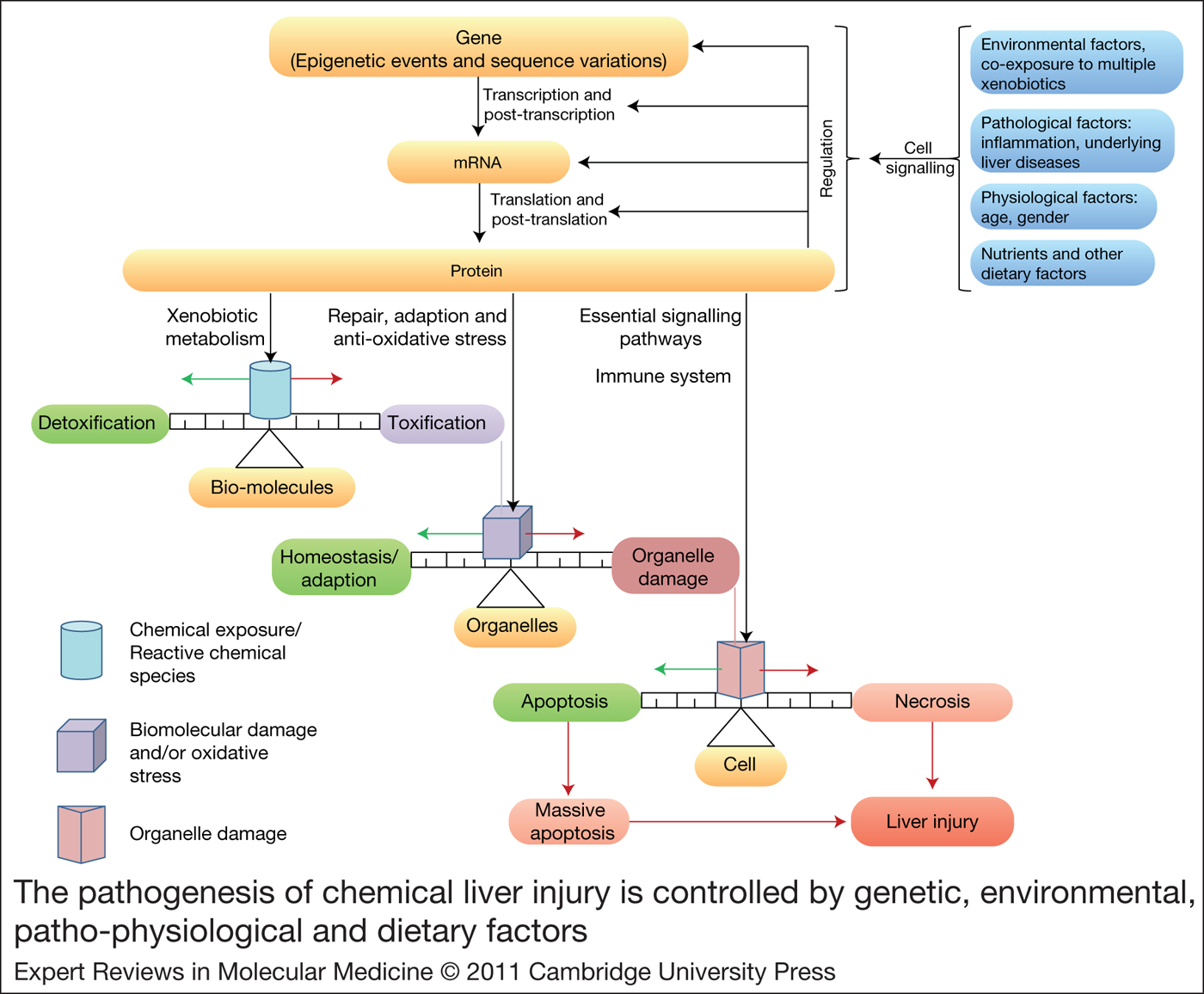
As stated previously, the pathogenesis of most chemical-induced liver injuries is initiated by metabolic activation of the chemical or an imbalance between bioactivation (toxification) and detoxification processes. The propensity of a molecule to form either toxic or chemically reactive metabolites is simply a function of its chemistry. Certain toxicophores present in drugs can be biotransformed into reactive species (Refs Reference Williams and Naisbitt31, Reference Kalgutkar32). Exposure to high doses of a potential toxicant increases the amount of reactive metabolites formed, thus increasing the risk for liver injury (Refs Reference Smith and Obach134, Reference Uetrecht135). In addition, drugs can either inhibit or induce the expression and function of drug-metabolising enzymes, as seen in the case of human cytochrome P450 3A4. Xenobiotic enzyme induction could lead to altered metabolism of chemicals (Ref. Reference Zhou136), which could greatly affect the toxicity outcome. The progression of chemical liver injury involves competition between cell-damaging events caused by reactive chemical species and protective responses aimed at quenching oxidative stress, repairing macromolecular damage or, ultimately, preserving the integrity of tissue by committing ‘self-sacrificing’ apoptotic cell death. Clinical manifestations of chemical-induced liver injury occur with extensive necrotic cell death and the triggering of an immune response. Therefore, the pathogenesis of chemical-induced liver injury involves many competing processes, shaped by the interplay among genetic determinants, environmental factors and potential underlying pathological processes (Fig. 3).
The pathogenesis of chemical liver injury is controlled by genetic, environmental, patho-physiological and dietary factors. Other factors such as gender, race, age and lifestyle choices can be risk factors for certain forms of chemical liver injury. The interplay between these factors controls gene transcription and cell signalling events, leading to a myriad of interconnected outcomes such as imbalances between detoxification and toxification metabolic pathways, impaired organelle function, compromised ability to repair molecular damage and alterations in overall cellular homeostasis. With prominent shifts in normal cellular homeostasis, the ability of compensatory and adaptive responses to correct the detrimental effects of toxicants can be hampered. Similarly, repair mechanisms can be overwhelmed and incapable of correcting the cellular chemical injury. Green arrows indicate pathways leading to cell recovery; red arrows show pathways to cell damage or death; black arrows indicate pathways that contribute to chemical-induced liver injury.

Genetic factors have a key role in the initiation of the pathogenesis of chemical-induced liver injury. The toxicant action of a specific xenobiotic chemical in the body is determined by its tissue dosimetry at target sites. The dose of the ultimate toxicant at the site of toxicant action is controlled by the coordination of Phases I–III of xenobiotic metabolism and disposition. The in vivo capacity for xenobiotic metabolism is determined by the combined function of different types of xenobiotic-metabolising enzymes in the body. There is a wild diversity of drug-metabolising enzymes to catalyse the Phase I and II reactions, along with transport proteins (Phase III) involved in drug disposition. For example, there are 57 different active genes encoding CYP enzymes and 58 pseudogenes present in the human genome (Ref. Reference Nelson137). The expression levels of these CYPs are not the same among individual subjects. In addition, individual enzymes show great variation among different individuals. The same is true for Phase II enzymes and drug transporters. Drug-metabolising enzymes and transporters are also known to have a differential and overlapping spectrum of substrates. Therefore, the capacity for metabolism and disposition of chemicals can differ widely from one individual to another.
The catalytic activity of an individual enzyme is determined by its abundance in a given tissue and its intrinsic activity. The amount of xenobiotic-metabolising enzymes in a particular tissue is also dependent on epigenetic regulation, gene transcription, and post-transcriptional, translational and post-translational events. Gene transcription is the most important control among these regulatory steps. Interindividual variation in the expression of xenobiotic-metabolising enzymes could result from polymorphic forms of regulatory systems for gene expression. For example, although the mechanism for controlling constitutive expression of CYP3A4 remains unknown, its gene expression is regulated by a number of transcription factors such as C/EBPα (CCAAT/enhancer binding protein, alpha), C/EBPβ, HNF4α (hepatocyte nuclear factor 4, alpha), HNF3γ, CAR and PXR. These transcription factors recognise regulatory modules in the human CYP3A4 gene (Ref. Reference Martinez-Jimenez138). Changes in PXR expression levels and its amino acid sequence are known to produce alterations in the expression of CYP3A4 (Refs Reference Liu139, Reference Koyano140, Reference Tompkins, Sit and Wallace141). Similarly, expression of the Phase I and II enzymes and transporters is coregulated by a set of nuclear receptors, such as PXR. Therefore, all aspects of xenobiotic metabolism and disposition are regulated by certain transcription factors simultaneously in a coordinated manner. However, the intrinsic activity of individual drug-metabolising enzymes is determined by the amino acid sequences of the peptide and variations thereof. These variations or polymorphic forms have been widely identified and studied. Many polymorphic variants of drug-metabolising enzymes are well known to contribute to the interindividual variations in intrinsic activity, and thus drug biotransformation, even when protein expression levels are the same as for the normal form of the enzyme. About 40% of CYP-dependent metabolic reactions are catalysed by polymorphic enzymes (Refs Reference Liu139, Reference Ingelman-Sundberg142, Reference Zanger143). A collection of genetic information on CYP variants, along with their effects on function, can be found in a web-based resource (Ref. Reference Sim and Ingelman-Sundberg144). Sequence variations are also found in conjugative enzymes and drug transporters (Ref. Reference Pauli-Magnus and Meier125). In addition, the availability of cofactors and allosteric modificators for xenobiotic-metabolising enzymes and transporters also have an important role in their function and activity. For example, haem is a key cofactor for CYP enzymes. Haem also regulates CYP protein synthesis, assembly, repair and disposal. Cellular haem content itself is tightly controlled by genetic factors (Ref. Reference Correia, Sinclair and De Matteis145).
Several environmental factors have a role in chemical-induced liver injury by regulating xenobiotic metabolism and disposition through various signalling pathways. Transcription factors are cellular mediators of gene regulation. Some of these sense the presence of exogenous stressors and regulate gene expression accordingly as an attempt to counteract any of their potential damaging effects. For example, a variety of structurally diverse endobiotics and xenobiotics, such as bile acids, pharmaceuticals, and environmental, dietary and occupational chemicals, activate PXR and CAR to induce Phase I and II drug-metabolising enzymes and transporters (Refs Reference Hernandez, Mota and Baldwin146, Reference Zollner147). Induction or suppression of expression of drug-metabolising enzyme results in changes in the normal metabolism and disposition of drugs, which might in turn promote adverse responses (Ref. Reference Lynch and Price148).
Predisposition by pathological and physiological factors also has a role in the initiation of pathogenesis of chemical liver injury. The induction and downregulation of CYP3A4 under pathophysiological conditions, such as inflammatory responses, are key processes involved in determining whether the toxic or therapeutic effects of drugs predominate (Ref. Reference Pelkonen149). Significant changes in the expression of drug-metabolising enzymes occur during ontogeny. The CYP3A subfamily is the major drug-metabolising CYP enzyme expressed at all stages of development. The expression of different members of this subfamily, however, undergoes significant switches from the fetal predominant form CYP3A7 to the major adult form CYP3A4 (Refs Reference Hakkola, Tanaka and Pelkonen150, Reference Hines151). A higher expression of CYP3A4 mRNA in women has also been reported (Ref. Reference Wolbold152). In addition to affecting drug metabolism and disposition capacity, genetic determinants, environmental factors and pre-existing pathological conditions can also modulate the function of repair systems for damaged proteins, DNA and lipids, and also immune responses in the liver. For example, human leukocyte antigen (HLA)-B*5701 genotype is a major determinant of antimicrobial agent flucloxacillin-induced liver injury (Ref. Reference Daly153).
In summary, the initiation of pathogenesis of chemical liver injury is the outcome of interactions between xenobiotics and genetic factors, environmental factors, pre-existing pathobiological conditions and physiological factors. The dynamic changes, uncertainty and diversity of these factors pose significant difficulties in predicting and managing liver injury induced by xenobiotics.
Perspective
Integrity of the function and structure of the liver is necessary for preserving human health. Notably, the liver is particularly susceptible to chemical injury because of its strategic location, prominent blood supply and prevalent role in biotransformation of xenobiotics. Xenobiotic-induced liver diseases represent a major health care problem and a significant source of morbidity and mortality. Unfortunately, the underlying pathophysiological mechanisms of most hepatocellular forms of chemical-induced hepatic injury are still poorly understood. Without a proper understanding of the underlying cellular and molecular mechanisms involved, diseases such as alcoholic liver disease and idiosyncratic drug responses remain a challenge for both basic research scientists and clinicians. A better understanding of the molecular mechanisms of drug-induced liver injury and the interplay among genetic determinants, environmental factors and pre-existing pathological conditions will help in the prevention and development of new forms of treatment for acute and chronic liver diseases.
Chemical-induced liver injury is an acquired disease, and therefore it can be prevented. The ideal approach, in the case of therapeutic drugs, would be to avoid the use of medications with a hepatotoxic liability in patients prone to develop idiosyncratic reactions. This would require a thorough knowledge of genetic traits and environmental factors that render certain individuals vulnerable to drug liver toxicity. Another approach would be the use and development of new drugs that lack known toxicophores, while retaining the desired pharmacological and therapeutic activity (Refs Reference Evans154, Reference Coles155, Reference Kalgutkar156, Reference Thompson157).
Proper diagnosis is critical for the correct management of chemical liver disease. Early identification of hepatotoxicity may avert serious complications associated with liver injury. Accurate diagnosis of chemical-induced liver injury is based on a combination of appropriate history, clinical presentation, biochemical tests and exclusion of other potential causes (Ref. Reference Halegoua-De Marzio and Navarro158). The information required for accurate diagnosis may not always be available or might not be complete. Certain subclinical statuses, such as an adaptive response, insidiously developing liver tumours or hepatic fibrosis, might not be detected by conventional biochemical tests of liver function (Ref. Reference Carithers, Mcclain, Sleisenger and Feldman4). Sometimes liver diseases can result from exposure to unknown chemical species. However, liver toxicity by certain individual chemicals tends to have a characteristic clinical signature (Ref. Reference Gunawan and Kaplowitz8). Macromolecular adducts can provide reliable indicators of exposure to certain chemicals and can help in predicting toxicological outcomes (Refs Reference Rubino71, Reference Chang159). Future research endeavours on the molecular mechanisms of chemical liver injury, including the identification of reactive intermediates, macromolecular adducts and altered cell signalling pathways, could lead to the development and standardisation of novel clinical or subclinical biomarkers of liver toxicant exposure, initiation of injury, progression of disease and overall prognosis.
Collectively, the susceptibility to chemical liver injury is thought to be determined in great part by genetic factors. The Human Genome Program, the HapMap Program (Ref. Reference Consortium160), the 1000 Genomes Project (Ref. Reference Durbin161) and the ENCODE program (Ref. Reference Birney162) have been instrumental in decoding the role of genetics and gene regulation networks in many pathological conditions, as well as adverse reactions induced by drugs. The accomplishment of these efforts will help in understanding the susceptibility and responses of individuals to chemical exposure in order to develop or choose appropriate therapies for specific patients (personalised medicine) while minimising the possibility of adverse reactions.
Acknowledgements
We recognise many outstanding published contributions of investigators in the field that could not be included in this review owing to space limitations. We express our thanks to the peer reviewers of the article. J.E.M. was the Marlene L. Cohen and Jerome H. Fleisch Scholar in the Department of Pharmaceutical Sciences at the University of Connecticut from 2007 to 2010. X.G., postdoctoral fellow in José Manautou's laboratory, was supported by the National Institutes of Health Grant DK069557.