Introduction
Acanthocephala is a parasitic phylum including a group of intestinal worms parasitizing all classes of vertebrates as definitive hosts. Currently, it includes four classes (Archiacanthocephala Meyer, 1931; Palaeacanthocephala Meyer, 1931; Eoacanthocephala Van Cleave, 1936; Polyacanthocephala Amin, Reference Amin1987), 26 families, 157 genera and about 1298 species (see Amin, Reference Amin2013). Palaeacanthocephala is the largest class and includes most fish acanthocephalans.
To date, little is known about the details of most acanthocephalan life cycles, since complete life cycles are known for only about 25 species of acanthocephalans from those parasitizing fish, amphibians, reptiles and birds (Ribas & Casanova, Reference Ribas, Casanova, Morand, Krasnov and Poulin2006). Most of these cycles have been completed experimentally. In these cycles, adult acanthocephalans lay their embryonated eggs in the intestine of their vertebrate definitive hosts, eggs pass in the host's faeces to the environment and are ingested accidentally by an arthropod intermediate host. After ingestion, an acanthor hatches from the egg and penetrates the gut wall to enter the arthropod haemocoel, where it develops into an acanthella, then into the infective cystacanth stage and awaits ingestion by an appropriate vertebrate definitive host. In this host, the cystacanth excysts as an active juvenile, attaches to the intestinal wall with its proboscis and develops into an adult worm. Thus, in most known life cycles, acanthocephalans utilize a single arthropod intermediate host and a vertebrate definitive host, but some acanthocephalans utilize one or more species as a paratenic, or rarely as a post-cyclic, host (Amin et al., Reference Amin, Heckmann and Van Ha2004). In paratenic hosts, the worm penetrates the intestinal wall and usually encapsulates in the mesenteries or viscera, where it remains in the infective cystacanth stage and exhibits no further development, i.e. paratenic hosts move the parasite to the next trophic level to complete its life cycle or act as ecological bridges between the intermediate and definitive hosts. A post-cyclic host is a predator vertebrate feeding on a prey fish harbouring the worm, and in which the worm can re-establish (Nickol, Reference Nickol2003; Amin et al., Reference Amin, Heckmann and Van Ha2004).
Currently (see Amin, Reference Amin2013), the acanthocephalan genus Sclerocollum Schmidt & Paperna, 1978 (Palaeacanthocephala: Rhadinorhynchidae) includes three species: S. rubrimaris Schmidt & Paperna, 1978 (type species) (from the siganid fish Siganus rivulatus Forsskål & Niebuhr, 1775 and S. rostratus Valenciennes, 1835 in the Red Sea); S. robustus (Edmonds, 1964) Schmidt & Paprena, 1978 (from Siganus lineatus Linnaeus, 1758 on the southern Great Barrier Reef); and S. saudii Al-Jahdali, Reference Al-Jahdali2010 (from Siganus rivulatus in the Red Sea). These species constitute a unique genus deferring from all other acanthocephalans by infecting purely herbivorous fish hosts. However, S. rubrimaris and S. saudii are still known only from the Red Sea, and differ from all other acanthocephalans in that they inhabit a tropical marine ecosystem where water temperature fluctuations are less than 6°C year-round. The life cycles of all Sclerocollum spp. are still completely unknown. In the present study, the authors describe the complete life cycle of S. saudii under semi-natural conditions.
Materials and methods
Collection and examination of amphipods and fish
In a small lagoon (c. 60 m in diameter) within the mangrove swamps found on the Egyptian coast of the Gulf of Aqaba (between 28°7′N and 28°18′N), the rabbitfish S. rivulatus (Teleostei, Siganidae) is permanently resident and is highly parasitized by S. saudii (Acanthocephala: Rhadinorhynchidae) (see Al-Jahdali, Reference Al-Jahdali2010), the only acanthocephalan species found in this lagoon. Larval forms (acanthors, acanthellae and cystacanths) of this parasite were only found in the amphipod Megaluropus agilis Hoek, 1889 (Crustacea: Gammaridea: Megaluropidae), the most abundant benthic amphipod found on algae and seagrasses in the same lagoon. Other crustaceans examined include amphipods (Eriopisella sechellensis Chevreux, 1901; Urothoe elegans Bate, 1857), copepods (Stenhelia longifurca Sewell, 1934; Peltidium hawaiiense Pesta, 1935) and isopods (Apanthura motasi Negoescu, 1980; A. sandalensis Stebbing, 1900; Haliophasma palmatum Wägele, 1981), but were not infected with any acanthocephalan species. Fish identification was based on Randall (Reference Randall1983) and their names follow Froese & Pauly (2012). Amphipod identification was based on Walker (Reference Walker1904) and Ledoyer (Reference Ledoyer1975).
Ten random samples, each containing 25–40 specimens of the amphipod M. agilis, were collected from the coast of Tur-Sinai (180 km south of the mangrove swamps), and examined carefully to make sure that they were not naturally infected with any acanthocephalan larvae. All of these specimens were uninfected. Therefore, 2500 specimens of this amphipod were transferred alive in January 2012 to special aquaria near the lagoon to follow their experimental infection with S. saudii from its beginning. Aquaria were maintained at the same water temperature as in the lagoon (24–28°C). Similarly, ten random samples, each containing 10–18 specimens of the fish S. rivulatus from the same location, were found to be uninfected with any acanthocephalan species. Therefore, 550 specimens of this fish were starved for 3 days in aquaria containing sea water, tagged and transferred alive to the lagoon to allow their natural infection with S. saudii to occur, and to estimate the approximate time taken by this acanthocephalan to reach maturity in its definitive host. To prevent fish from escaping from the lagoon, a plastic net with narrow mesh size was used as a barrier at the lagoon mouth; the lagoon can therefore be considered as a natural aquarium. Some uninfected fish were kept alive in aquaria during the study.
Experimental infections
Fully gravid females of S. saudii were taken alive from the intestine of naturally infected S. rivulatus inhabiting the lagoon; each contained 2734–3228 embryonated eggs. One female was placed in a Petri dish containing filtered sea water and, when gently ruptured with a fine needle, eggs were released from its pseudocoel. The contents of the Petri dish were gently poured into an aquarium (length × width × height = 120 × 50 × 60 cm) containing 500 uninfected amphipods (taken from Tur-Sinai). To detect egg hatching in the amphipod intestine, six amphipods were taken from the aquaria at 1-h intervals and their intestines were examined microscopically for newly hatched acanthors. To elucidate the successive development of the next stages (acanthellae and cystacanths) within the amphipod haemocoel, six amphipods were taken from the aquaria every 2 days and, if these stages were present, they were examined carefully. Cystacanths are bright orange and clearly visible through the translucid cuticle of infected amphipods. To detect their excystment in the intestine of S. rivulatus, 12 fish were each infected with six amphipods. Every 4 h the entire alimentary tract of one of the fish was dissected and examined for newly excysted juvenile worms. To estimate the development of the next stages of S. saudii and the approximate time at which they reached sexual maturity in their definitive host, six tagged fish from those transferred to the lagoon were taken every 2 days to examine the different developmental stages of the parasite found in their intestines.
Examination and identification of larval, juvenile, immature and mature stages
In the amphipod haemocoel, S. saudii larvae were considered to be still in the acanthella stage if the proboscis apparatus was not fully developed (not yet functional), and considered as cystacanths if this apparatus had completed its development and the proboscis was functional and able to invert and to evert. In the fish hosts, male worms of S. saudii were considered as juveniles if the testes were little developed, the seminal vesicle was scarcely seen and, together with cement glands, appeared to be shrivelled (not yet functional); as immature if the testes were moderately developed, the seminal vesicle was clearly seen and, together with the cement glands, appeared to be shrivelled (not yet functional); and as mature if the testes were fully developed, the seminal vesicle was enlarged and cement glands were turgid. Female worms were considered as juveniles if the ovary was entirely within the ligament sac and other genital organs were little developed; as immature if the ovary was entirely within the ligament sac and other genital organs were fully developed; as mature if the ligament sac was ruptured and floating ovarian balls were present in the pseudocoel; as recently copulated if they had cement plugs remaining attached to their posterior ends and no embryonated eggs in the pseudocoel; as partially gravid if the pseudocoel contained some embryonated eggs; and as fully gravid if the pseudocoel was fully filled with embryonated eggs.
Larval acanthocephalans were examined alive and some were cleared with glycerine and examined in temporary whole-mounts in glycerine. The stages, from immature to adults, were fixed in hot 10% formalin under slight coverslip pressure, stored in 75% ethanol, stained in acid carmine, cleared in terpineol and mounted in Canada balsam. Specimens were examined by a fully motorized Nikon Eclipse 90i microscope, equipped with differential interference contrast optics (Nomarski), a DS-5M Nikon digital camera, Nikon ACT-2U measuring software and a drawing tube attachment (Nikon, Florida, USA). Measurements of eggs, acanthors, acanthellae, cystacanths, newly excysted juveniles, immature worms and mature worms were based on 30, 15, 15, 20, 20, 30 and 30 specimens, respectively; all are quoted as the mean with the range in parentheses, and are given in micrometres. Embryological terms follow those of Cable and Dill (Reference Cable and Dill1967) and Schmidt (Reference Schmidt, Crompton and Nickol1985).
To confirm our results, this work was completely repeated from July to October 2012.
Results
During 2012, the population of both male and female S. rivulatus inhabiting the studied lagoon was heavily parasitized by both sexes of the acanthocephalan S. saudii Al-Jahdali, Reference Al-Jahdali2010 (Rhadinorhynchidae); unisexual infection was never observed in the infected fish. In this year, regular monthly samples of 40 fish were collected from the lagoon and examined for infection with S. saudii. In all months, 34–40 fish (85–100% of the sample) were found to be infected, with a high mean intensity fluctuating slightly between 143.23 and 158.28 worms/fish. However, monthly infrapopulations of S. saudii collected from the infected fish mostly contained all the developmental stages and adults, including gravid females. Moreover, infected amphipods and infecting cystacanths were consistently available, and monthly food items in the intestine of S. rivulatus were mainly composed of algae and seagrasses on which the amphipod intermediate host is commonly found. Therefore, no significant seasonal periodicity in prevalence, intensity or maturation of this parasite was observed. Although a large number of fully gravid females was present in each infrapopulation, their embryonated eggs were not observed among the intestinal contents of S. rivulatus, and some dead fully gravid females and adult male worms were seen hanging out of the anus of many fish.
Embryonated eggs and development of larval stages
Embryonated eggs
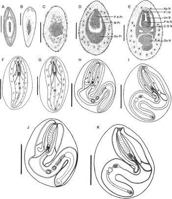
Oval- to elongate-shaped embryonated eggs within the uterus of gravid females measured 24 (20–28) × 9 (8–10) (fig. 1A). Each egg was surrounded by a thin outer membrane, fibrillar coat, a fertilization membrane with distinct polar elongations and a thin inner membrane directly surrounding the body surface of the embryo (acanthor).
Developmental stages of S. saudii in the amphipod host M. agilis up to 34 days post-infection: (A) embryonated egg; (B) acanthor; (C) early acanthella at 10 days; (D) acanthella at 14 days; (E) acanthella at 18 days; (F) late male acanthella at 24 days; (G) late female acanthella at 24 days; (H) early male cystacanth at 26 days; (I) early female cystacanth at 26 days; (J) male cystacanth at 34 days; (K) female cystacanth at 34 days. Ap N, apical nuclei; C G N, cerebral ganglion nuclei; Go N, gonad nuclei; Go Pr, gonadal primordium; M Pr, muscle primordium; P A Pr, proboscis apparatus primordium; P I N, proboscis invertor nuclei; P N R, proboscis nuclear ring, Un B, uncinogenous bands. Scale bars: (A) 10 μm, (B) 20 μm, (C, D) 40 μm, (E) 50 μm, (F, G) 200 μm, (H, I) 250 μm, (J, K) 300 μm.

Acanthor
Hatching of embryonated eggs took place 1 h after ingestion, to release spindle-shaped acanthors, each armed with two prominent rows of minute hooks on its aclid organ (fig. 1B). A central nuclear mass or ‘entoblast/syncytium’ was surrounded by a cortical area with small nuclei which developed into the giant nuclei of the acanthella. Using their hooks, the acanthors penetrated the intestinal epithelium in 12–18 h to reach the connective tissue serosa, where they remained for up to 3 days, by which time they had increased in size to 66 (6–72) × 24 (20–28) and their cortical nuclei appeared more differentiated. Acanthors at 4 days post-infection penetrated the intestinal wall of the amphipod, remained attached to the outer surface of the intestine for 4 days, then detached and dropped free into the amphipod haemocoel, where they lost their hooks and spindle shape and transformed into oval acanthellae. Thus, the acanthor stage lasted for about 8 days.
Acanthellae
Early acanthellae (10 days post-infection) were oval, measured 92 (79–105) × 45 (37–53) and were surrounded by a thin, transparent and acellular membranous envelope with partially or completely disintegrated amphipod haemocytes adhering to its outer surface (fig. 1C). This envelope persisted until and throughout the cystacanth stage. In the acanthella, the entoblast was larger than that of the acanthor, the cortical nuclei differentiated into 24 clearly visible giant nuclei and numerous protoplasmic strands appeared in the cortical area. Infected amphipods contained 3–5 of these acanthellae in their haemocoel and, consistently, one or sometimes two of them were large in size, colourless and appeared in good condition, while the others were smaller, dark and appeared weak, moribund or dead.
Acanthellae at 14 days post-infection measured 137 (130–144) × 86 (80–92). The entoblast (starting point for organogenesis) began to differentiate into the proboscis apparatus, muscle and gonadal primordia (fig. 1D). Therefore, the cortical giant nuclei, which significantly increased in size, were pushed to the periphery.
At 18 days post-infection acanthellae measured 186 (174–198) × 117 (104–130) and contained primordia differentiated into the definite masses that produce the future organs (fig. 1E). The proboscis apparatus primordium successively differentiated into five masses separated from each other by thin membranes: an apical mass containing four large nuclei – ‘apical nuclei’; a narrow transverse mass containing the proboscis invertor nuclei; a longitudinal mass containing a syncytium – ‘uncinogenous bands’; a narrow mass forming a ring – ‘proboscis nuclear ring’; and a posterior mass containing the cerebral ganglion nuclei. These masses eventually produce the apical sense organ of the proboscis, the proboscis invertor muscles, the proboscis hooks, the proboscis wall and the cerebral ganglion, respectively. By this time, four cortical giant nuclei arranged to form a ring (the ‘lemniscal ring’) at the level of the proboscis invertor nuclei, and will eventually migrate to the developing lemnisci. However, gonad nuclei began to differentiate from those of the other genital organs. During this rearrangement, some cavities appeared in the entoblast and seemed to be the developing pseudocoel.
In acanthellae at 24 days post-infection, the worm sex could be determined and sexual size dimorphism was clear. Male acanthellae measured 491 (435–548) × 179 (165–193), while female acanthellae were distinctly larger, 620 (596–644) × 203 (190–216). Male and female acanthellae were nearly parallel in their development (fig. 1F, G). In both, the proboscis apparatus was not fully developed and grew in an everted position, and its developing hooks appeared as blunt conical projections from the uncinogenous bands. Although the neck retractor muscles and the proboscis invertor and retractor muscles were distinct, the proboscis never seen inverted (not yet functional). However, the cerebral ganglion was clearly visible and the two lemnisci started to appear as lateral evaginations of the body wall directly behind the region of the lemniscal ring. Moreover, gonad anlage appeared within a ligament sac and most of the associated genital organs were distinctly visible.
Cystacanths
Early cystacanths (26 days post-infection) tended to be orange in colour. Male cystacanths measured 1021 (962–1080) × 128 (110 − 146), and female cystacanths 1260 (1166–1354) × 142 (122–162). In both sexes, the proboscis hooks completed their development and the proboscis was entirely invaginated within the proboscis receptacle (fig. 1H, I), i.e. the proboscis at that time was functional and was able to invert and to evert. However, the two lemnisci appeared well differentiated and the four lemniscal nuclei moved into them, two into each lemniscus. At this time, the reproductive structures were more differentiated within the ligament sac, and cortical giant nuclei were broken down into nuclear fragments and moved to their proper locations in the hypodermis. Feeding experiments revealed that all of these cystacanths were non-infective, i.e. they failed to excyst in the intestine of S. rivulatus.
Cystacanths at 34 days post-infection were bright orange in colour and readily visible through the translucid cuticle of their amphipod hosts (fig. 1J, K). Male cystacanths measured 1059 (998–1120) × 136 (115–157), and female cystacanths 1205–1397 × 126–168 (1300 × 147). In both sexes, the opacity of the body wall started to increase, and lemniscal giant nuclei seemed broken down into nuclear fragments which moved to their proper locations. In cystacanths at 38 and 42 days post-infection, the body progressively increased in size to reach 1140 (1074–1205) × 146 (124–168) in males and 1404 (1301–1507) × 157 (134–180) in females. All of these cystacanths were non-infective.
Fully developed cystacanths (46 days post-infection) were closely similar to those at 34 days post-infection, but were slightly larger in size. Male cystacanths measured 1207 (1135–1279) × 155 (131–179), and female cystacanths 1488 (1382–1594) × 167 (143–191). Most of these cystacanths were infective, and therefore were considered to be fully developed. Infected amphipods contained one, or sometimes two, of these cystacanths. Thus, S. saudii in the amphipod M. agilis develops into infective cystacanths by 46 days post-infection.
Development of S. saudii in the fish host
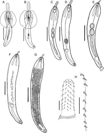
In the intestine of S. rivulatus, fully developed cystacanths excysted from the envelope to become juvenile worms during the first 4 h after infection. This occurred in the middle region of the intestine, where juveniles were found firmly attached by their probosces to the intestinal wall. Excystment seemed to be mainly mechanical, involving the proboscis, which released strongly to penetrate the envelope at one side, and the posterior region of the body, which propelled strongly to penetrate the opposite side. At this time, the envelope appeared as a frustum-shaped ring around the anterior half of the newly excysted juvenile (fig. 2A, B). The ring moved backward and slipped off over the posterior end of the juvenile.
Developmental stages of S. saudii in the fish host S. rivulatus: (A) newly excysted male juvenile; (B) newly excysted female juvenile; (C) immature male; (D) immature female; (E) mature male; (F) mature female; (G) fully gravid female; (H) proboscis. Scale bars: (A, B) 500 μm, (C, D) 1 mm, (E, F, G,) 2 mm, (H) 100 μm.

Newly excysted juveniles
General. Trunk elongate. Proboscis relatively short, cylindrical, armed with ten alternating longitudinal rows of sharp recurved hooks; each row includes 8–9 hooks of variable length (basal hooks alternate with each other). Neck short. Proboscis receptacle cylindrical, double walled. Cerebral ganglion at the middle of the posterior half of proboscis receptacle. Lemnisci claviform, distinctly longer than the proboscis receptacle.
Male juveniles (fig. 2A). Trunk 1307 (1210–1404) × 289 (260–318). Proboscis 329 (290–368) × 120 (102–138). Proboscis hooks: first three hooks, 29 (26–32); next three 33 (30–36); the remaining two or three 23 (21–25) in length. Neck 41 (36–46) in length. Proboscis receptacle 342 (290–394) × 132 (118–146). Lemnisci 496 (482–510) × 40 (31–49). Two testes, oval, tandem, slightly separated, little developed, near middle of trunk, sub-equal, 86 (68–104) × 53 (44–62). Seminal vesicle hardly seen. Four cement glands, long, in two tandem pairs, shrivelled (not yet functional); anterior pair 93 (82–104); posterior pair 108 (96–120) in length. Säfftigen's pouch and copulatory bursa little developed. Genital pore terminal.
Female juveniles (fig. 2B). Trunk 1566 (1429–1702) × 315 (269–361). Proboscis 336 (310–362) × 121 (110–132). Proboscis hooks closely similar in size to those of male juveniles. Neck 47 (42–52) in length. Proboscis receptacle 345 (298–392) × 122 (108–136). Lemnisci 501 (460–542) × 45 (38–52). Ovary little developed, entire, within a ligament sac. Uterine bell 63 (52–74) × 46 (42–50), its base 501 (462–540) from posterior end. Selector apparatus little developed. Uterus tubular, 406 (362–450) in length. Vagina guarded by a sphincter muscle on each side. Genital pore terminal.
Immature worms
Immature males and females first appeared 9 and 14 days post-infection, respectively.
General. Trunk elongate–fusiform. Conspicuous sclerotized plaques embedded in the tegument of the extreme anterior region of the trunk. Proboscis relatively short. Neck short. Proboscis receptacle cylindrical, double walled. Cerebral ganglion at the middle of the proboscis receptacle. Lemnisci claviform, much longer than the proboscis receptacle.
Immature males (fig. 2C). Trunk 3707 (3160–4254) in length; greatest width 586 (530–642) at the anterior region. Proboscis 377 (366–388) × 144 (135–153). Proboscis hooks: first three hooks 32 (30–34); next three 35 (32–38); the remaining two or three 25 (23–28) in length. Neck 70 (62–78) in length. Proboscis receptacle 433 (410–456) × 154 (142–166). Lemnisci 990 (898–1082) × 96 (78–114). Two testes, elongate–oval, tandem, contiguous, nearly at middle of trunk, moderately developed; anterior testis 305 (264–346) × 169 (140–198), posterior testis 324–410 × 132–172 (367 × 152). Four cement glands, tubular, in two tandem pairs, shrivelled (not yet functional); anterior pair 571 (490–652); posterior pair 472 (394–550) in length. Seminal vesicle clearly seen, long, shrivelled (not yet functional). Säfftigen's pouch and copulatory bursa moderately developed. Genital pore terminal.
Immature females (fig. 2D). Trunk 5287 (4461–6113) in length; greatest width 674 (598–750) at the level of the lemnisci. Proboscis 370 (358–382) × 149 (140–158). Proboscis hooks closely similar in size to those of males. Neck 73 (62–84) in length. Proboscis receptacle 443 (416–470) × 157 (138–176). Lemnisci 1005 (946–1164) × 101 (88–114). Ovary entire within a ligament sac, moderately developed. Uterine bell 186 (162–210) × 169 (154–184), its base 915 (694–1136) from posterior end. Uterus tubular, 792 (622–962) in length. Selector apparatus well developed. Vagina guarded by a sphincter muscle on each side. Genital pore terminal.
Mature worms
Mature males and females first appeared 17 and 23 days post-infection, respectively.
General. Body moderate in size. Trunk elongate–fusiform. Females larger than males. Trunk thick-walled, without spines. Conspicuous sclerotized plaques embedded in the tegument of the extreme anterior region of the trunk. Proboscis relatively short, armed with ten alternating rows of 8–9 sharp recurved hooks; basal hooks alternate with each other (fig. 2H). Neck short. Proboscis receptacle double walled. Cerebral ganglion near middle of proboscis receptacle. Lemnisci claviform, equal in length, much longer than proboscis receptacle.
Mature males (fig. 2E). Trunk 9032 (7624–10,440) in length; greatest width 1176 (874–1478) near the anterior region. Proboscis 384 (374–394) × 148 (138–158). Proboscis hooks: first three hooks 36 (33–39); next three 38 (36–40); the remaining two or three 27 (24–30) in length. Neck 79 (63–94) in length. Proboscis receptacle 725 (632–818) × 187 (164–210). Lemnisci 1552 (1158–1946) × 138 (98–178). Lemnisci length/proboscis receptacle length is about 1:2.1. Two testes, elongate–oval, tandem, contiguous, in middle of the trunk or slightly anterior; anterior testis 693 (510–876) × 538 (492–584); posterior testis 848 (734–962) × 607 (574–640). Seminal vesicle claviform, relatively large, swollen. Four cement glands, tubular, in two tandem pairs, turgid; anterior pair 1453 (1201–1705) × 134 (106–162); posterior pair 1287 (1050–1524) × 128 (92–164). Säfftigen's pouch pyriform, 717 (543–891) in length. Copulatory bursa hemispherical, well developed, 494 (462–626) in diameter. Genital pore terminal. In copulating male, the extrusion of the bursa appears to induce a slight displacement of the sexual organs toward the posterior end.
Mature females (fig. 2F). Trunk 12,806 (8295–17,318) in length; greatest width 1278 (908–1648) in near its anterior region. Proboscis 397 (386–408) × 154 (142–166). Proboscis hooks closely similar in size to those of males. Neck 93 (76–110) in length. Proboscis receptacle 736 (642–848) × 200 (172–228). Lemnisci 1586 (1186–1986) × 141 (98–184). Lemnisci length/proboscis receptacle length is about 1:2.1. Ligament sac ruptured. Floating ovarian balls 58(42–74) in diameter. Uterine bell well developed, 344 (292–396) × 475 (410–540), its base 2463 (1832–3094) from posterior end. Selector apparatus 901 (744–1067) × 152 (122–182). Uterus tubular, relatively short, 2133 (1425–2841) in length. Vagina short, guarded by sphincter muscle. Genital pore terminal. Embryonated eggs small, elongate–oval (fig. 1A), with polar swelling of middle membrane; uterine eggs 24 (20–28) × 9 (8–10).
Recently copulated females first appeared 26 days post-infection. In the next 2 days all females were found to have copulated. Partially gravid females first appeared 31 days post-infection, while fully gravid females first appeared 35 days post-infection (fig. 2G). Mature males and fully gravid females started to die off naturally 31 and 43 days post-infection, and were totally expelled from the fish intestine 42 and 52 days post-infection, respectively. Thus, fully gravid females remain alive in the intestine for about 12 more days after the death of males. Some dead worms were seen hanging out of the anus of many fish.
Discussion
The prevalence, intensity and development of acanthocephalans in their intermediate and definitive hosts have mostly shown seasonal periodicities throughout the year (Awachie, Reference Awachie1965; Chubb, Reference Chubb1982; Kennedy, Reference Kennedy2006). Such periodicities may possibly be related to oscillations in water temperature, fluctuations in the availability of intermediate infected hosts and infecting larvae, and to alterations in diet composition and feeding behaviour of the hosts (E.R. Kennedy, Reference Kennedy, Crompton and Nickol1985; Amin, Reference Amin1987; C.R. Kennedy, Reference Kennedy2006). If these conditions are more stable and allow recruitment, maturation and mating of acanthocephalans to occur in all seasons of the year, no seasonal periodicity is evident (Hine & Kennedy, Reference Hine and Kennedy1974; E.R. Kennedy, Reference Kennedy, Crompton and Nickol1985; C.R. Kennedy, Reference Kennedy2006). In the present study, infrapopulations of S. saudii in the intestine of S. rivulatus were relatively large throughout the year, contained all the developmental stages and adults, including gravid females. However, infected amphipods and infecting cystacanths were available, and food items in the intestine of S. rivulatus were mainly composed of algae and seagrasses on which the amphipod intermediate host is commonly found. Thus, occurrence of S. saudii in the intestine of S. rivulatus appeared to be in a state of dynamic balance between recruitment and loss of adults throughout the year. Therefore, no significant seasonal periodicity in prevalence, intensity and maturation was observed.
Although fully gravid females were present in most infrapopulations of S. saudii, their embryonated eggs were never observed among the intestinal contents of S. rivulatus, and some gravid female and male worms were seen hanging out of the anus of many fish. Thus, S. saudii females may not lay eggs, but pass out of the intestine of S. rivulatus, where they degenerate in the environment and the released embryonated eggs are ingested by the amphipod intermediate host. This suggestion was used by Muzzall & Rabalais (Reference Muzzall and Rabalais1975) and Hassanine (Reference Hassanine2006) to explain similar situations in Acanthocephalus jacksoni Bullock, 1962 and Diplosentis nudus Harada, 1938, respectively. This strategy, in which eggs are carried into the environment by gravid females, could increase transmission success by dispersing eggs closer to the bottom of the lagoon, where the target crustacean hosts are possibly found (Kopp et al., Reference Kopp, Elke, Caddigan, Raj, Rodriguez, Young and Sparkes2011). Egg transmission in the present life cycle follows this strategy, since S. saudii eggs are ingested by a benthic amphipod intermediate host.
Like the eggs of other palaeacanthocephalans, such as those of Echinorhynchus truttae Schrank, 1788, E. lageniformis Ekbaum, 1938 and Acanthocephalus parksidei Amin, 1975 (Awachie, Reference Awachie1966; Olson & Pratt, Reference Olson and Pratt1971; Amin, Reference Amin1982, respectively), embryonated eggs of S. saudii are surrounded by four membranes, having a fertilization membrane with distinct polar elongations and containing an armed acanthor. These eggs hatched in the intestine of the amphipod M. agilis during the first hour after ingestion, and hooks of the released acanthors appeared more prominent. These hooks assist in penetration of the egg membranes (Schmidt & Olsen, Reference Schmidt and Olsen1964; Uglem, Reference Uglem1972). The acanthor stage lasted for about 8 days before reaching the amphipod haemocoel, where the acanthor loses its hooks and transforms into an oval acanthella.
Acanthocephalan larvae in the haemocoel of their arthropod intermediate hosts are surrounded by a thin, transparent and acellular membranous envelope, protecting them from the arthropod immune cells or haemocytes (Lackie & Holt, Reference Lackie and Holt1988; Holt, Reference Holt1989; Dezfuli & Giari, Reference Dezfuli and Giari1999). This envelope appeared around early acanthella of S. saudii and, as in all studied acanthocephalan larvae, the envelope persisted throughout the larval stages. Dezfuli et al. (Reference Dezfuli, Simoni, Duclos and Rossetti2008) concluded that the envelope is formed by detached microvilli from the acanthella tegument and residues from the arthropod haemocytes contribute to its thickness. The presence of the envelope around early acanthella of S. saudii and the adherence of partially or completely disintegrated amphipod haemocytes on its outer surface greatly supports this conclusion.
Some early acanthellae become weak or moribund following their penetration of the arthropod intestine to reach its haemocoel (Robinson & Strickland, Reference Robinson and Strickland1969; Lackie, Reference Lackie1972). The growth rate of such acanthellae is slow and increases the time of their exposure to arthropod haemocytes (Nickol & Dappen, Reference Nickol and Dappen1982; Gleason, Reference Gleason1989). The envelope surrounding each of these acanthellae is probably not a rigid barrier against the haemocytes, which, in this case, are able to cross the envelope and release their contents (e.g. melanin) within the vicinity of the acanthella tegument, making their colour dark due to melanization (Dezfuli et al., Reference Dezfuli, Simoni, Duclos and Rossetti2008). Such cases were noticed in the present study, since infected amphipods contained 3–5 early acanthellae of S. saudii in their haemocoel and, consistently, one or sometimes two of them were large, colourless and appeared to be in good condition, while the others were smaller, moribund or dead and distinctly dark. These acanthellae appeared dysfunctional, were probably attacked by haemocytes (melanized) and could not reach the cystacanth stage, since in the latter stage only one or two cystacanths were found in the infected amphipods.
Giant nuclei first appear in early acanthellae and their numbers have been reported to differ from species to species; for example, 10, 18 and 30 were observed in E. truttae, E. lageniformis and Acanthocephalus anguillae Mueller, 1780 (Awachie, Reference Awachie1966; Olson & Pratt, Reference Olson and Pratt1971; Leuckart, Reference Leuckart1876, respectively). In early acanthellae of S. saudii, 24 giant nuclei were distinct and, similar to most palaeacanthocephalans, four of these nuclei were involved in the formation of lemnisci, and the others were broken down into hypodermal nuclear fragments during the cystacanth stage.
Generally, S. saudii acanthellae are similar to those of other palaeacanthocephalans that use amphipod intermediate hosts, and are very similar those of E. truttae, E. lageniformis and N. cristatus (see Awachie, Reference Awachie1966; Olson & Pratt, Reference Olson and Pratt1971; Uglem, Reference Uglem1972, respectively), but have some significant differences. They are similar to those of E. truttae in having a proboscis that develops in an everted position, then inverts in the cystacanth stage; in all other palaeacanthocephans, the opposite is true. However, precocious development was not evident in S. saudii acanthellae, but was clear in E. truttae acanthellae, where ovarian balls were distinct in females and the copulatory bursa was protruded in males. Sclerocollum saudii acanthellae are similar to those of E. lageniformis and N. cristatus, but mainly differ in having a proboscis developing in an everted position. Moreover, late acanthellae of S. saudii showed a clear sexual size dimorphism, which is common in acanthocephalans (Poulin & Morand, Reference Poulin and Morand2000; Sasal et al., Reference Sasal, Jobet, Faliex and Morand2000) and mostly begins to appear during the cystacanth stage (Amin et al., Reference Amin, Burns and Redlin1980; Steinauer & Nickol, Reference Steinauer and Nickol2003). This is thought to be a consequence of sexual selection which affects the adult body size (Andersson, Reference Andersson1994; Benesh & Valtonen, Reference Benesh and Valtonen2007), and favours a larger body size in females than males, probably because larger females usually have greater fecundity than smaller ones (Poulin & Morand, Reference Poulin and Morand2000).
In early cystacanths of S. saudii, the proboscis apparatus completed its development, the proboscis was functional, and all other organs appeared to be well developed and in place, except the lemniscal giant nuclei, which were entire. In cystacanths at 34 days post-infection, these nuclei were broken down into nuclear fragments and moved to their proper locations. These cystacanths and those at 38 and 42 days post-infection were non-infective, while most cystacanths at 46 days post-infection were infective, and therefore considered as fully developed. The combination of these results greatly supports the suggestion of Nickol (Reference Nickol, Crompton and Nickol1985) that not all acanthocephalan species are infective to vertebrates immediately upon reaching the cystacanth stage, but some require a period of development as a cystacanth before attaining infectivity. Thus, S. saudii in the amphipod M. agilis develops into infective cystacanths 46 days post-infection. In other palaeacanthocephalans, such as N. cristatus, E. lageniformis and E. truttae, 16, 30 and 82 days are required in the amphipod host to reach a cystacanth stage infective to the fish host (Uglem, Reference Uglem1972; Olson & Pratt, Reference Olson and Pratt1971; Awachie, Reference Awachie1966, respectively).
In their crustacean intermediate hosts, cystacanths of several acanthocephalan species are bright yellow to orange in colour (Crompton & Nickol, Reference Crompton and Nickol1985) due to the carotenoid pigments that are taken from the host haemolymph (Barrett & Butterworth, Reference Barrett and Butterworth1968; Taraschewski, Reference Taraschewski2000). Acanthocephalans with coloured cystacanths mostly use crustacean amphipods as intermediate hosts. Some authors (e.g. Bakker et al., Reference Bakker, Mazzi and Zala1997; Moore, Reference Moore2002) suggested that the clear visibility of coloured cystacanths through the translucid cuticle of their amphipod host alters its phenotype in ways that make it more susceptible to predation by the fish predators. This mechanism is known as ‘oddity selection’ (Bakker et al., Reference Bakker, Mazzi and Zala1997). It has also been suggested (e.g. Barrett & Butterworth, Reference Barrett and Butterworth1973; Bakker et al., Reference Bakker, Mazzi and Zala1997) that carotenoids provide photoprotection for acanthocephalan larvae against the threat of UV radiation passing through the translucid cuticle of their amphipod hosts. Sclerocollum saudii cystacanths are bright orange in colour and are readily visible through the translucid cuticle of infected amphipods, while S. rivulatus is a purely herbivorous fish feeding mainly by grazing benthic algae and seagrasses, but is not a predator and does not feed selectively on infected or uninfected amphipods. Thus, S. saudii is trophically transmitted to the fish when the latter accidentally ingests an infected amphipod entangled in its food. This greatly supports the experimental finding of Kaldonski et al. (Reference Kaldonski, Perrot-Minnot, Dodet, Martinaud and Cezilly2009) that yellow-orange colouration of cystacanths plays no adaptive role in the trophic transmission of cystacanths to their final fish hosts. However, uninfected amphipods or those infected or with S. saudii tend to hide in benthic algae or among seagrasses. The lagoon is also shallow and thus amphipods are likely to swim away and avoid the potential effects of UV radiation near the surface waters of the lagoon. With these observations, the suggestion that carotenoids provide photoprotection for acanthocephalan cystacanths becomes highly questionable.
So far, excystment of acanthocephalan cystacanths as active juveniles in the intestine of their vertebrate hosts has been examined in only four species (see Graff & Kitzman, Reference Graff and Kitzman1965; Awachie, Reference Awachie1966; Lackie, Reference Lackie1974; Kennedy et al., Reference Kennedy, Broughton and Hine1978). This is thought to be mainly stimulated superficially by the osmotic pressure of the surrounding medium or by the bile of the host. The exact role of bile is not fully understood (Khanna, Reference Khanna2004). Sclerocollum saudii cystacanths excysted as active juveniles in the middle region of the intestine of S. rivulatus during the first 4 h after infection, probably because this region is usually rich in simple nutrients that can be absorbed by the body wall of acanthocephalans (Petrochenko, Reference Petrochenko1956). However, excystment seemed to be mainly mechanical via the proboscis and the posterior region of body, which penetrate the envelope at two definite, opposite sides. Male and female juveniles reached maturity 17 and 23 days post-infection, respectively. Thus, male juveniles reached maturity 6 days before the females. Early maturation of males, as suggested by several authors (e.g. Ghiselin, Reference Ghiselin1974; West Eberhard, Reference West Eberhard1983), is probably favoured by sexual selection (male–male competition for access to females), which was described as intense in S. saudii (Al-Jahdali & Hassanine, Reference Al-Jahdali and Hassanine2012), or by sexual size dimorphism (Morand & Hugot, Reference Morand and Hugot1998). Delayed maturation of females suggests that females invest more in growth than males (Benesh, Reference Benesh2007). Recently copulated females first appeared 26 days post-infection and all females seemed to have copulated by 28 days post-infection. Thus, females copulated 3 days after they reached sexual maturity in a mating period extending for about 2 days. So, a period of about 9 days is required for mature males before mating. During this period mature males probably continue to produce and store more sperm and cement in their seminal vesicles and cement reservoirs, since mature worms of S. saudii, as in many acanthocephalan species, are polygamous and form a sexual congress (Al-Jahdali & Hassanine, Reference Al-Jahdali and Hassanine2012), i.e. aggregate within groups for mating in the intestine of their host (Richardson et al., Reference Richardson, Martens and Nickol1997), and a single male can mate with several females (Crompton, Reference Crompton1974; Abele & Gilchrist, Reference Abele and Gilchrist1977). A short time after mating (about 3 days, or at day 32 after infection), mature males started to die off naturally, probably due to the intense male–male competition for access to females (Sasal et al., Reference Sasal, Jobet, Faliex and Morand2000), and during the next 10 days passed out of the intestine of S. rivulatus, i.e. were totally expelled from the fish by 42 days post-infection. Females appeared partially gravid and then fully gravid 3 and 6 days after mating, i.e. 31 and 35 days post-infection. After about 8 days, or at day 44 after infection, fully gravid females started to die off naturally, and during the next 8 days passed out of the intestine of S. rivulatus, i.e. were totally expelled from the fish by 52 days post-infection. Thus, fully gravid females remain alive in the fish for about 12 more days after the death of males. This length of time is probably necessary for embryonic development within the eggs. Fully gravid females expelled from the fish degenerate in the environment and the released embryonated eggs are ingested by the amphipod intermediate host. Generally, the life cycle of S. saudii extended for 89 days and is similar to other known palaeacanthocephalan life cycles, but has its own characteristics.
Acknowledgements
We should like to extend our appreciation to the staff of Rabigh-Faculty of Science and Arts, King Abdulaziz University, for their help during the collection of the material.
Financial support
We are very grateful to King Abdulaziz University, Saudi Arabia, for continued encouragement and support.
Conflict of interest
None.