Introduction
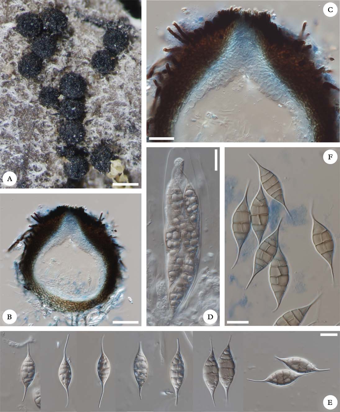
The genus Capronia Sacc. is characterized by black, usually setose, perithecioid ascomata with periphysate ostioles, an absence of pseudoparaphyses, fissitunicate, usually 8- to multi-spored asci of the Herpotrichiella-type and septate to muriform, hyaline to pale grey or brown ascospores (Halici et al. Reference Halici, Hawksworth, Candan and Türk2010). It comprises c. 70 species, which mostly grow on decaying wood, bark and leaves, but some are also associated with fungi or lichens (Untereiner et al. Reference Untereiner, Gueidan, Orr and Diederich2011; Friebes Reference Friebes2012). So far, 17 species (two still unnamed; see Etayo Reference Etayo2002: 92 and Etayo Reference Etayo2010: 35) obligately growing on lichens have been recognized (e.g., Etayo & Sancho Reference Etayo and Sancho2008; Halici et al. Reference Halici, Hawksworth, Candan and Türk2010; Flakus & Kukwa Reference Flakus and Kukwa2012a ; Zhurbenko Reference Zhurbenko2012). In this paper, we introduce a new species of the genus Capronia with very unusual ascospores, with both ends protruding into long appendages (caudae).
This work is a further contribution to the knowledge of the lichenicolous fungi of Bolivia, which is the result of our current study focusing on the taxonomy and diversity of lichens and allied fungi in that country (e.g., Flakus & Wilk Reference Flakus and Wilk2006; Flakus & Kukwa Reference Flakus and Kukwa2007, Reference Flakus and Kukwa2012a , Reference Flakus and Kukwa b ; Flakus et al. Reference Flakus, Ahti, Kukwa and Wilk2008, Reference Flakus, Etayo, Schiefelbein, Ahti, Jabłońska, Oset, Bach, Rodriguez Flakus and Kukwa2012; Flakus Reference Flakus2009; Kukwa & Flakus Reference Kukwa and Flakus2009; Kukwa et al. Reference Kukwa, Etayo and Flakus2012; Etayo et al. Reference Etayo, Flakus and Kukwa2013).
Material and Methods
The material is deposited in KRAM, LPB and hb. Etayo. Morphology and anatomy were examined using Nikon SMZ800 and Nikon Eclipse 80i (DIC) microscopes. The anatomy was studied on material mounted in water, KOH solution (K), lactophenol cotton blue (LPCB), and for ascus structure Lugol's iodine solution without (I) or with pretreatment with KOH (K/I). Measurements of ascospores and other structures were made in water.
The New Species
Capronia paranectrioides Etayo, Flakus & Kukwa sp. nov.
MycoBank No.: MB 803885
Differs from other Capronia species by pale brown, submuriform and bicaudate ascospores, and I+ red, K/I+ pale blue hamathecial gelatine.
Type: Bolivia, Dept. La Paz, Nor Yungas Province, Parque Nacional y Área Natural de Manejo Integrado Cotapata, 30′ walking of Unduavi by Sillu Tincara pre-Columbian route, transition Páramo Yungueño–Yungas cloud forest, on epiphytic Erioderma leylandii, 3518 m, 16°17′22″S, 67°53′29″W, 25 May 2011, J. Etayo 28054, A. Flakus, M. Kukwa, O. Plata & U. Schiefelbein (LPB—holotype; KRAM, hb. Etayo—isotypes).
Capronia paranectrioides (holotype). A, habit of perithecioid ascomata on the upper surface of Erioderma; B, cross-section of ascoma showing pseudoparenchymatous exciple composed of several cell layers (mounted in LPCB); C, periphysoids (mounted in LPCB); D, 8-spored ascus; E, ascospores (mounted in tap water); F, ascospores (mounted in LPCB). Scales: A=200 µm; B=50 µm; C=25 µm; D–F=10 µm. In colour online.
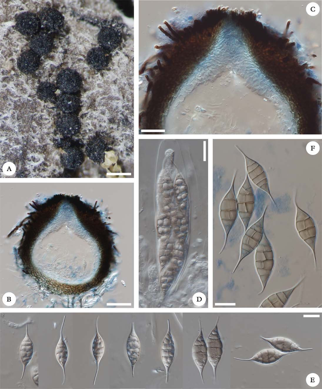
Ascomata perithecioid, ostiolate, dispersed on the host thallus to aggregated, superficial, black, subglobose to barrel-shaped with slightly applanate apex, habitually not collapsing, when dry, with abundant setae in upper half to almost glabrous, 150–220 µm diam. Setae simple, non or rarely septate, smooth-walled, dark brown, obtuse to slightly pointed at the apex and with thickened base, sometimes with constrictions near septa, 30–95×3·0–4·5 µm. Exciple 25–35 µm thick, pseudoparenchymatous, composed of c. 6–11 layers of thin-walled cells, the outermost layers brown to dark brown, of angular to elongate cells (c. 5–9×3–4 µm), the inner layers pale brown to hyaline (I+ yellow-orange), of more elongate cells in cross-section. Hamathecium gelatinized, hyaline, I+ red, K/I+ pale blue, pseudoparaphyses absent. Periphysoids slightly clavate, hyaline, 1–4-septate, apically usually faintly branched, 11–25×3–6(–9) µm. Asci 8-spored, clavate, fissitunicate, wall apically thickened, ascus apex I−, K/I−, ascus wall K/I+ faintly pale blue, 70–80×14–15 µm. Ascospores pale brown with pigment uniformly distributed, broadly ellipsoidal with acuminate apices prolonging into (3–)5–9(–14) µm long and often curved caudae, submuriform, mature 8–11-celled, with 3–5 transverse and 1–2 longitudinal septa with slight constriction near the septa, without perispore, smooth-walled, 15–29×8–10(–11) µm (measured without caudae); ascospores measured together with cilia 35–50 µm long.
Conidiomata not observed.
Host
The species inhabits the upper thallus surface of corticolous Erioderma leylandii. As the fungus does not cause damage to the host thallus, we assume it is a parasymbiont.
Etymology
The new species is named after the characteristic submuriform ascospores with both ends prolongated in long caudae, thus resembling the hypocrealen genus Paranectria Sacc. (e.g. Hawksworth Reference Hawksworth1982; Ertz Reference Ertz, Nash, Ryan, Diederich, Gries and Bungartz2004; Zhurbenko Reference Zhurbenko2009).
Distribution and habitat
So far, the species is known from high Andean forests in Bolivia and Peru. The type locality, where it grows in the transitional vegetation between Páramo Yungueño and Yungas cloud forest is very diverse in terms of lichenicolous fungi, and other species will be treated elsewhere.
Notes
Capronia paranectrioides is the first known member of the genus growing on Erioderma. It differs from all other species predominantly by its bicaudate ascospores (Hawksworth Reference Hawksworth1980, Reference Hawksworth1990; Kondratyuk & Galloway Reference Kondratyuk and Galloway1995; Etayo Reference Etayo1996, Reference Etayo2002, Reference Etayo2003, Reference Etayo2010; Aptroot et al. Reference Aptroot, Diederich, Sérusiaux and Sipman1997; Etayo & Diederich Reference Etayo and Diederich1998; Etayo & van den Boom Reference Etayo and van den Boom2006; Etayo & Sancho Reference Etayo and Sancho2008; Halici et al. Reference Halici, Hawksworth, Candan and Türk2010; Flakus & Kukwa Reference Flakus and Kukwa2012a ; Friebes Reference Friebes2012; Zhurbenko Reference Zhurbenko2012). Another of the unusual features characterizing the new species is the positive amyloid reaction of the hymenium (I+ red and K/I+ blue). So far only two lichenicolous taxa, C. leopoldiana Etayo and C. thamnoliae Zhurb., share this character with C. paranectrioides, but their ascospores are not caudate (Etayo & Sancho Reference Etayo and Sancho2008; Zhurbenko Reference Zhurbenko2012). The reaction, although not always tested, has also been noticed among non-lichenicolous species (Winka et al. Reference Winka, Eriksson and Bang1998; Untereiner Reference Untereiner2000; Friebes Reference Friebes2012). Those species may represent a separate genus due to the atypical reaction of the hamathecial gelatine, but other characters fit the concept of Capronia well, thus we have decided to keep them within this genus. Nevertheless, further molecular data could show the taxa with amyloid hamathecial gelatine to represent a separate phylogenetic lineage, as Capronia is already known to be polyphyletic (Untereiner et al. Reference Untereiner, Gueidan, Orr and Diederich2011).
This type of ascospore is very similar to that of Paranectria, but that hypocrealean genus can easily be distinguished by its pinkish to orange peridium and hyaline ascospores (e.g., Hawksworth Reference Hawksworth1982; Zhurbenko Reference Zhurbenko2009).
Additional specimen examined. Peru: Cuzco: Prov. Paucartambo, road Paucartambo-Pillcopata, 1 km NE of the turn to Tres Cruces, on Erioderma, c. 13°7′S, c. 71°37′W, c. 3500 m, 1981, R. Santesson, A. Tehler & G. Thor P106: 6 (UPS).
We are greatly indebted to Rosa I. Meneses Q., the Director of Herbario Nacional de Bolivia, Instituto de Ecología, Universidad Mayor de San Andrés, La Paz, for generous cooperation, and also to Paul Diederich (Luxemburg) and an anonymous reviewer for valuable and constructive comments on the manuscript. We also thank U. Schiefelbein (Rostock) and O. Plata (La Paz) for their help during fieldwork and Professor Rolf Santesson (Uppsala) for making the Peruvían speciman available for our study. This research received support from the National Centre for Research and Development (NCBiR) in Poland under the LIDER Programme for the years 2010–2013 (no. 92/L–1/09).