Introduction
Marine mammals harbour a diverse fauna of parasites, with some species causing actual or potential threats to human health (Lehnert & Hernández-Orts, Reference Lehnert, Hernández-Orts, Smit and Sures2025). The family Brachycladiidae (Digenea) is among them. This is the only species-rich, globally distributed digenean taxon restricted to marine mammals (Fraija-Fernández et al., Reference Fraija-Fernández, Olson, Crespo, Raga, Aznar and Fernández2015). Maritae (sexual adults) of this family are well-known to cause a severe pathogenic impact on their definitive hosts (Lehnert et al., Reference Lehnert, Seibel, Hasselmeier, Wohlsein, Iversen, Nielsen, Heide-Jørgensen, Prenger-Berninghoff and Siebert2014; Andersen-Ranberg et al., Reference Andersen-Ranberg, Lehnert, Leifsson, Dietz, Andersen, Siebert, Measures and Sonne2018; Díaz-Delgado et al., Reference Díaz-Delgado, Fernández, Sierra, Sacchini, Andrada, Vela, Quesada-Canales, Paz, Zucca, Groch and Arbelo2018; Nakagun et al., Reference Nakagun, Shiozaki, Ochiai, Matsuda, Tajima, Matsuishi, Watanabe, Horiuchi and Kobayashi2018; Nakagun & Kobayashi, Reference Nakagun and Kobayashi2020; de Amorim et al., Reference de Amorim, Piva, Ribeiro, Perosa, Gris, Perin, Nunes, Cony, Hoppe, Soares and Sonne2024), but otherwise brachycladiids remain poorly studied. The systematics of the family is controversial, as most studies are based on badly preserved maritae from stranded and deceased hosts (Gibson & Bray, Reference Gibson and Bray1997). Molecular genetic data failed to resolve the relationships within the Brachycladiidae unambiguously, and a robust phylogenetic reconstruction for this group is still lacking (Fernández et al., Reference Fernández, Aznar, Latorre and Raga1998; Fraija-Fernández et al., Reference Fraija-Fernández, Aznar, Fernández, Raga and Fernández2016; Kim et al., Reference Kim, Youn, Lee, Lee, Kim, Kang, Choe and Georgieva2023). Intraspecific genetic diversity in brachycladiids is also barely studied, and factors shaping their population structure remain uncertain (Marigo et al., Reference Marigo, Cunha, Bertozzi, Souza, Rosas, Cremer, Barreto, de Oliveira, Cappozzo, Valente, Santos and Vicente2015). Finally, the circulation pathways of the Brachycladiidae are mostly unknown since the life cycle has been elucidated just for a single species (Kremnev et al., Reference Kremnev, Gonchar, Krapivin, Knyazeva and Krupenko2020).
Studies involving intermediate hosts can provide various insights into the biology of the Digenea, including questions on species recognition, population genetics, transmission routes and life cycles, ecological and evolutionary consequences of the host-parasite interactions, and human health-related issues (Fredensborg & Poulin, Reference Fredensborg and Poulin2006; Bates et al., Reference Bates, Poulin and Lamare2010; Galaktionov et al., Reference Galaktionov, Solovyeva and Miroliubov2021; Gonchar & Galaktionov, Reference Gonchar and Galaktionov2021; Krupenko et al., Reference Krupenko, Kremnev, Gonchar, Uryadova, Miroliubov, Krapivin, Skobkina, Gubler and Knyazeva2022, Reference Krupenko, Gonchar, Krapivin, Kremnev, Skobkina and Efeykin2025; Bennett et al., Reference Bennett, Presswell and Poulin2023; Agustina et al., Reference Agustina, Saichua, Laha, Tangkawatana, Prakobwong, Laoprom, Kamphasri, Chareonchai, Blair and Suttiprapa2024). An additional benefit for research focusing on parasites of marine mammals is that intermediate hosts are much easier to collect and dissect than the definitive ones. The only requirement is that the life cycle of a parasite has been elucidated. Within the Brachycladiidae, Orthosplanchnus arcticus Odhner, 1905, is a suitable model. It is a common hepatic parasite known from several true seal species and the walrus in the Arctic and North Pacific (Odhner, Reference Odhner1905; Delyamure et al., Reference Delyamure, Yurakhno, Popov, Shults and Fay1984; Adams & Rausch, Reference Adams and Rausch1989; Andersen-Ranberg et al., Reference Andersen-Ranberg, Lehnert, Leifsson, Dietz, Andersen, Siebert, Measures and Sonne2018; Walden et al., Reference Walden, Bryan, McIntosh, Tuomi, Hoover-Miller, Stimmelmayr and Quakenbush2020). The life cycle of O. arcticus involves Cryptonatica affinis (Gmelin, 1791), a moon snail from the family Naticidae, as the first intermediate host, and cardiid bivalves as the second intermediate hosts (Kremnev et al., Reference Kremnev, Gonchar, Krapivin, Knyazeva and Krupenko2020). However, the genuine range of the second intermediate hosts of this parasite can include more bivalve species, as some previously described metacercariae share morphological similarities with O. arcticus (Kremnev et al., Reference Kremnev, Gonchar, Krapivin, Knyazeva and Krupenko2020).
In the present study, our primary objective was to verify the phylogenetic position of small ocellate metacercariae reported from the Barents Sea bivalves by Chubrik (Reference Chubrik1966). We also examined other marine bivalves that could potentially host metacercariae of the Brachycladiidae in the sub-Arctic Seas, and identified such parasitic larvae only in three cardiid species and the Iceland scallop Chlamys islandica (O. F. Müller, 1776). Additionally, we observed an accidental infection with brachycladiid metacercariae in a common whelk, the carnivorous gastropod Buccinum undatum Linnaeus, 1758. As the obtained molecular genetic data showed that all these life cycle stages belong to O. arcticus, we wondered if there are any preferences for the second intermediate host in this brachycladiid. Two additional questions were addressed: (1) which factor has more influence on infection patterns, bivalve size or species; and (2) do the metacercariae from different bivalve species vary in morphology. We applied statistical analysis to resolve these issues.
Material and methods
Sampling was conducted in 2020–2023 from intertidal and subtidal zones of the White Sea (Kandalaksha Bay: Keret Archipelago, 66°18′N 33°38′E; Velikaya Salma Strait, 66°33′N 33°06′E), the Barents Sea (Kola Peninsula: Dalniye Zelentsy, 69°06′N 36°03′E), and the Pechora Sea (Vaygach Island: Lyamchina Bay, 69°50′N 59°24′E). Moon snails and various species of bivalves (Table 1) were collected, measured, and checked for infection. Gastropods were removed from the shell and visually inspected under a stereomicroscope. Bivalves were cut into small pieces, which were squeezed between two glass plates to detect metacercariae. For each infected bivalve specimen, the number of metacercariae was counted.
List of bivalves inspected for infection with brachycladiids metacercariae

The putative life cycle stages of brachycladiids were fixed with 96% ethanol. Metacercariae were removed from cyst and heat-killed before fixation (Cribb & Bray, Reference Cribb and Bray2010). For morphological analysis, metacercariae were stained with acetocarmine (Sigma Aldrich, Germany), destained in 0.1 M HCl in 70% ethanol, dehydrated in a graded alcohol series, clarified in xylol, and mounted in Canada balsam or BioMount medium (Bio Optica, Italy). Pieces of infected molluscs were fixed in Zenker’s solution with 40% formaldehyde (10:1) to make histological sections. After a two-hour fixation and a two-hour wash in tap water, the specimens were incubated in 70% ethanol with iodine for one hour. Next, they were transferred to 70% ethanol, dehydrated through a graded series of alcohols, and embedded in Paraplast medium (Leica Microsystems, Germany). Sections were cut at 5 μm and stained with Mallory’s trichrome stain.
Slides and whole mounts were studied with a Leica DM 2500 compound microscope (Leica Microsystems, Germany). Photographs were taken under the same microscope with a Nikon DS Fi3 camera (Nikon, Japan). Encysted metacercariae in tissues of the intermediate hosts were photographed alive using an MBS-10 stereomicroscope (LOMO, Russia) equipped with a Canon EOS 70D camera (Canon, Japan). Measurements were made using Fiji software (Schindelin et al., Reference Schindelin, Arganda-Carreras, Frise, Kaynig, Longair, Pietzsch, Preibisch, Rueden, Saalfeld, Schmid, Tinevez, White, Hartenstein, Eliceiri, Tomancak and Cardona2012). All measurements are in micrometres. Whole mounts with metacercariae used for generating morphometric data (Table 4) were deposited in the Helminths collection of the Zoological Institute of the Russian Academy of Sciences (ZISP), section Trematoda (non-type material), under accession numbers 2025.07.12.001–33.
We used standard methods of biological statistics (Zuur et al., Reference Zuur, Ieno and Smith2007). Statistical analyses were performed using the R 4.4.1 statistical programming environment (R Core Team, 2024). P-values less than 0.05 were considered significant. Mean values with 95% confidence intervals (CIs) for variables are provided in the text and on the plots (unless otherwise stated). For mean intensities, mean abundances, and Poulin’s D, bootstrap CI were counted using the boot R package (Davidson & Hinkley, Reference Davidson, Hinkley, Davidson and Hinkley1997; Canty & Ripley, Reference Canty and Ripley2024). Exact CIs are provided for percentages (Clopper & Pearson, Reference Clopper and Pearson1934). Generalized linear models (GLMs) with a negative binomial distribution were fitted using the MASS package (Venables & Ripley, Reference Venables and Ripley2002). Generalized least squares linear models (GLS) were fitted using the nlme package (Pinheiro et al., Reference Pinheiro, Bates, DebRoy, Sarkar, authors, Heisterkamp, Willigen, Ranke and Team2023). All models were validated with the DHARMa package (Hartig, Reference Hartig2024). McFadden’s pseudo-R2 statistics were obtained using the performance package (Lüdecke et al., Reference Lüdecke, Ben-Shachar, Patil, Waggoner and Makowski2021). Non-metric multidimensional scaling (NMDS) ordination and permutational multidimensional analysis of variance (PERMANOVA) were performed using the vegan package (Oksanen et al., Reference Oksanen, Simpson, Blanchet, Kindt, Legendre, Minchin, O’Hara, Solymos, Stevens, Szoecs, Wagner, Barbour, Bedward, Bolker, Borcard, Carvalho, Chirico, De Caceres, Durand, Evangelista, FitzJohn, Friendly, Furneaux, Hannigan, Hill, Lahti, McGlinn, Ouellette, Ribeiro Cunha, Smith, Stier, Ter Braak and Weedon2024). Plots were generated with the ggplot2 package (Wickham, Reference Wickham2016). The raw datasets used for statistical analyses are available as Supplementary Tables S1 and S2, and the R code is provided as Supplementary File S3.
We used three conventional estimates that describe infection patterns (Bush et al., Reference Bush, Lafferty, Lotz and Shostak1997): prevalence (the percentage of infected hosts), mean intensity (the sum of parasite individuals divided by the number of all infected hosts), and mean abundance (the sum of parasite individuals divided by the total number of examined hosts). We also provided Poulin’s D-index as a measure of the metacercariae aggregation (Poulin, Reference Poulin1993). The analysis of infection patterns was based on the data of four bivalve species across two regions, the White and Barents Seas (Table 5). A single infected Iceland scallop from the Pechora Sea (Table 1) was omitted.
GLM with binomial distribution and logit link function was used to model the infection probability (i.e., prevalence) dependence on the host species and host shell length. GLM with a negative binomial distribution and a log link function was used to assess the impact of host species and shell length on the parasite abundance. LM with a log-transformed response variable was used to model the effect of host species and shell length on infection intensity. The attempts to fit a negative binomial GLM to the analysis of infection intensity were unsuccessful due to the absence of zeros in the data. The initial models included the following explanatory variables: host species, host shell length, and their interaction. Then the choice was made between the full model and the model without the interaction term, based on the Akaike information criterion (AIC). All models were checked for patterns in residuals, overdispersion, and zero inflation/truncation if needed.
NMDS was performed to visualize the morphological differences between metacercariae obtained from different host species. We used 33 measured metacercariae from 14 bivalve specimens of 4 species, with 1 to 6 metacercariae taken from an individual host. The matrix of Euclidean distances between the parasite specimens was based on 13 morphological variables and visualized with 2-dimensional NMDS. 95% ellipses for factor centroids for host species were plotted on the graph. Original variables significantly correlated with the NMDS axes (tested with randomization tests with p-value correction for multiple tests) were used to produce a biplot diagram. We also plotted host shell lengths on the diagram as smooth surfaces using the ordisurf function from the vegan package. The same matrix of Euclidean distances was used in PERMANOVA to test if the impact of host species on metacercarial morphological traits was significant.
We used a piece of a redia or whole metacercariae for molecular analysis. A list of the isolates and their hosts is provided in Table 2. For DNA extraction, samples from 96% ethanol were dried completely and incubated in 200 μL of 5% Chelex® 100 resin (Bio-Rad, Hercules, California, USA) with 0.2 mg/mL proteinase K (Evrogen, Moscow, Russia) at 56°C for 4 h, then heated for 8 min at 90°C to inactivate proteinase K, and next centrifuged for 10 min at ~16,000 g. The supernatant with DNA was transferred into a new tube and stored at −20°C.
Isolates of Orthosplanchnus arcticus putative life cycle stages, their origin, and GenBank accession numbers for sequences. The species identification for all new isolates is provided based on the information presented in the Results section (see below)

We amplified fragments of the 28S rDNA (domains D1–D3), fragments containing ITS1, and fragments of the cytochrome oxidase I (cox1) and NADH dehydrogenase 3 (nad3) genes with primers and conditions listed in Table 3. The PCR mixture contained 4 μL of ScreenMix-HS (Evrogen), 0.5 μL of each primer (10 pmol μL−1), 2 μL of DNA matrix, and 13 μL of PCR-grade water (Evrogen). PCRs were run on a T100 Thermal Cycler (Bio-Rad, California, USA). PCR products were stained with ethidium bromide and visualized through electrophoresis on a 1% agarose gel. Sequencing was performed with PCR primers on an AB3500xL genetic analyser (Applied Biosystems, MA, USA). Geneious Prime 2025.1.1 software (https://www.geneious.com) was used to analyse chromatograms and build alignments. The Basic Local Alignment Search Tool (BLAST) was used to find similar sequences. The relevant data were obtained for comparison from GenBank. Pairwise genetic distances were calculated in MEGA 11 (Tamura et al., Reference Tamura, Stecher and Kumar2021) using the Maximum Composite Likelihood Method. Haplotype networks were constructed in PopART 1.7 (Leigh & Bryant, Reference Leigh and Bryant2015) using the TCS network algorithm (Clement et al., Reference Clement, Snell, Walke, Posada and Crandall2002).
Primers and PCR temperature profiles used in this study

Results
General observations
Among 498 dissected Cryptonatica affinis from the White Sea, a single specimen was infected with daughter rediae and cercariae identical to the previously described intramolluscan life-cycle stages of Orthosplanchnus arcticus (Kremnev et al., Reference Kremnev, Gonchar, Krapivin, Knyazeva and Krupenko2020). All 247 dissected C. affinis from the Barents Sea were free from O. arcticus infection. Other inspected species of moon snails, including 44 Amauropsis islandica (Gmelin, 1791) from the White Sea, 71 Euspira pallida (Broderip & G. B. Sowerby I, 1829) from the White Sea, and 32 E. pallida from the Barents Sea, were also uninfected.
We found metacercariae similar to the larvae of O. arcticus identified earlier (Kremnev et al., Reference Kremnev, Gonchar, Krapivin, Knyazeva and Krupenko2020) in several cardiid species from the White and Barents Seas, as well as in the scallops C. islandica from the Barents and Pechora Seas. All the other dissected bivalves were free from infection with brachycladiids (Table 1). Additionally, numerous metacercariae resembling O. arcticus were found in a single whelk specimen, B. undatum, from the White Sea (of 3022 dissected).
Molecular analysis
To verify our morphological identification of the O. arcticus life-cycle stages, we analysed fragments of the nad3 gene sequenced for 14 new isolates (Table 2), including a redia from C. affinis, metacercariae from bivalves, and metacercaria from B. undatum. Sequenced fragments were trimmed to a length of 304 bp and uploaded to GenBank under accession numbers PV891904–17. Four previously published nad3 sequences of O. arcticus (Table 2) were also included in the analysis. After trimming, the final alignment was 291 bp long and contained 6 polymorphic sites. The maximal variation within the dataset was 0.017 ± 0.008 or five substitutions (Supplementary Table S4). We visualized these results as a haplotype network (Figure 1a). Seven haplotypes were present, with one central dominant haplotype comprising seven isolates from the White, Barents, and Pechora Seas. Other specimens from these seas differed from the central haplotype by 1–2 substitutions. Isolates from the Baffin Bay formed a separate haplotype which differed from the central one by three substitutions.
Haplotype network of Orthosplanchnus arcticus isolates based on (a) partial nad3 (n = 18) and (b) cox1 (n = 12) gene sequences. The circle size represents the haplotype frequency. The black dot indicates a missing haplotype. The number of hatch marks corresponds to the number of substitutions between haplotypes. Isolates from the definitive host are coloured in black, from the first intermediate host are in blue, and from the second intermediate host are in orange. The isolate IDs are given as in Table 2, with host species abbreviated. Abbreviations: Buc_und, Buccinum undatum; Cer_edu, Cerastoderma edule; Chl_isl, Chlamys islandica; Cil_cil, Ciliatocardium ciliatum; Cry_aff, Cryptonatica affinis; Pus_his, Pusa hispida; Ser_gre, Serripes groenlandicus.

We also analysed fragments of the cox1 gene generated for nine isolates (Table 2) from all the sampled areas. Sequenced fragments were trimmed to a length of 753–793 bp and submitted to GenBank under accession numbers PV880121–9. Three available cox1 sequences of O. arcticus maritae from GenBank (Table 2) were also included in the analysis. After trimming, the final alignment was 285 bp to match short sequences of O. arcticus from GenBank. The alignment contained eight polymorphic sites. The maximal variation within the dataset was 0.014 ± 0.007 or four substitutions (Supplementary Table S5). We visualized these results as a haplotype network (Figure 1b). Seven haplotypes were present, with one central dominant haplotype comprising four isolates from the White, Barents, and Pechora Seas. Other specimens from these seas differed from the central haplotype by 1–2 substitutions. Isolates from the Baffin Bay formed two separate haplotypes, which differed from the central one by 2–3 substitutions.
To assess the variation in the nuclear ribosomal markers of O. arcticus over a wide geographic range, we sequenced a fragment of the 28S rDNA and ITS1 for two new remote isolates, one from the Barents Sea and the other from the Pechora Sea (Table 2). The two generated 28S rDNA sequences were 1276 bp long and deposited in GenBank under accession numbers PV879473–4. These sequences were identical to the 28S rDNA fragment of the O. arcticus isolate from the White Sea (MT153593; Kremnev et al., Reference Kremnev, Gonchar, Krapivin, Knyazeva and Krupenko2020), aside from ambiguities in positions 2 and 1273 in the latter. Newly generated ITS1 sequences for the same two isolates were 579 bp and submitted to GenBank under accession numbers PV916034–5. We compared them to a previously published ITS1 sequence from the White Sea (MT184875; Kremnev et al., Reference Kremnev, Gonchar, Krapivin, Knyazeva and Krupenko2020). In a 579 bp fragment, the sequences differed in a single substitution in position 236: the isolate 45.45.c (MT184875) had A, the isolate O1.9 (PV916035) had G, and the isolate D12.11 (PV916034) had a double peak (R).
Localization and morphology of metacercariae
Metacercariae can be located in almost every part of the bivalves: adductor muscles, digestive gland, foot, gills, gonad, intestinal wall, labial palps, mantle, and the muscles of the visceral mass at the foot base. In cardiids, the muscles at the foot base are preferred as an encystment site; metacercariae can sometimes be found there in large numbers close to each other, forming a patch (Figure 2a, c). Such aggregation of metacercariae was observed 17 times in the infected Serripes groenlandicus specimens and documented twice in Ciliatocardium ciliatum. In the Iceland scallop, the most preferred site for encystment is the mantle fold at the base of the gills. Dead metacercariae were uncommon. We encountered them in one infected C. ciliatum (four dead and three alive larvae) and in two infected S. groenlandicus specimens (six dead and six alive larvae; a single dead metacercaria).
Infection site of Orthosplanchnus arcticus metacercariae in Serripes groenlandicus (a, c) and Buccinum undatum (b, d). (a) A patch of live metacercariae in the muscles of the visceral mass at the foot base of S. groenlandicus. (b) Live metacercariae in the digestive gland of an infected B. undatum specimen. (c) Histological section through a patch of metacercariae in the muscles of the visceral mass at the foot base of S. groenlandicus. (d) Histological section through the stomach and digestive gland of an infected B. undatum specimen. Scale bars are 200 μm. Abbreviations: ct, connective tissue; dg, digestive gland; mv, muscles of the visceral mass; st, stomach wall epithelium. Arrowheads indicate individual metacercaria.

Once infection with O. arcticus metacercariae was found in a common whelk, B. undatum, from the White Sea (of 3022 dissected). This gastropod contained several hundred individuals (the exact number had not been calculated). All metacercariae were alive. They were located exclusively in the whelk’s digestive system, specifically in the connective tissue beneath the stomach wall and within the digestive gland (Figure 2b, d).
Measurements of metacercariae are given in Table 4 for each bivalve host species separately. We recommend referring to these data rather than previously published (Kremnev et al., Reference Kremnev, Gonchar, Krapivin, Knyazeva and Krupenko2020), as the latter were based on non-heat-killed metacercariae. Regardless of the bivalve host species, the metacercariae demonstrated a similar level of organ development (Figure 3). All specimens had tegumental spines covering the whole body, remnants of pigmented eyespots, oral and ventral suckers, a pouch in the prepharynx, pharynx, oesophagus, anterior and posterior caeca, and excretory bladder (Figure 3). Within the primordium of the reproductive system, primordia of gonads and all ducts were visible.
Morphometric data for Orthosplanchnus arcticus metacercariae from different bivalve host species. Means are followed by the range given in parentheses

Whole mounts of Orthosplanchnus arcticus metacercariae from different bivalve host species: (a) Cerastoderma edule, (b) Chlamys islandica, (c) Ciliatocardium ciliatum, and (d) Serripes groenlandicus. Scale bars are 50 μm. Abbreviations: ac, anterior caecum; eb, excretory bladder; os, oral sucker; pc, posterior caecum; pe, remnants of pigmented eyespot; ph, pharynx; pr, primordium of the reproductive system; vs, ventral sucker.

Infection patterns analysis
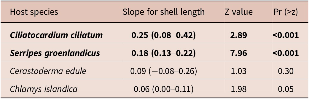
Infection data on O. arcticus metacercariae from four bivalve species are in Table 5. To assess which factor, host size or species, has more effect on infection patterns with metacercariae, we used statistical analysis. When the host shell length was included in the model, the host species did not significantly affect the infection probability, log-transformed infection intensity, and metacercariae abundance (Figure 4; Table 6). The final models for the infection probability and the logarithm of infection intensity did not include the interaction term. Only the final model for metacercariae abundance included the interaction between the host species and host shell length (Table 6). The simple slopes analyses (Table 7) showed that metacercariae abundance depended significantly on host shell length in only two host species (C. ciliatum and S. groenlandicus). The model for the infection probability explained 30% of the variation, and the models for the metacercariae abundance and the logarithm of intensity both explained 15% of the variation (Table 6).
Infection data on Orthosplanchnus arcticus metacercariae from four bivalve species. 95% confidence intervals are given in parentheses

Abbreviations: BS, Barents Sea; WS, White Sea.
Modelled effect of the host shell length and host species on the infection probability (a) and log-transformed infection intensity (b) with Orthosplanchnus arcticus metacercariae in the four bivalve species. Lines are predictions from binomial GLM (a) and LM (b) with 95% CI. Coloured dots represent (a) the prevalence in different size classes, the numbers indicate the quantity of bivalves in the size class, and (b) the infection intensity of individual hosts.

The results of the models’ analyses on the effect of the host species and host shell length on infection patterns with Orthosplanchnus arcticus metacercariae in the four bivalve species

Abbreviations: DF, degrees of freedom; F, F-statistic; LR Chisq, likelihood ratio chi-square statistic; SS, sum of squares.
The simple slopes analysis of the model, describing the effect of host species and host shell length on the metacercariae abundance of Orthosplanchnus arcticus in the four bivalve species. 95% confidence intervals are given in parentheses

Comparison of metacercariae morphological traits
We did not observe considerable morphometric differences between metacercariae obtained from different host species (Figure 5). On the nMDS ordination plot, the 95% confidence intervals for host species centroids overlapped with each other (Figure 5). Morphometric variation of metacercariae collected from different specimens of the same bivalve species (and sometimes even from a single specimen) was comparable to the variation between those obtained from other host species. PERMANOVA results did not show a significant effect of bivalve host species (df = 3, F = 1.4, p = 0.20) and shell length (df = 1, F = 2.0, p = 0.13) on the morphometry of metacercariae.
NMDS ordination of individual metacercariae of Orthosplanchnus arcticus from different hosts based on morphometric data (Euclidean distance matrix). In (a), ellipses are 95% CI for the host species centroids, and numbers indicate individual host specimens (dots with identical numbers are parasites from the same host specimen). In (b), arrows represent the original measured traits significantly correlated with the NMDS axes, and iso-lines represent host shell length. Abbreviations: CC, Ciliatocardium ciliatum; CE, Cerastoderma edule; CI, Chlamys islandica; SG, Serripes groenlandicus.

Discussion
Intermediate hosts range and genetic variation
Rediae and cercariae of O. arcticus were initially described by Marasaev (Reference Marasaev1984), who assigned them to Neophasis sp., a fish parasite belonging to the Acanthocolpidae, a family closely related to the Brachycladiidae. In our previous work, we demonstrated genuine species affiliation of these life cycle stages using molecular genetic methods (Kremnev et al., Reference Kremnev, Gonchar, Krapivin, Knyazeva and Krupenko2020). Additionally, we highlighted the morphological similarity between metacercariae of O. arcticus identified in the White Sea cardiids (C. ciliatum and S. groenlandicus) and small ocellate metacercariae from the Barents Sea bivalves (Astarte crenata (Gray, 1824) and C. edule), which Chubrik (Reference Chubrik1966) attributed to Neophasis oculata. We proposed that the metacercariae described by Chubrik (Reference Chubrik1966) might belong to the Brachycladiidae as well (Kremnev et al., Reference Kremnev, Gonchar, Krapivin, Knyazeva and Krupenko2020), but their exact affinity remained uncertain. In this study, we sampled C. edule in the same area where Chubrik (Reference Chubrik1966) conducted her research (Dalnezelenetskay Bay of the Barents Sea). We also searched for metacercariae similar to O. arcticus in other bivalve species from the White, Barents, and Pechora Seas (Table 1). We found metacercariae in cardiids (the edible cockle C. edule, the Greenland cockle S. groenlandicus, the Iceland cockle C. ciliatum) and in a pectinid, the Iceland scallop C. islandica. Surprisingly, we also discovered numerous similar metacercariae in a single specimen of the gastropod B. undatum from the White Sea.
The analysis of the partial cox1 and nad3 genes for the obtained isolates showed that they indeed belong to O. arcticus (Figure 1). Thus, we expanded the molecularly proven range of the second intermediate hosts for this species. An updated diagram of the O. arcticus life cycle is in Figure 6. Variation in nuclear ribosomal markers (28S rDNA and ITS1) was consistent with the mitochondrial data, further confirming the integrity of the species over a wide geographic range in the Arctic and sub-Arctic. Finally, we also provided solid evidence that ocellate metacercariae from the Barents Sea edible cockles, formerly assigned to N. oculata by Chubrik (Reference Chubrik1966), represent the life cycle stages of O. arcticus.
Updated diagram of the Orthosplanchnus arcticus life cycle.

Previously, interspecific genetic variation of mitochondrial genes was studied only for one species of the brachycladiids, Synthesium pontoporiae Raga, Aznar, Balbuena & Dailey, 1994; Marigo, Vicente, Valente, Measures & Santos, 2008 (Marigo et al., Reference Marigo, Cunha, Bertozzi, Souza, Rosas, Cremer, Barreto, de Oliveira, Cappozzo, Valente, Santos and Vicente2015). On a sample of more than 30 sequences, the authors identified 13 polymorphic sites in the nad3 and 30 polymorphic sites in the cox1 analysed fragments (Marigo et al., Reference Marigo, Cunha, Bertozzi, Souza, Rosas, Cremer, Barreto, de Oliveira, Cappozzo, Valente, Santos and Vicente2015). Compared to it, the variation of mitochondrial genes in O. arcticus is substantially lower, with no more than 8 and 6 polymorphic sites in the partial cox1 and nad3 genes sequenced for specimens from the Baffin Bay in the West to the Pechora Sea in the East (Figure 1). We suggest that an intriguing story is likely hidden behind the low genetic variation observed in O. arcticus. A separate study is necessary to untangle this question, involving more comprehensive sampling and further analysis of longer genetic fragments.
Preferences for the second intermediate hosts
Infection patterns with O. arcticus metacercariae vary between the studied bivalve species (Table 5) and are largely affected by the host shell length (Table 6). The host size indeed is an important factor affecting the infection patterns of bivalves with the digenean metacercariae (Bowers & James, Reference Bowers and James1967; de Montaudouin et al., Reference de Montaudouin, Wegeberg, Jensen and Sauriau1998, Reference de Montaudouin, Jensen, Desclaux, Wegeberg and Sajus2005; Poulin et al., Reference Poulin, Steeper and Miller2000; Nikolaev et al., Reference Nikolaev, Sukhotin and Galaktionov2006; Leung & Poulin, Reference Leung and Poulin2008; Fermer et al., Reference Fermer, Culloty, Kelly and O’Riordan2009; Presta et al., Reference Presta, Cremonte and Ituarte2014; Bommarito et al., Reference Bommarito, Thieltges, Pansch, Barboza, Pranovi and Wahl2021; Mahony et al., Reference Mahony, Lynch, de Montaudouin and Culloty2022; Richard et al., Reference Richard, Maire, Daffe, Magalhães and de Montaudouin2022). Overall, the larger the mollusc, the greater its chance of being infected and hosting more metacercariae. Statistical analysis of our dataset showed that the mollusc species has no effect on infection probability (i.e., prevalence) with O. arcticus metacercariae when size is considered (Figure 4a; Table 6). Infection intensity exhibited a similar trend, with larger bivalves, irrespective of species, tending to host more metacercariae (Figure 4b; Table 6). Metacercariae abundance was significantly affected by host size and the interaction between host size and host species, but the effect of the host species alone was insignificant (Table 6). The explanatory power of our models for infection intensity and metacercariae abundance of O. arcticus was relatively low (Table 6), and the inclusion of some overlooked factors could have increased it. Host age is likely the first to consider, as it is a good predictor explaining variation in both of these infection patterns, as shown for other bivalve-metacercariae systems (Nikolaev et al., Reference Nikolaev, Sukhotin and Galaktionov2006; Fermer et al., Reference Fermer, Culloty, Kelly and O’Riordan2009; Mahony et al., Reference Mahony, Lynch, de Montaudouin and Culloty2022).
Not all hosts provide a similar environment for the parasite development, resulting in size variations observed for metacercariae of the same species obtained from different host species (Rangel & Santos, Reference Rangel and Santos2009; Francisco et al., Reference Francisco, Almeida, Castro, Pina, Russell-Pinto, Rodrigues and Santos2011; Peoples & Poulin, Reference Peoples and Poulin2011; Agustina et al., Reference Agustina, Saichua, Laha, Tangkawatana, Prakobwong, Laoprom, Kamphasri, Chareonchai, Blair and Suttiprapa2024). A host harbouring larger metacercariae could be considered a higher-quality one and potentially more important for parasite transmission (Peoples & Poulin, Reference Peoples and Poulin2011). We found no significant differences in the size and development of metacercariae obtained from the four studied bivalve species (Figures 3 and 5; Table 4). Together with infection patterns analyses, this leads to the following conclusions: (1) O. arcticus likely does not exhibit strict specificity for a particular second intermediate host species, but rather shows higher infection rates in larger bivalve specimens, and (2) all four studied bivalve species are equally suitable for the development of O. arcticus metacercariae.
The occurrence of numerous O. arcticus metacercariae in a common whelk B. undatum highlights the broad specificity for the second intermediate host. Low prevalence (one of 3022 individuals) indicates that this was an uncommon accidental infection, and the site of encystment, the digestive system, suggests a trophic infection pathway. Carnivorous gastropods may act as paratenic hosts of digeneans if they prey on infected invertebrates containing encysted metacercariae (McCarthy et al., Reference McCarthy, Irwin and Fitzpatrick1999; McFarland et al., Reference McFarland, Mouritsen and Poulin2003). Typically, metacercariae stay alive in the gut for several days before eventually exiting with the faeces. Unlike these, metacercariae of O. arcticus in the infected B. undatum were located in the connective tissue beneath the stomach wall and within the digestive gland (Figure 2b, d). It would be impossible for encysted metacercariae, consumed with one or even more infected bivalves by a whelk, to undertake such a migration. Together, these argue against the fact that the source of infection for B. undatum was a bivalve or multiple bivalves. Consumption of an infected moon snail C. affinis, the first intermediate host, would explain the infection pattern observed in the common whelk (Figure 6). Infected moon snails contain numerous infective cercariae (Kremnev et al., Reference Kremnev, Gonchar, Krapivin, Knyazeva and Krupenko2020), which could have penetrated the stomach wall of a whelk and encysted afterwards. Additionally, this would account for the presence of so many metacercariae in the infected whelk specimen. The data on feeding preferences of the common whelk support our assumption. Despite B. undatum typically hunting bivalves, it can also feed on gastropods (Nielsen, Reference Nielsen1974; Taylor, Reference Taylor1978). Another major food source for the common whelk is carrion (Himmelman & Hamel, Reference Himmelman and Hamel1993), and it is possible that the infection occurred while the mollusc feasted on a dead infected C. affinis.
Causes underlying the patterns of the second intermediate hosts infection, and their advantages for transmission
Why is O. arcticus so unselective for the second intermediate hosts? The answer likely lies in the moderate development of metacercariae in this species. Indeed, metacercariae of O. arcticus are ~100 μm larger compared to cercariae, and their digestive and reproductive systems are developed just a little further (Figure 3; Table 4; Kremnev et al., Reference Kremnev, Gonchar, Krapivin, Knyazeva and Krupenko2020). Such modest morphogenesis reduces the host requirements and explains the broad specificity for the second intermediate host in the studied species.
Why do larger molluscs show higher infection rates with O. arcticus metacercariae? Generally, larger bivalves filter more water (Zaklan & Ydenberg, Reference Zaklan and Ydenberg1997), thus increasing their chances of inhaling cercariae into the mantle cavity (de Montaudouin et al., Reference de Montaudouin, Wegeberg, Jensen and Sauriau1998; Leung & Poulin, Reference Leung and Poulin2008). Infection with O. arcticus likely occurs similarly, explaining both the low chances of infection in small individuals of cockles and scallops, as well as the absence of metacercariae in small filter-feeding bivalves such as Musculus discors and in deposit-feeding protobranchs (Table 1).
A final question to address is how the life cycle of O. arcticus benefits from the combination of broad specificity and higher infection rates in larger bivalve individuals. First, the likelihood of successful transmission increases as the definitive hosts prefer larger bivalve specimens for their prey (Fukuyama & Oliver, Reference Fukuyama and Oliver1985). Second, small species and small specimens of large bivalves serve as important food items for marine invertebrates, such as moon snails and sea stars (Fukuyama & Oliver, Reference Fukuyama and Oliver1985; Kabat, Reference Kabat1990). Since small molluscs have low infection chances with O. arcticus metacercariae, the parasite evades a potential population sink due to invertebrate predation pressure, including that of its own first intermediate host, C. affinis. Lastly, the broad specificity for the second intermediate host suggests that metacercariae of O. arcticus could potentially be found in other large filter-feeding bivalves overlooked in this study, such as Arctica islandica (Linnaeus, 1767) and Mya spp. If so, even more species could be involved in the circulation pathways of this parasite.
Transmission, dead-end hosts, and human health implications
The presence of parasite larvae in the invertebrate host can indirectly indicate a trophic link between the intermediate and definitive hosts (Valtonen et al., Reference Valtonen, Marcogliese and Julkunen2010; Trani et al., Reference Trani, Medina, Cremonte and Gilardoni2025; Viriyautsahakul et al., Reference Viriyautsahakul, Poulin, Johnson and Bennett2025). Feeding habits, however, also require consideration, as some naturally infected second intermediate hosts may take a minimal to zero part in the circulation pathways of digeneans (Leung & Poulin, Reference Leung and Poulin2008; Koppel et al., Reference Koppel, Leung and Poulin2011). For O. arcticus, transmission through C. ciliatum and S. groenlandicus is certain, as these cockles are an important food source for some of the definitive hosts (Lowry et al., Reference Lowry, Frost and Burns1980; Fay, Reference Fay1982; Fukuyama & Oliver, Reference Fukuyama and Oliver1985; Hjelset et al., Reference Hjelset, Andersen, Gjertz, Lydersen and Gulliksen1999; Svetocheva & Svetochev, Reference Svetocheva and Svetochev2015). Transmission via an accidental host, B. undatum, is also plausible, as some true seal species and the walrus prey on whelks as well (Lowry et al., Reference Lowry, Frost and Burns1980; Fay, Reference Fay1982; Gjertz & Wiig, Reference Gjertz and Wiig1992; Svetocheva & Svetochev, Reference Svetocheva and Svetochev2015).
The role of the Iceland scallop and the edible cockle in the O. arcticus life cycle is not that clear. There is no solid evidence of trophic interactions between the definitive hosts of this parasite and these bivalve species. Some researchers, however, suggest that bearded seals and walruses may consume scallops (Gjertz & Wiig, Reference Gjertz and Wiig1992; Svetochev et al., Reference Svetochev, Kavtsevich and Svetocheva2024), which makes the participation of C. islandica in the transmission of O. arcticus possible. On the other hand, if the Iceland scallop and the edible cockle are absent from the diet of true seals and walruses, then they are dead-end hosts.
Marine mammal-associated zoonoses pose a persistent challenge to human health (Lehnert & Hernández-Orts, Reference Lehnert, Hernández-Orts, Smit and Sures2025), including notorious foodborne diseases such as anisakiasis and trichinellosis (Tryland et al., Reference Tryland, Nesbakken, Robertson, Grahek-Ogden and Lunestad2014; Thompson, Reference Thompson2023). Bivalves hosting O. arcticus metacercariae, especially edible cockles and Iceland scallops, are widely exploited by humans for food across their range (Pedersen, Reference Pedersen1994; Maia et al., Reference Maia, Barroso and Gaspar2021). As the studied parasite species has already passed the encounter filter successfully (Combes, Reference Combes2001), it could be considered as a potentially zoonotic one (Figure 6). But can O. arcticus challenge the compatibility filter to become a true zoonotic species? The specificity for the definitive hosts in the Digenea is generally broad (Gibson & Bray, Reference Gibson and Bray1994; Cribb et al., Reference Cribb, Bray and Littlewood2001), and brachycladiids are no exception. The family is largely restricted to marine mammals (Fraija-Fernández et al., Reference Fraija-Fernández, Olson, Crespo, Raga, Aznar and Fernández2015), but one species can also use the thresher shark as a definitive host (Adams et al., Reference Adams, Hoberg, McAlpine and Clayden1998). Furthermore, although zoonotic foodborne digeneans primarily circulate in freshwater and terrestrial environments (Chai & Jung, Reference Chai and Jung2022), some examples are known from the marine and brackishwater realm, including species that infect humans through bivalves containing metacercariae (Lee et al., Reference Lee, Chai and Hong1993, Reference Lee, Choi, Seo and Chai1995; Chai et al., Reference Chai, Han, Park, Guk and Lee2001). No cases of human infection with O. arcticus have been reported yet. However, a clear recommendation to avoid eating cockles and scallops raw seems reasonable, considering the severe liver and bile duct damage reported in infected ringed seals, some of the natural definitive hosts of O. arcticus (Andersen-Ranberg et al., Reference Andersen-Ranberg, Lehnert, Leifsson, Dietz, Andersen, Siebert, Measures and Sonne2018).
Supplementary material
The supplementary material for this article can be found at http://doi.org/10.1017/S0022149X25100746.
Acknowledgements
We are grateful for the fieldwork infrastructure provided by the Educational and Research Station ‘Belomorskaia’ of Saint Petersburg University (SPbU), the White Sea Biological Station (WSBS) ‘Kartesh’ of the Zoological Institute of the Russian Academy of Sciences (ZIN RAS), the N. A. Pertsov WSBS of Lomonosov Moscow State University, and the Biological Research Station of Murmansk Marine Biological Institute of RAS in Dalnie Zelentsy. We extend our sincere thanks to all who contributed to the sampling, especially Alexandra Uryadova, Dr. Alexey Miroliubov, Prof. Kirill V. Galaktionov, Olga Skobkina, Sergei Bagrov, and Dr. Valeriia Khabibulina. During research, we used the equipment of the resource centre ‘Molecular and Cell Technologies’ of the Research Park of SPbU. We particularly thank Dr. Anna Romanovich for excellent sequencing. Georgii Kremnev acknowledges Margarita Kobeletskaya for her inspiration while writing the manuscript draft. Finally, we express our gratitude to two anonymous reviewers for their valuable comments, which helped improve the initial version of the manuscript.
Competing interests
The authors declare none.
Ethical standard
This study was conducted in compliance with all institutional, national, and international guidelines on the care and use of animals.
Financial support
The study was performed with the financial support of the Russian Science Foundation, project No 25-24-00386, https://rscf.ru/project/25-24-00386/.