Pancreatic ductal adenocarcinoma (PDA) is the fourth leading cause of cancer mortality in the USA, surpassing breast and prostate cancer, with ~37 000 new cases and ~33 700 deaths in 2007 (Ref. Reference Jemal1). PDAs originate from the exocrine gland ducts of the pancreas (which in the normal pancreas transport pancreatic juice containing digestive enzymes from the pancreatic acinar cells to the duodenum) and account for ~95% of all cases of pancreas cancer.
The majority of PDAs (>90%) are sporadic; <10% report a family history (Ref. Reference Klein2). Established aetiological factors for sporadic PDA are cigarette smoking (Ref. Reference Lowenfels3), alcohol-related chronic pancreatitis (Ref. Reference Lowenfels4) and new-onset glucose intolerance with or without overt diabetes mellitus (Ref. Reference Gullo, Pezzilli and Morselli-Labate5). A meta-analysis of 20 epidemiological studies found a relative risk of PDA of 2.1 in those diagnosed with diabetes (Ref. Reference Everhart and Wright6). There is evidence that PDA may cause glucose intolerance, with diabetes occurring within two years of diagnosis (Ref. Reference Gullo, Pezzilli and Morselli-Labate5) and improved diabetic control following pancreaticoduodenectomy (Ref. Reference Permert7). However, the prevalence of PDA in new-onset diabetes is <1%, which does not warrant screening of the diabetic population.
Several hereditary syndromes, associated with defects in a variety of genes, predispose to PDA, including hereditary pancreatitis (PRSS1), Peutz–Jehgers (STK11), familial atypical multiple mole melanoma (CDKN2A), breast cancer (BRCA2), nonpolyposis colorectal cancer (MLH1) and cystic fibrosis (CFTR) (Ref. Reference Canto8). In familial PDA, the risk of developing the disease increases as the number of first-degree relatives with pancreas cancer increases: kindreds with three or more, two and one affected first-degree relative have a 32-, 6.4- and 4.5-fold increased risk of developing pancreas cancer, respectively (Ref. Reference Klein2).
PDA has a high mortality as a result of advanced stage at presentation, complete resection being noncurative due to micrometastases, and adjuvant therapy at best being only palliative (Ref. Reference Neoptolemos9). There is no screening biomarker of high sensitivity and specificity for sporadic PDA. Approximately 9% of PDA patients undergo surgical resection, but of these only 22% have localised disease, and the overall 5-year survival rate after a pancreaticoduodenectomy is ~20% (Ref. Reference Alexakis10). Neoadjuvant chemotherapy followed by surgery is likely to improve survival rates (Ref. Reference Kim11), while adjuvant gemcitabine appears to have improved median survival over fluorouracil (5-FU)-based chemoradiation (Ref. Reference Oettle and Neuhaus12). Gemcitabine (an inhibitor of DNA replication) has been the standard palliative therapy for unresectable and metastatic disease for the past 12 years (Ref. Reference Burris13). Unfortunately, the median survival rates of 6–12 months have not been significantly enhanced with the addition of other chemotherapy agents (Ref. Reference Cartwright, Richards and Boehm14) or targeted therapies to gemcitabine (Refs Reference Cartwright, Richards and Boehm14, Reference Von Hoff15).
There has been and continues to be a deep void in translational research due to lack of a precise animal model for PDA where critical biological pathways could be interrogated in detail to ascertain their relevance to human disease. Mouse xenografts of human cell lines and human PDA tissue (Ref. Reference Rubio-Viqueira16) as well as genetically engineered mice (Ref. Reference Olive and Tuveson17) have been developed, although the latter do not completely recapitulate human disease observed in the clinic. The recent whole-genome sequencing of PDA patients has identified a dozen mutated signalling pathways (Ref. Reference Jones, Zhang and Parsons18), which should provide relevant leads for target discovery and development of drugs to better control the disease. However, genome sequencing for driver mutations is unlikely to provide a comprehensive understanding of PDA unless linked to gene expression and epigenetic profiling. Many gene expression profiling studies (Refs Reference Han19, Reference Crnogorac-Jurcevic20, Reference Iacobuzio-Donahue21, Reference Iacobuzio-Donahue22, Reference Campagna23) have attempted to address the desmoplastic reaction in PDA – a dysregulation of the extracellular matrix (ECM) and stromal cells surrounding the tumour (Ref. Reference Mahadevan and Von Hoff24). However, there is no clear understanding of this process except identification of some of the key players, including pancreas stellate cells (PSCs) (Ref. Reference Apte25), and changes in production of growth factors and ECM (Refs Reference Menke and Adler26, Reference Infante, Matsubayashi and Sato27).
The successful development of effective current and future therapies will depend on research focusing on appropriate preclinical models, with corroborative translational clinical studies that translate accurately to the clinical situation. This review summarises current and future trends in understanding of the biological mechanisms of PDA and in management of PDA that should raise some hope in patients and their oncologists.
Molecular and cellular abnormalities in PDA
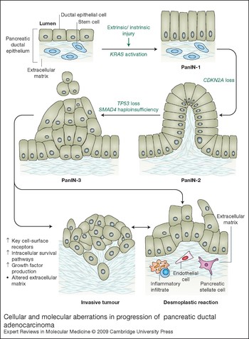
Similar to many common epithelial malignancies, the progression of PDA from preinvasive lesion to unresectable cancer appears to occur in several steps (Ref. Reference Hruban28). There is convincing evidence to support the origin of invasive ductal adenocarcinomas from three distinct lesions: intraductal papillary mucinous neoplasm (IPMN), pancreatic intraepithelial neoplasias (PanINs) and mucinous cystic neoplasm (MCN) (Ref. Reference Maitra29). In sporadic PDA, several genetic lesions are established – activation of the oncogene KRAS (>90%), and loss of tumour suppressor genes encoding the cell cycle regulators p16INK4a (CDKN2A) (>90%) and p53 (TP53) (75%), and the transforming growth factor β (TGF-β) signalling mediator SMAD4/DPC4 (SMAD4) (55%) (Ref. Reference Hruban28) – and a model has been proposed for progression through PanIN1 (KRAS activation), PanIN2 (CDKN2A inactivation) and PanIN3 (TP53, SMAD4 and BRCA2 inactivation) (Fig. 1). By contrast to these early stages, the progression to invasive ductal adenocarcinoma from preinvasive lesions has a lack of defined genetic aberrations. A recent whole-genome sequencing study identified several additional mutated genes associated with PDA (Ref. Reference Jones, Zhang and Parsons18) (see below).
Cellular and molecular aberrations in pancreatic ductal adenocarcinoma progression. Invasive pancreatic ductal adenocarcinoma (PDA) evolves through three preinvasive lesions (PanINs) of increasing aggressiveness. Normal ductal epithelial cells have an intact Ras signalling pathway with normal p53 and CDKN2A expression (the G1 cyclin-dependent kinase inhibitor p16INK4A and the tumour suppressor ARF are both encoded at this locus). Transforming growth factor β (TGF-β) activation of its receptor (TGFBR1/2) signalling through SMAD4 and E-cadherin provides a growth-suppressive effect. In the figure, mutations in the genes encoding KRAS (KRAS), p16INK4 (CDKN2A), p53 (TP53) and SMAD4 (SMAD4) are shown occurring in a sequential manner, leading to an aggressive invasive PDA phenotype. Mutated KRAS is considered the major driver of initiation and progression, but the timing of the loss of each tumour suppressor (TP53, CDKN2A, SMAD4) is unknown. The changes culminate in fibroblast proliferation, immune suppression and increased paracrine and autocrine growth factors with modification of the extracellular matrix, which in turn activates several oncogenic signalling pathways that lead to genomic instability and drug resistance. Several key cell-surface receptors (NOTCH, uPAR, PATCH, SMO, RON, MET, TGFBR1/2, CTGFR, CEACAM6 and α5β1/αvβ5) are overexpressed and activated. Intracellular activation of the survival (PI3K–AKT–mTOR), antiapoptotic (BCL2, XIAP) and cell cycle (CDKs, CHKs, Aurora kinases, polo-like kinases) pathways drive the malignant phenotype. In addition, an altered ECM (collagen I/III, fibronectin, laminin, SPARC, byglycan, MMPs, TIMPs) coupled to elaboration of several growth factors (SHH, TGF-β, HGF, PDGF, IGF1, CXCL12, WNT) and an abnormal vasculature (VEGF) is the basis of the desmoplastic reaction in PDA. See main text for further discussion of these changes and full versions of abbreviations.

PanIN1, PanIN2 and PanIN3 are asymptomatic and not visible on conventional imaging (<5 mm in size) (Ref. Reference Canto8), although pancreatic ductal dilatation and obstruction may be important radiological signs (Ref. Reference Ishikawa30). Early pancreatic neoplasia are morphologically well-defined noninvasive lesions or small (<2 cm) ductal adenocarcinomas that are also generally asymptomatic, and are surgically curable. However, although tumours are resectable if identified early, metastasis has usually occurred from tumours beyond 1 cm in size. The transition from resectable to unresectable tumour has been reported to arise over a period of 6 months prior to diagnosis (Ref. Reference Gangi31). Most symptomatic PDA patients have tumours >2 cm in size, with regional metastasis via angiolymphatic and perineural spread and involvement of adjacent veins and arteries. The imaging and staging of PDAs in clinical management is discussed further below.
Most PDAs are mucin-producing tumours associated with a dense desmoplastic reaction, and a predisposition to vascular thrombosis due to secretion of procoagulants (Ref. Reference Benson and Pazdur32). Desmoplasia is defined as proliferation of fibrotic tissue with an altered ECM conducive to tumour growth and metastasis. Histopathological analyses of human PDA in comparison with normal pancreas depict dense collagen (type I and III) bundles associated with fibroblasts, with loss of basement membrane integrity and invasion of malignant cells into the interstitial matrix and exposure to collagens (Ref. Reference Mahadevan and Von Hoff24).
Preclinical models, genomics and stem cell approaches
Several cell lines and mouse models have been developed to glean insight into the biology of PDA. Here we discuss pros and cons of these models and their relevance to human disease.
Cell lines
Given the difficulty in obtaining adequate biopsy material from PDA patients, numerous human PDA-derived cell lines of varying differentiation states (well, moderately and poorly differentiated) and genetic backgrounds (KRAS or TP53 wild-type or mutants) have been utilised. The most popular cell lines for functional and therapeutic studies are Panc-1 (poorly differentiated), MiaPaCa-2 (moderately differentiated) and BxPC-3 (well differentiated); other cell lines used are CaPan-1, CaPan-2 and HPAF-2. Together, studies on these cell lines have provided biological data of varying significance to the clinical situation. However, there are many limitations to cell culture models of PDA, including possible changes arising from long-term culture with multiple passages and derivation from metastases and/or propagation as xenografts (Ref. Reference Bissell and Radisky33).
Recently, three PDA cell lines (PP109, PP117, PP161) were established from primary human specimens and well characterised with respect to specific genetic alterations [KRAS, TP53, CDKN2A and SMAD4) in vitro growth characteristics, DNA profile by microsatellite fingerprinting and cytogenetics by karyotype analysis (Ref. Reference Chifenti34). Three genes were altered in all three PDA cell lines (KRAS, TP53, CDKN2A) but no mutations were detected in SMAD4. Furthermore, a complex karyotype with many chromosomal abnormalities – both structural (1p, 1q, 3p, 6q, 7q, 11p, 17p, 19q) and numerical (gains of 7 and 20; losses of 6, 12, 13, 17, and 18) – was identified. The PDA cell lines had a similar cytokeratin expression to normal ductal cells [CK7 (KRT7), CK8 (KRT8), CK18 (KRT18), CK19 (KRT19)]. The intermediate filament protein vimentin (VIM) was expressed only in PP117 and is generally associated with poorly differentiated tumours with high metastatic potential. This was confirmed by an in vitro migration assay, where PP117 had a higher motility compared with PP109 and PP161. In PP117 cells, expression of ezrin (EZR; one of the ERM protein family that ensures crosslinking between the actin cytoskeleton and the plasma membrane) was localised to membrane ruffles and short filopidia structures involved in the motility machinery, and, with coexpression of vimentin, characterises a poorly differentiated phenotype. Hence, despite a similar genetic profile in these cell lines, cellular differentiation appears to determine the aggressiveness of PDA. Therefore, dissecting the molecular mechanisms underlying the differentiation status is likely to provide insights into an improved prognostic and therapeutic signature.
Mouse models
The existence of human PDA cell lines has allowed the generation of subcutaneous flank and orthotopic (Ref. Reference Bruns35) xenograft mouse models. Despite the generally poor predictability of these mouse xenograft models they continue to be utilised as screening tools and/or proof of concept models for evaluating novel therapeutics. Although tumour growth inhibition (TGI) is an important endpoint in these studies, a better measure of activity is significantly enhanced Kaplan–Meier survival (e.g. >2 weeks) compared with control or standard therapy. For targeted therapy evaluations these models can provide a fairly robust readout of target inhibition/modulation and help develop biomarkers for Phase I clinical trials (Ref. Reference Riley36).
In addition, several genetically engineered pancreas-targeted mouse models have been generated (Refs Reference Hingorani37, Reference Aguirre38, Reference Hingorani39, Reference Izeradjene40, Reference Takaku41), based on the four most common genetic mutations (KRAS, TP53, CDKN2A, SMAD4) detected in human PDA (Ref. Reference Hruban28) (Box 1). Although these elegant mouse models appear to have histological features common to human PDA there are several drawbacks that highlight significant differences to human disease: the observed genetic changes do not reflect the chronology of mutations; the sites of metastasis do not match human disease; other histological subtypes of pancreas cancer are present; primary tumours develop in other organs (e.g. gastric cancer in KRas/Smad4 model); and numerous genes that are specific to higher vertebrates are absent in rodents [e.g. carcinoembryonic antigen-related cell adhesion molecule 6 (CEACAM6); Ref. Reference Riley36].
Box 1. Mouse models of pancreas cancer: predictors or detractors of biology?
Xenograft models
Mouse xenograft models of pancreatic ductal adenocarcinoma (PDA) continue to be utilised in creative ways. A transgenic mouse model with a KRas G12V mutation under the control of the cytokeratin 19 (Krt19) promoter develops hyperplastic lesions without overt PDA (Ref. Reference Deramaudt155). Pancreas cells isolated from these mice show oncogenic effects of KRas G12V but this is counterbalanced by upregulation of cell cycle inhibitors [p16INK4A/p19Arf (encoded by Cdkn2a) and p27KIP1 (encoded by Cdkn1b)], leading to a proliferator phenotype without induction of a senescence state. These KRas G12V pancreas cells become oncogenic when retrovirally transduced with Trp53 (p53) V143A, as seen in soft-agar assays and xenograft models (subcutaneous and orthotopic), and show increased cell migration, invasion and tissue architecture disruption compared with wild-type, KRas G12V or Trp53 V143A cells, respectively. Cellular events that govern this PDA model appear to be recapitulated by biochemical and genetic disruption of N-cadherin (Cdh2) or keratinocyte growth factor receptor (Fgfr2). These interactions preferentially induce AKT activation (Ref. Reference Deramaudt155).
Genetically engineered mouse models
Genetically engineered mouse models based on KRAS knock-in and tumour suppressor knockout highlight general concepts of PDA oncogenesis. Although the underlying pathobiology between mouse and human PDA is likely to be distinct, certain oncogenic signalling pathways common to human PDA are activated. In these models, PDA can arise following alterations of a subset of genes implicated in human PDA, and shows unique phenotypes and associated invasive behaviour, raising questions as to whether mutations in additional tumour suppressors and the relative timing of such changes alter PDA progression and/or each pathway constitutes a distinct genetic route to PDA with a unique phenotype.
KRasG12D model. The mutant KRas G12D model (expression of activating KRas mutation) induces the entire progression of PDA from preinvasive (PanIN-1 to -3) to invasive and metastatic disease. In mice that lived longer, the pancreas had extensive ductal lesions and the acinar parenchyma were replaced by an intense desmoplastic reaction comprising collagen, fibroblasts and inflammatory cells, reminiscent of human PDA (Ref. Reference Hingorani37). PanINs demonstrated that quiescent oncogenic signalling pathways are activated, including Notch (manifested by Hes1 transcription factor expression), as well as COX2 (a component of the prostaglandin pathway involved in inflammation, encoded by Ptgs2) and the matrix metalloproteinase MMP7 (involved in extracellular matrix modelling) (Ref. Reference Hingorani37). Unfortunately, clinical trials targeting KRAS, COX2 and MMP in human PDA were negative (Ref. Reference Mahadevan and Von Hoff24), indicating none of these targets and pathways by itself is sufficient to influence proliferation, particularly in advanced disease.
KRasG12DandCdkn2a deletion model. Mice expressing the activated KRas and with deletions of the Cdkn2a locus (affecting expression p16INK4A/p19Arf) showed early appearance of PanIN lesions and rapid progression to highly invasive and metastatic PDA. The tumours had a proliferative stromal component and ductal lesions with a propensity to advance to a poorly differentiated state. However, lung and liver metastases were rarely observed (Ref. Reference Aguirre38). This may be due to species-specific signalling pathways.
KRas G12DandTrp53mutation model. Mice carrying activated KRas and mutated p53 (Trp53) developed invasive and widely metastatic carcinoma with a high degree of genomic instability manifested by nonreciprocal translocations without telomere erosion, leading to chromosomal instability. However, no mutations were found in other tumour suppressors implicated in human PDA. A molecular analysis of the epidermal growth factor receptor (EGFR, HER1), ERBB2 (HER2), E-cadherin (CDH1) and sonic hedgehog (SHH) showed both EGFR and ERBB2 were absent in metastatic lesions, contrary to that observed in human PDA, and SHH and CDH1 were present in both preinvasive and invasive lesions in moderately well differentiated but not in poorly differentiated lesions (Ref. Reference Hingorani39). Clinical trials targeting EGFR and ERBB2 in advanced human PDA have been disappointing despite the approval of erlotinib, a small-molecule tyrosine kinase inhibitor of EGFR.
KRas (G12D)andSmad4haploinsufficiency. Haploinsufficiency of SMAD4, a downstream component in transforming growth factor β (TGF-β) signalling, in addition to activation of KRas in mouse progenitor cells of the pancreas (Ref. Reference Izeradjene40), generated mucinous cystic neoplasms (MCNs) that may progress to PDA. The pancreas of these mice had PanIN lesions (body and tail of the pancreas) but the primary abnormality was gastric cancer of squamous cell origin with metastasis, leading to early mortality. By contrast to the KRas/Trp53 mutant model, EGFR and ERBB2 expression persisted in invasive and metastatic lesions, SHH expression was diminished in metastatic lesions, and Notch pathway expression (Hes1) was observed in invasive/advanced disease. The latter may indicate a link between TGF-β and Notch signalling. Moreover, compared with the KRas/Trp53 model, there were fewer markers of genetic instability in the KRas/Smad4 model (Ref. Reference Hingorani39). A further model – KRas/Trp53/Smad4 – appears to develop classical PDA of the head of the pancreas with glandular histology as was observed in the KRas/Trp53 model (Refs Reference Hingorani39, Reference Izeradjene40).
Smad7overexpression. Constitutive deletion of Smad4 did not affect normal pancreas development or alter parenchymal architecture (Refs Reference Aguirre38, Reference Hingorani39), but pancreas-specific expression (under the elastase promoter) of SMAD7, a specific inhibitor of TGF-β signalling, produces early pancreas cancer. At age 6 months, most of the transgenic mice developed PanINs accompanied by increased proliferation of ductal and acinar cells and an increased fibrosis around the ductal lesions (Ref. Reference Kuang156). This study demonstrates that in vivo inactivation of the TGF-β signalling pathway leads to the development of premalignant pancreatic lesions, and provides a promising model for molecular dissection of this pathway (including the mechanism of fibrosis) and for early therapeutic intervention using small-molecule inhibitors to TGFBR1.
PancXenoBank model
This in vivo model provides a platform for integrating drug screening and biomarker discovery and is a perpetual live bank. Passages of tumours in successive cohorts of mice do not appear to change their susceptibility to anticancer agents but may change the tumour–stroma composition. Agents targeting EGFR (erlotinib), the mammalian/mechanistic target of rapamycin MTOR (temsirolimus), mitogen-activated protein kinase (MAPK) (CI-1040) and DNA/RNA (gemcitabine) individually evaluated in these 14 human xenograft mice showed a response rate of 0%, 7%, 14% and 43%, respectively, which appears to match those observed in clinical trials (Ref. Reference Rubio-Viqueira16). This model is thus useful for late-phase preclinical drug development and pharmacodynamic monitoring to fully predict antitumour activity of novel agents and rational combinations. The model can prospectively evaluate patient outcome while the patient is undergoing adjuvant gemcitabine and be used to guide treatment at the time of progression (Ref. Reference Jimeno78). The biggest limitation of this model is that ~80–90% of patients present with unresectable or metastatic disease, where the biology is likely to be distinct: in this setting, therapeutic screening in the PancXenoBank model with potential targeted agents may prove inconclusive.
In conclusion, the current mouse models of PDA can be useful predictors or misleading detractors of PDA biology. If utilised with the understanding of limitations and potential pitfalls, then meaningful data can be obtained to guide mechanism-based therapeutic targeting in patients with PDA.
Xenografts derived directly from patient biopsies have also been successfully generated for several solid malignancies (colon, lung, pancreas) in the hope that they are biologically more predictive than traditional xenografts and genetically engineered mouse models (Ref. Reference Jones, Zhang and Parsons18). In a pancreas-cancer-specific study, resected tumours from 14 PDA patients were xenografted and expanded in successive groups of nude mice to develop cohorts of tumour-bearing mice suitable for drug therapy (PancXenoBank model). These xenografted tumours appear to maintain their original genotype despite serial passages, and recapitulate the genetic heterogeneity of PDA.
A summary of findings from mouse models of PDA is given in Box 1. In conclusion, none of the mouse models completely recapitulates human PDA; however, each model system has its advantages that can be utilised with caution (without over interpretation) to help decipher biological principles that can be evaluated in human clinical trials.
Genomics
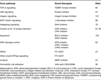
Altered gene function in human PDA has been studied in some depth at the DNA (genotype), mRNA (transcriptome), or protein (proteome) level in cells or tissue. Recently, a comprehensive genetic makeup of human PDA was determined by DNA sequencing of 24 patients with advanced disease. The tumours were first passaged in vitro as cell lines or in nude mice as xenografts to remove contaminating non-neoplastic cells, thus facilitating detection of mutations. DNA sequencing of mRNA transcripts (23 219), representing 20 661 protein-coding genes, revealed on average 63 genetic alterations (the majority of which were point mutations), which defined a core set of 12 partially overlapping cellular signalling pathways and processes (Ref. Reference Jones, Zhang and Parsons18) (Table 1). However, the mutated pathway components can vary widely between individual tumours. Each pathway is genetically altered in the majority (67–100%) of PDA samples with a predominance of low-frequency mutations, thus generating tumour heterogeneity. Mapping of these mutations to known three-dimensional protein structures showed that 55 of the 404 mutations were localised to domain interfaces or ligand-binding sites that are likely to interfere with normal function. Aberrant core pathways include: frequently altered genes such as KRAS and regulation of the G1–S cell cycle transition; pathways in which a few altered genes predominate, such as TGF-β signalling; and pathways in which many different genes are altered, such as in integrin signalling, regulation of invasion, homophilic cell adhesion, and small guanine triphosphatase (GTPase)-dependent signalling (Table 1).
Drugs targeting the core signaling pathways in pancreatic ductal adenocarcinoma

Abbreviations: ATM, ataxia telengiectasia mutated; BCL2, B cell lymphoma 2; CDK, cyclin-dependent kinase; CEACAM, carcinoembryonic antigen-related cell adhesion molecule; CHK, checkpoint kinase; FTI, farnesyl transferase inhibitor; GGTI, geranylgeranyl transferase inhibitor; JNK, Janus kinase; mAb, monoclonal antibody; MMP, matrix metalloproteinase; SHH, sonic hedgehog; TGF, transforming growth factor; TGRBR1, transforming growth factor β receptor 1; WNT, wingless; XIAP, X-linked inhibitor of apoptosis.
To analyse the transcriptome of the 24 PDA samples, SAGE (serial analysis of gene expression) (Ref. Reference Velculescu42) was performed in combination with massively parallel sequencing to provide a measure of differential gene expression. There were 541 genes that were tenfold overexpressed in >90% of the 24 PDAs [compared with normal pancreatic duct cells or the HPDE (human pancreatic duct epithelial) cell line]. Of the overexpressed genes, 54 encoded proteins predicted to be secreted or expressed on the cell surface (Ref. Reference Jones, Zhang and Parsons18). The findings indicated that expression changes contribute to dysregulation of the 12 core pathways in PDA pathogenesis.
There are several drawbacks to this analysis: alteration of gene expression when human PDA cells are cultured and/or grown in nude mice; a lack of attention to the prominent tumour–stroma interaction that appears to drive invasion and drug resistance in PDA; and no epigenetic analysis to understand the process of gene silencing in the setting of the 12 core oncogenic pathways. Nevertheless, this landmark paper (Ref. Reference Jones, Zhang and Parsons18) concludes that seeking agents that broadly target downstream mediators or key nodal points are preferable to targeting specific mutated genes. Pathways that appear to represent good targets include metabolic disturbances, neoangiogenesis, misexpression of cell-surface proteins, altered cell cycle proteins, cytoskeletal abnormalities and defective DNA repair. Table 1 highlights potential therapeutic agents that target each of the 12 core overlapping oncogenic pathways. The challenge for the future is to rationally combine these agents to obtain optimal cell kill without major off-target side effects.
Differential gene expression profiles associated with progression of PDA from localised to locally advanced to metastatic disease have also been reported (Ref. Reference Campagna23). This study showed significant gene expression changes (mRNA level) between localised and locally advanced disease (matched or unmatched samples). The genes identified to be overexpressed in locally advanced versus localised disease were pertinent to the mitogen-activated protein kinase (MAPK) and WNT pathway, cellular metabolism, immune regulation and cell–cell or cell–ECM interactions. Similar signalling pathways were identified when localised disease was compared with normal pancreas tissue (Ref. Reference Campagna23). However, this study did not evaluate differential gene expression between locally advanced versus metastatic gene expression profiles and surprisingly could not differentiate between localised and metastatic disease. The reasons for this discrepancy are not given. A second study evaluated differential gene expression profiles in lymph-node-positive versus -negative PDA and identified 194 genes with altered expression (66 overexpressed; 126 downregulated). Genes promoting tumour growth were highly expressed while antiapoptotic genes were downregulated (Ref. Reference Kim43).
Two abstracts presented at the American Society of Clinical Oncology meeting in 2008 (Refs Reference Collisson44, Reference Mori45) assessed prognostic information by gene expression profiling from resected PDA biopsies (with or without microdissection), in patients with long versus short survival. The data suggest that the disparity in outcomes seen after intended curative resection is largely intrinsic to the tumour rather than due to host factors and/or surgical technique and more likely due to tumour–stroma interactions.
The four studies discussed in this section (Refs Reference Jones, Zhang and Parsons18, Reference Campagna23, Reference Collisson44, Reference Mori45) highlight major problems and discrepancies in the study of human PDA by gene expression profiling.
Stem cells
Cancer stem or progenitor cells (CSCs) were initially identified in acute myeloid leukaemia (Ref. Reference Bonnet and Dick46). Subsequently, the first epithelial CSCs were discovered in breast cancer, with a cell-surface marker phenotype of CD44+, CD24+ and ESAlow; these cells were tumourigenic and formed tumours that recapitulated the patient's tumour in severe combined immunodeficient (scid) mice (Ref. Reference Al-Hajj47). Since then, CSCs have been identified in several solid tumours (brain, colon, head/neck, liver, melanoma and prostate) (Ref. Reference Simeone48). Pancreas-specific CSCs were recently identified in primary pancreas cancer and cell lines on the basis of CD44+, CD24+ and ESAlow surface markers, and found to constitute 0.2–0.8% of all pancreatic cancer cells (Ref. Reference Li49). A second study isolated CSCs by CD133 expression and found overlap by 14% between these cells and CD44+, CD24+ and ESAlow cells (Ref. Reference Hermann50). These pancreatic CSCs are highly tumourigenic and possess the ability to self-renew and produce differentiated progeny that reflects the heterogeneity of the patient's primary tumour. Several developmental signalling pathways are upregulated in these CSCs, including hedgehog, BMI1 (polycomb ring finger oncogene) and Notch (Ref. Reference Simeone48). Identification of drugs targeting these and other pathways in CSCs is an active area of research (see below), with Phase I trials imminent.
In general, CSCs are resistant to conventional chemotherapy and radiation except in testicular cancer, childhood leukaemias and good-prognosis Hodgkin lymphoma, where long-term cure rates are high. Treatment of pancreas cancer cells with gemcitabine with or without radiation leads to the enrichment of a CSC population (Refs Reference Li49, Reference Hermann50), providing a mechanism of escape and resistance to conventional therapy. Gene expression profiling and small interfering RNA (siRNA) technology are being used to identify genes important in pancreas CSC self-renewal in ‘tumourspheres’ (Ref. Reference Simeone48). In order to evaluate potential CSC therapeutics, primary pancreas cancer orthotopic xenograft models would be optimal, with or without cotransfer of human pancreatic stromal cells. This approach would preserve tumour heterogeneity and/or tumour–stroma desmoplasia and provide stringent evaluation of drug response (Ref. Reference Simeone48). Novel imaging agents and techniques would be valuable in this setting as would be the discovery of biomarkers of response.
Clinical management of PDA
Diagnosis and staging
PDA is a ‘silent’ killer in that symptoms are generally of a nonspecific nature, and at presentation the disease is often advanced. Nonspecific symptoms are fatigue, abdominal pain, heartburn, weight loss and poor appetite; specific signs are jaundice, back pain, pruritis, pale stool and dark urine. Occasionally PDA is discovered accidentally on imaging studies or through increased bilirubin (jaundice), although this is rare with localised disease and more frequent with metastatic disease, especially when the tumour is located in the body and/or tail of the pancreas. Approximately 65% of PDAs arise in the head, and the rest in the body (20%) and tail (15%) of the pancreas. Patients presenting with painless jaundice due to a mass at the head of the pancreas may be fortunate to have resectable disease. Common sites of metastasis are lymph nodes, liver, lung and bone, while rare sites are bone marrow, brain and skin (Ref. 51).
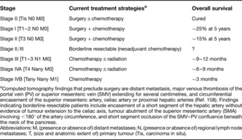
As with any solid tumour, staging is very important in making disease-management decisions and for prognosis in patients with PDA. The tumour, nodal, metastases (TNM) staging system was recently modified by the American Joint Committee on Cancer (AJCC) (Ref. 51). Table 2 describes TNM classification of PDA, with currently assigned treatment modality per staging. In the current AJCC system, patients with PDA are divided into distinct prognostic and clinical groups based on resectability: stage I and II are resectable; stage III (locoregional spread) and stage IV (distant metastasis) are deemed unresectable (Ref. Reference Katz52). For all stages, overall 1-year survival is 23% and 5-year overall survival from diagnosis is 4%. Median survival for patients with locally advanced disease is 9–12 months and, for patients with metastatic disease, the median survival is 3–6 months (Ref. 51). A recent study proposed borderline resectable PDA, a newly emerging stage, with favourable prognosis in a subset of patients treated neoadjuvantly (Ref. Reference Katz53). For patients with tumours confined to the head of the pancreas (2–5 cm) (localised), pancreaticoduodenectomy with lymphadenectomy and venous resection is the optimal surgical procedure (Whipple or modified Whipple) (Ref. Reference Glanemann54).
Staging, treatment strategies and outcome in pancreatic ductal adenocarcinoma

aComputed tomography findings that preclude surgery are distant metastasis, major venous thrombosis of the portal vein (PV) or superior mesenteric vein (SMV) extending for several centimetres, and circumferential encasement of the superior mesenteric artery, celiac artery or proximal hepatic arteries (Ref. Reference Fuhrman158). Findings indicating borderline resectable patients include encasement of a short segment of the hepatic artery without evidence of tumour extension to the celiac axis, tumour abutment of the superior mesenteric artery (SMA) involving <180° of the artery circumference, and short segment occlusion of the SMV–PV confluence beneath the neck of the pancreas.
Abbreviations: M, (presence or absence of) distant metastases; N, (presence or absence of) regional lymph node metastases; T, (size and anatomic extent of) primary tumour (Tis, carcinoma in situ).
Accurate staging is achieved by noninvasive and invasive imaging methods. Contrast-enhanced computed tomography (CT) accurately predicts resectability in 80–90% of patients (for further details see Table 2 footnote). Magnetic resonance imaging (MRI) is useful in identifying small hepatic metastasis but is not superior to CT. Positron emission tomography (PET) is an evolving technique, as discussed further below. Endoluminal ultrasonography (EUS) is sensitive at detecting small pancreatic tumours, and useful for clarifying suspected vascular invasion, evaluating peripancreatic adenopathy and ascites, and, importantly, obtaining a biopsy prior to neoadjuvant therapy (Ref. Reference Ujiki and Talamonti55). Endoscopic retrograde cholangiopancreatography (ERCP) is useful for stent placement for biliary and/or pancreatic duct obstruction, brush biopsies, detection of mucin production in the PDA, and localising the extent of disease (Ref. Reference Tanaka56). For biopsies, fine-needle aspiration, EUS and ERCP may give insufficient material, and ‘tru-cut’ biopsies may be more useful for evaluation of pancreas pathology (Ref. Reference Levy57).
Chemotherapy
Chemotherapy has been used as palliative therapy for unresectable PDA, and alongside surgery to improve survival following resection. Tumours deemed borderline resectable and/or unresectable (locally advanced) may be resectable after neoadjuvant chemotherapy. These patients will also benefit from postsurgical adjuvant chemotherapy. Patients with unresectable or metastatic disease are treated similarly with standard gemcitabine alone (Ref. Reference Burris13) or gemcitabine plus erlotinib (an inhibitor of epidermal growth factor receptor tyrosine kinase activity) (Ref. Reference Moore58). Other agents that may be used as front-line therapy with gemcitabine are taxols (microtubule destabilisers), including abraxane (paclitaxel), cisplatin or oxaliplatin, and xeloda. These agents may also be used in second-line treatments if patients fail gemcitabine treatment either alone or in combination with erlotinib (Ref. Reference O'Reilly and Abou-Alfa59). Third-line therapies include oxaliplatin plus 5-FU (DNA replication inhibitors) (Ref. Reference Pelzer60) or clinical trials. Thus far, none of the clinical trials of various chemotherapy combinations has resulted in meaningful responses that have significantly improved overall survival. The value of radiation therapy in PDA treatment is discussed in Box 2.
Box 2 Is there a role for radiation therapy in combination with chemotherapy for pancreatic ductal adenocarcinoma treatment?
Radiation therapy (X-ray therapy; XRT) in combination with chemotherapy has been utilised for over 25 years for the treatment of pancreatic ductal adenocarcinoma (PDA), mainly in patients with unresectable disease or as adjuvant therapy after surgery.
In a study in 1981, the Gastrointestinal Tumor Study Group (GITSG) found median survival of unresectable PDA patients was ~9.0 months for XRT with fluorouracil (5-FU) treatment compared with 5.2 months for XRT (Ref. Reference Moertel159); however, treatment of unresectable PDA with gemcitabine alone provided a median overall survival superior (13 months) to that of 5-FU-based chemoXRT (8.6 months) without the major side effects of chemoXRT (Ref. Reference Chauffert160).
The role of neoadjuvant chemoradiation for resectable PDA is not well established. Studies by the GITSG (Ref. Reference Kalser and Ellenberg161) showed overall survival of 20 months versus 11 months, while the more recent RTOG 9704 (Ref. Reference Regine162) showed that 5-FU-based chemoXRT had a better overall survival (3 year: 18.9 months versus 16.7 months) in resected patients on the gemcitabine arm compared with the 5-FU arm (Ref. Reference Oettle163) only for tumours of the head of the pancreas. ESPAC-1 (Ref. Reference Neoptolemos9) showed that 5-FU-based chemotherapy was superior to chemoXRT or XRT alone, with 2-year survival of 41% versus 29%, favouring no chemoradiotherapy. However, the CONKO-001 trial (Ref. Reference Neuhaus164) showed adjuvant gemcitabine (six cycles) had a superior overall survival at 3 years (36.5%) and 5 years (21%) compared with observation (19.5% and 9.0%, respectively). Taken together, these studies provide indisputable evidence for use of adjuvant gemcitabine; however, the role of XRT is in doubt despite current practice in the USA.
Hence, gemcitabine is a ‘gold standard’ for resected, unresectable and metastatic PDA. The addition of erlotinib (epidermal growth factor receptor inhibitor) to gemcitabine was approved by the US Food and Drug Administration (FDA) for unresectable and metastatic disease but no studies are published for this combination in the adjuvant setting. The addition of XRT to gemcitabine in unresectable PDA patients (E4201) was recently completed by ECOG (early termination due to poor accrual), with a median survival of 11 months for gemcitabine and XRT and 9.2 months for gemcitabine alone (Ref. Reference Loehrer165). However, the FFCD/SFRO study (Ref. Reference Mueller154) clearly revealed a superior survival with gemcitabine alone (13 months) compared with the 11 months with gemcitabine and XRT in unresectable patients in E4201 (Ref. Reference Loehrer165).
A cell biology perspective appears to corroborate the ESPAC-1 study (Ref. Reference Neoptolemos9): XRT treatment of stromal fibroblasts increased the invasiveness of PDA cells compared with nonradiated fibroblasts (Ref. Reference Ohuchida86), highlighting the detrimental effect of XRT in the therapy of PDA. Therefore, gemcitabine alone continues to be the most effective agent in all settings based on Phase III trials.
Improvements to chemotherapeutic treatment: ongoing trials
Phase III trials
In patients with advanced PDA, gemcitabine is given as a 30 min infusion. A fixed dose rate (FDR) may improve its clinical activity, but the recent ECOG E6201 study showed no survival advantage to FDR therapy (Ref. Reference Poplin61). Fifteen Phase III clinical trials for advanced PDA patients have been conducted with gemcitabine in combination with other chemotherapeutic agents [exatecan, irinotecan, 5-FU with or without leucovorin (two studies), capecitabine (three studies), cisplatin, oxaliplatin] or targeted therapy (marimistat, tipifarnib, pemetrexed, cetuximab, bevacizumab, erlotinib) (Ref. Reference Cartwright, Richards and Boehm14). The progression-free survival and overall survival range from 3.3–5.7 months and 5.5–9.0 months, respectively, with virtually no added survival advantage for all agents except for erlotinib, where overall survival was 6.24 versus 5.91 months in favour of combination therapy (Ref. Reference Moore58). A recent Phase III study in newly diagnosed metastatic PDA patients found the addition of bevacizumab to gemcitabine plus erlotinib did not significantly increase overall survival, but there was a trend towards improved survival (6.0 versus 7.1 months) and progression-free survival (3.6 versus 4.6 months) (Ref. Reference Vervenne62) compared with gemcitabine plus erlotinib. However, the possible improvements are modest, with expensive drugs and the potential for extra adverse effects. Despite these results, the addition of gemcitabine to capecitabine (7.4–9.0 months) or cisplatin (7.5 months) provides a higher survival than gemcitabine plus erlotinib.
A plethora of nongemcitabine regimens have also been explored without any significant improvement over standard gemcitabine (Refs Reference Von Hoff, Mahadevan and Bearss63, Reference Burtness64). Also, there is no consensus regarding second-line therapy after disease progression on gemcitabine plus another agent. The CONKO-003 Phase III study randomised gemcitabine-resistant PDA patients to 5-FU plus folinic acid (FA) (FF, N = 84) versus oxaliplatin plus FF (OFF, N = 76). The median progression-free survival was 13 weeks versus 9 weeks and overall survival 20 weeks versus 13 weeks for OFF versus FF, respectively (Ref. Reference Pelzer60). Unfortunately, progression-free survival and overall survival are modest for this combination, with likely compromised quality of life. Participation in appropriate targeted therapy trials are encouraged prior to considering second- and third-line therapies for advanced PDA.
Promising Phase II trials
Two new formulations of paclitaxel have given promising results in Phase II trials. The first is nab-paclitaxel (nab-pac/abraxane, Abraxis BioScience), a novel albumin nanoparticle formulation of paclitaxel that is approved by the US Food and Drug Administration (FDA) for the treatment of metastatic breast cancer. It appears to promote preferential delivery of active drug to the site of the tumour, as demonstrated in several preclinical tumour models (Ref. Reference Infante, Matsubayashi and Sato27). In a Phase I study, the combination of nab-pac and gemcitabine in PDA patients was evaluated. Pancreas cancer patients with no prior chemotherapy except a radiation sensitiser received nab-pac (100 mg/m2) plus gemcitabine (1000 mg/m2) on days 1, 8 and 15 every 28 days. Twenty patients were enrolled, with endpoints of toxicity and response. Responses by RECIST (‘response evaluation criteria in solid tumours’) (N = 16) showed partial response in nine patients, stable disease in six patients and progressive disease in one patient. The median cycles to response was 2 (1–5) and no grade 3/4 sensory neuropathy was observed (Ref. Reference Drengler65). The preliminary data suggest that nab-pac plus gemcitabine is very active in patients with advanced PDA, and based on these promising data a randomised Phase III study has been initiated. Moreover, investigation of the expression of SPARC (‘secreted protein and rich in cysteine’ – an ECM protein secreted by peritumoural stromal fibroblasts, which may bind albumin) in this cohort of PDA patients suggested improved response to nab-pac correlates with SPARC overexpression (Ref. Reference Smith66) and appears to be associated with a CA19.9 tumour marker response too.
A second formulation of paclitaxel known as endoTAG-1, developed by MediGene, comprises cationic liposomes that encapsulate taxol and apparently bind preferentially to proliferating endothelial cells and thus interfere with the tumour vasculature. In a Phase II study, 200 advanced PDA patients received three different doses of endoTAG-1 (11, 22, 44 mg/m2) in combination with one weekly gemcitabine dose (1000 mg/m2) for 7 weeks. The 1-year survival with EndoTAG-1 plus gemcitabine nearly doubled compared with gemcitabine alone (33–36% versus 17.5%). The median survival for the combination was 9.4 months (44 mg/m2) and 8.7 months (22 mg/m2) compared with 6.8 months for gemcitabine (Ref. Reference Löhr67). It is very likely that this combination will be studied in a Phase III randomised clinical trial of advanced PDA.
Novel agents for PDA therapy
Several mutated and/or dysregulated oncogenes in combination with gene silencing (deletion, mutation or epigenetic modulation) of tumour suppressors lead to aberrant activation of core oncogenic signalling pathways, ultimately resulting in PDA. Current technology is able to identify, validate and target overexpressed/mutated enzymes (by using small-molecule inhibitors; SMIs) and surface receptors [via monoclonal antibodies (mAbs) and peptibodies]. However, targeting protein–protein interaction at the transcription and translation level as well as within cellular signalling pathways (e.g. apoptosis) remains a challenge. Gene targeting with siRNA technology can potentially overcome the hurdle of direct inhibition of protein–protein interaction, but first- and second-generation antisense therapies (e.g. directed at the apoptosis inhibitors – see below) have not met their expectations as effective therapeutic agents. Although there are multiple targets and drugs described as potential therapies for PDA, here we highlight a few promising examples where the underlying mechanisms are better understood.
Small-molecule inhibitors
Numerous investigational SMIs have been evaluated in large Phase III clinical trials and have shown no survival benefit alone and/or when combined with gemcitabine. These inhibitors are not discussed here and have been reviewed elsewhere (Ref. Reference Cartwright, Richards and Boehm14). Instead, we review here novel targets and agents that are/or should be evaluated alone and/or in combination with gemcitabine.
Hedgehog inhibitors
The hedgehog pathway is critical to normal development (e.g. morphogenesis, tissue patterning, stem cell maintenance) in metazoans. It is aberrantly overexpressed and activated in PDA and other gastrointestinal malignancies (Ref. Reference Berman68). Sonic hedgehog (SHH) binds to patched (PTCH), a 12-transmembrane receptor, and de-inhibits or activates smoothened (SMO), a G-protein-coupled seven-transmembrane receptor of the frizzled (WNT-binding) family. Activated SMO subsequently activates and induces the translocation of GLI, a cytoplasmic transcription factor, to the nucleus, with consequent elevation of GLI target genes (e.g. PTCH1, GLI1). Mutations (inactivating PTCH, activating SMO) or overexpression (GLI1) of genes in this pathway have been shown to be tumour promoting (e.g. in basal cell carcinoma) (Ref. Reference Taipale and Beachy69).
SHH signalling may play a role in early- as well as late-stage PDA progression, since misexpression of SHH in ‘Pdx-SHh mice’ (SHH misexpression driven by the pancreatic-specific Pdx1 promoter) was sufficient to induce early pancreatic lesions (PanINs) during mouse development (Refs Reference Thayer70, Reference Morton71), with associated overexpression of SMO and GLI target genes (Refs Reference Thayer70, Reference Prasad72). However, in PDA it is likely that the SHH pathway is activated by oncogenic KRAS (Ref. Reference Pasca di Magliano and Hebrok73) via the Raf–MEK–MAPK (mitogen-activated protein kinase) pathway, since MEK-specific SMIs (U0126, PD98059) abrogate GLI activity (Ref. Reference Ji74). Furthermore, PDA-prone mice (‘KRas/Trp53’; Box 1) with homozygous genetic knockout of the Smo gene showed multistage development of pancreas cancer, implying that SHH–PTCH–SMO autocrine signalling is not required for PDA progression; however, GLI target gene expression continued to be maintained and appears to be regulated through activated KRAS and TGF-β pathways (Ref. Reference Nolan-Stevaux75).
Cyclopamine, a steroidal alkaloid, is a direct SMO inhibitor and induces TGI of ~50–60% in L3.6sl and Panc 05.04 cell mouse xenograft models (Ref. Reference Thayer70), but its activity was not evaluated in the Pdx–SHh mice with pancreatic cancer. There is a clear disparity between the murine models mentioned above (Refs Reference Thayer70, Reference Nolan-Stevaux75) with regard to the role of SMO and its promotion of malignant ductal epithelial cells. Recent work has shown that SHH fails to activate signalling in tumour epithelial cells, but elaboration of SHH by epithelial cells supports SMO activation in stromal fibroblasts (Ref. Reference Yauch76). Oral administration of a small-molecule hedgehog antagonist (GDC-0449, Genentech) to mice harbouring primary human PDA xenografts resulted in an average TGI of 29%; furthermore, in accordance with the notion that paracrine activation of the hedgehog pathway may contribute to tumour growth, specific inhibition of hedgehog signalling using a neutralising antihedgehog antibody or genetic deletion of Smo in the mouse stroma also resulted in growth inhibition in xenograft tumours (Ref. Reference Yauch76). Thus, depending on the cell line and mouse model used, the data support both autocrine and paracrine activation of hedgehog signalling in PDA tumourigenesis.
Since PDA is a disease with early metastasis, investigators studied the cell line E3LZ10.7, established from a liver metastasis of a PDA patient, which grows orthotopically in mice accompanied by spontaneous metastasis. Treatment of these cells with cyclopamine downregulates hedgehog target genes and mRNA for the transcription factor ‘Snail’ but upregulates E-cadherin protein (CDH1) (Ref. Reference Feldmann77). These results suggest inhibition of hedgehog signalling inhibits epithelial-to-mesenchymal transition (which is thought to be involved in invasion), but only minor morphological changes were observed, with the majority of cells ending in apoptosis. The dose of cyclopamine used in these studies (6 µm) is high, and likely off-target effects (Ref. Reference Jimeno78) may explain the discrepancy observed. Cyclopamine treatment did not significantly inhibit orthotopic tumours in comparison with control, and the combination with gemcitabine was no different to gemcitabine alone. However, the degree of metastasis was markedly reduced in the cyclopamine arm compared with the control arm (spleen, liver, lymph nodes, lung) and absent in the combination arm (Ref. Reference Feldmann77). Although cyclopamine has no TGI activity at the primary site, the combination with gemcitabine could be promising in patients with unresectable disease with hedgehog inhibitors to prevent metastasis.
Since pancreatic CSCs have been isolated and characterised (Ref. Reference Li49) and the hedgehog pathway is associated with CSC signalling, the PancXenoBank mouse model could be used as to target CSCs with hedgehog inhibitors, with a view to increasing the efficacy of gemcitabine (Ref. Reference Jimeno78). PDA tumours from gemcitabine-sensitive xenografts were treated with gemcitabine until tumour regression and then randomised to no gemcitabine, gemcitabine, cyclopamine, or cyclopamine plus gemcitabine. Gemcitabine alone enriched the CSCs [based on detection of the markers CD24 and aldehyde dehydrogenase (ALDH)], while cyclopamine plus gemcitabine decreased CSC markers and hedgehog signalling with an associated improved TGI over gemcitabine alone (Ref. Reference Jimeno78). However, this study did not evaluate gemcitabine-resistant tumours and the effect of combination therapy on tumour response. As mentioned in Box 1, the PancXenoBank mouse is useful only in the resected and not in the unresectable/metastatic setting. Furthermore, the immunohistochemistry studies for CSC markers (CD24, CD44 and ALDH) are inconclusive since CD44 levels were the same in all treatment arms and cytoplasmic CD24 detection is not the same as cell-surface detection.
Several small-molecule hedgehog inhibitors have now entered Phase I clinical trials. The first compound, GDC-0449, had excellent responses in metastatic basal cell carcinoma (Ref. Reference LoRusso79). The second compound, IPI-926 (Infiniti Pharmaceuticals), is an oral hedgehog pathway inhibitor that showed significant downregulation of hedgehog signalling in tumour stroma in PDA, thereby disrupting the communication process between tumour and stroma, leading to TGI (Ref. Reference Travaglione80).
TGFBR S/T kinase inhibitors
The TGF-β–SMAD signalling pathway has pleiotropic effects that regulate cell proliferation, angiogenesis, metastasis, and immune suppression through its serine/threonine (S/T) kinase TGF-β type I and II receptor complex (TGFBR1/TGFBR2). Dysregulation of the TGF-β pathway often leads to resistance to the antiproliferative effects of TGF-β, while supporting other cellular processes that promote tumour invasiveness and growth through effects on the tumour microenvironment and cancer cells. A study evaluated SMAD4 (DPC4) staining in 76 PDA patients at autopsy (30% died of locally destructive disease and 70% of metastatic disease) and correlated these findings to stage at initial presentation, patterns of failure and status of KRAS and TP53. Loss of SMAD4 immunolabelling correlated with the presence of widespread metastasis but not with locally destructive disease (Ref. Reference Iacobuzio-Donahue81). Activation of the TGF-β signalling pathway as a result of loss of SMAD4 (a tumour suppressor), leading to invasion and metastasis, is amenable to modulation with SMIs to the S/T kinase domain of TGFBR1, several of which are now in early- to late-stage clinical trials (Ref. Reference Singh82).
LY2109761, a TGFBR1/2 kinase SMI, significantly inhibited L3.6pl/GLT soft-agar growth, suppressed basal and TGF-β1-induced cell migration and invasion, and induced anoikis in a mouse model of PDA metastasis to liver. When combined with gemcitabine, LY2109761 resulted in a significant reduction in the tumour burden, enhanced survival and reduced spontaneous abdominal metastases (Ref. Reference Melisi83). SD-208, a 2,4-disubstituted pteridine, ATP-competitive inhibitor of TGFBR1, inhibited cellular activities and tumour progression of PANC-1 cells by blocking SMAD2 phosphorylation and expression of TGF-β-inducible proteins in cell culture. SD-208 also inhibited TGF-β-regulated genes involved in metastasis, angiogenesis, cell proliferation, survival and apoptosis, as assessed by cDNA microarray analysis and functional gene clustering. An orthotopic mouse xenograft model revealed that SD-208 reduced primary tumour growth and decreased the incidence of metastasis (Ref. Reference Gaspar84). Therefore, inhibition of TGFBR1 with a SMI may be effective as a therapeutic approach (Ref. Reference Ottaviano85) in the invasive stages of localised and unresectable rather than metastatic PDA. However, adverse effects caused by TGF-β signalling inhibition, including the induction of cancers by the repression of TGF-β-mediated growth inhibition, and cardiac valve fibrosis are a major concern (Ref. Reference Gaspar84).
MET tyrosine kinase inhibitors
Overexpression and activation of MET (c-Met), the receptor tyrosine kinase for hepatocyte growth factor (HGF), is a common event in human PDA in the context of invasion associated with the desmoplastic reaction. Irradiation of stromal fibroblasts activates MET on pancreas cancer cells (Ref. Reference Ohuchida86), implicating a paracrine mechanism of activation. A soluble MET receptor (decoy MET) interferes with HGF binding and inhibits MET activation by homodimerisation (Ref. Reference Michieli87). Lentiviral-vector-based delivery of local or systemic decoy MET in mice inhibited tumour cell proliferation and survival, impaired tumour angiogenesis by preventing host vessel arborisation, inhibited formation of spontaneous metastases, and synergised with radiotherapy in inducing tumour regression without affecting housekeeping functions (Ref. Reference Michieli87). Therefore, targeting the HGF–MET axis in PDA and its microenvironment may be an effective therapy. A humanised mAb to HGF (AMG102) and several MET small-molecule tyrosine kinase inhibitors are undergoing clinical evaluation (MP470, XL880, ARQ197, JNJ-38877605, PF-2341066, MK2461, SGX523; http://www.cancer.gov/clinicaltrials).
Notch inhibitor
Notch genes (NOTCH1, NOTCH2, NOTCH3, NOTCH4) encode membrane proteins activated by a family of membrane-bound ligands (DLL1, DLL3, DLL4, JAG1, JAG2) that regulate cell-fate determination. Once activated, Notch is cleaved, releasing intracellular Notch (NICD), which forms a transcriptional activator complex with RBP-J kappa (RBPJ) and activates genes of the enhancer split locus that plays important roles in development and cell growth. Aberrant Notch signalling prevents differentiation, ultimately guiding undifferentiated cells towards malignant transformation (e.g. cervical, lung, colon, head and neck, renal carcinoma, acute myeloid, Hodgkin and large-cell lymphomas and PDA). Also, NOTCH1 overexpression inhibits apoptosis, suggesting an oncogenic role (Ref. Reference Garber88). Genistein, a natural isoflavonoid found in soybean, downegulated the expression of NOTCH1 and its downstream molecules in PDA cells (BxPC-3, HPAC and PANC-1) and induced apoptosis (Ref. Reference Wang89). NOTCH1 downregulation also increased the cell population in the G0–G1 phase. NOTCH1-siRNA-transfected cells showed decreased expression of cyclin A, cyclin D1, and cyclin-dependent kinase 2 (CDK2), with associated upregulation of p21 and p27 and corresponding cell cycle changes (Ref. Reference Wang89). NOTCH1 downregulation may promote apoptosis through reduced nuclear factor NF-κB activity (Ref. Reference Wang89).
Several novel Notch pathway inhibitors are in development. The first (MK-0752), developed by Merck, is an inhibitor of the γ-secretase that cleaves all Notch receptors at the membrane to release NICD. These inhibitors are not specific to Notch subtypes, and inhibiting Notch shifts the balance of cell types in the gut from nutrient-absorbing cells to mucus-secreting cells, leading to profound diarrhoea in clinical trials. Specific inhibitors to Notch receptor subtypes or Notch ligands (e.g. antibody to DLL4) are likely to target cancer stem cells or angiogenesis, respectively, and thereby provide a direct therapeutic benefit with minimal off-target side effects (Ref. Reference Garber88).
Cell cycle kinase inhibitors
The cell cycle is frequently dysregulated at the G1–S cell cycle transition in PDA (Ref. Reference Von Hoff15). Transition from G1 to S phase of the cell cycle requires activation of CDK2, which is inhibited by several S/T kinase inhibitors, such as AT7519, flavopiridol, olomoucine and roscovitine. Olomoucine and roscovitine inhibit CDK2 activity and proliferation of four human pancreatic cancer cell lines (BxPC-3, PANC-1 Capan-2, CAV) (Ref. Reference Iseki90). Roscovitine was at least threefold more potent than olomoucine for both CDK2 activity and cell proliferation, regardless of mutations in KRAS, TP53 or CDKN2A genes. In combination with gemcitabine, these compounds may have potential therapeutic benefits in PDA (Ref. Reference Iseki90).
Aurora kinases A, B and C (AURKA, AURKB and AURKC) are a family of S/T protein kinases that regulate segregation of sister chromatids at mitosis. Overexpression of Aurora kinases induces spindle defects, chromosome mis-segregation and malignant transformation; conversely, downregulation causes mitotic arrest and apoptosis in a variety of tumour cell lines (breast, colon, pancreatic, ovarian, gastric, leukaemia) (Ref. Reference Mahadevan and Von Hoff24). Dual Aurora-A and Aurora-B inhibitors demonstrate a cellular phenotype related to Aurora-B inhibition, characterised by rapid inhibition of Ser10 phosphorylation on histone H3 and aberrant mitosis, leading to failed cytokinesis and endoreduplication. Aurora-inhibitor-treated cells adopt a polyploid morphology and ultimately undergo apoptosis. Several potent inhibitors of Aurora kinases – VX-680/MK-0457, PHA739358, AZD1152 (Aurora-B selective), MLN8054 (Aurora-A selective), AT9283 (Aurora-A/Aurora-B) – have progressed into clinical trials in both refractory solid and haematological malignancies (Ref. Reference Howard91). However, no data are available for activity in PDA either alone or in combination with gemcitabine.
Checkpoint kinases (CHEK1 and CHEK2) are a family of S/T kinases that are part of the DNA-damage recognition and response pathways and represent attractive targets to be combined with established cancer therapies. SMIs that target checkpoint kinases have demonstrated impressive preclinical activity by sensitising tumours to a variety of DNA-damaging agents in preclinical mouse models. The most advanced agents are now in Phase I clinical trials (XL-844, AZD7762 and PF-477736) (Ref. Reference Ashwell, Janetka and Zabludoff92).
MTOR inhibitors
The mammalian (mechanistic) target of rapamycin (MTOR), an S/T kinase downstream of the Ras–PI3K (phosphoinositide 3-kinase)–AKT pathway, regulates the cell cycle, growth and proliferation. Increased activation of the PI3K–AKT–MTOR pathway has been noted in ~50% of PDAs and is associated with a poor prognosis. In preclinical models of PDA, an MTOR inhibitor by itself or in combination with gemcitabine or erlotinib showed good antitumour activity (Ref. Reference Ito93). Based on the promising preclinical results, several clinical trials with MTOR inhibitors are under way in metastatic PDA. In a multi-institutional, single-arm Phase II study, RAD001 (Everolimus) was well tolerated in patients with gemcitabine-refractory, metastatic PDA. The study reported stable disease in 21% of patients, with a median progression-free survival of 1.8 months and overall survival of 4.5 months (Ref. Reference Wolpin94). Currently, a Phase I/II study is actively recruiting patients to investigate tolerability and activity of the mTOR inhibitor RAD001 in combination with gemcitabine (http://www.cancer.gov/clinicaltrials).
Monoclonal antibodies
As with SMIs, mAbs to growth factor ligands and cell-surface receptors targeting PDA, alone and/or in combination with gemcitabine, are currently not approved by the US FDA and are reviewed elsewhere (Ref. Reference Engelhardt95). Here, we review novel mAbs that may be more promising and are being evaluated alone and/or in combination with gemcitabine.
Anti-CTGF
Connective tissue growth factor (CTGF) is overexpressed in human PDA: on average its protein level is 59-fold higher than in normal pancreas tissue (in comparison, in chronic pancreatitis its level is only 4.5 fold higher than normal tissue) (Ref. Reference Wenger96). CTGF was identified as an invasion-specific gene that promotes epithelial-to-mesenchymal transition. It is secreted by PDA cells and stromal cells, including PSCs, and may have autocrine and paracrine activity (Ref. Reference Ryu97). Overexpression of CTGF in human PDA cells enhanced tumour growth in mice and anchorage-independent proliferation in cell culture (Ref. Reference Dornhöfer98). Furthermore, the extent of fibrotic connective tissue stroma correlates with CTGF levels (Ref. Reference Ryu97). The fully human mAb FG-3019 selectively binds to and inhibits the action of CTGF. A potential role for anti-CTGF therapy in treating PDA is supported by studies in mouse xenograft models demonstrating that FG-3019 inhibits the growth of human pancreatic tumours cells and prevents metastasis (Ref. Reference Burns99). FibroGen, Inc. has announced an open-label, Phase I clinical trial designed to evaluate the safety and tolerability of FG-3019, in combination with gemcitabine and erlotinib, in patients with locally advanced or metastatic PDA (http://www.cancer.gov/clinicaltrials). The study is expected to enrol up to 18 patients and to test three dose levels of FG-3019 administered every two weeks. After the second administration of FG-3019, patients will begin treatment with gemcitabine and erlotinib.
Anti-CEACAM6
CEACAM6 is composed of three immunoglobulin-like (Ig-like) extracellular domains linked to the plasma membrane via a glycophosphoinositide linker. It is not shed and is overexpressed in human PDA independent of stage and grade of disease. Dysregulated overexpression of CEACAM6 manifests a state of anoikis resistance. Therefore, CEACAM6 is an attractive novel therapeutic target for mAb therapy in PDA (Ref. Reference Riley36). The murine mAb 13-1 has target-specific cytotoxic activity in human PDA cell lines. Novel humanised anti-CEACAM6 single-chain variable fragments (scFvs) based on mAb 13-1, with a Gly-Ser linker incorporating a cysteine residue for polyethylene glycol attachment to enhance plasma half-life, bound to CEACAM6 with high affinity, exhibited similar cytotoxic activity to mAb 13-1 and induced dose-dependent PARP [polyADP-ribose) polymerase] cleavage (an assay of apoptosis) (Ref. Reference Riley36). Murine PDA xenograft models treated with humanised scFv alone elicited TGI, and when combined with gemcitabine further enhanced TGI. Tumours evaluated by immunohistochemistry showed significant apoptosis induction, inhibition of angiogenesis and decreased proliferation with preservation of the target. Collectively, these results have important implications for the development of novel antibody-based therapies to CEACAM6 in PDA (Ref. Reference Riley36).
Anti-RON
The RON receptor tyrosine kinase, which shows significant homology to MET and is activated by HGF-like (HGFL) protein (also known as macrophage-stimulating protein), is overexpressed during PDA progression (Ref. Reference Thomas100). IMC-41A10, an IgG1 mAb generated using a phage display library, binds with high affinity to RON, effectively blocking interaction with its ligand HGFL, and it is a potent inhibitor of cell migration and tumourigenesis through inhibition of HGFL-induced phosphorylation of RON, MAPK and AKT. In BXPC-3 PDA xenograft tumour models, IMC-41A10 inhibited tumour growth by ~50–60% (Ref. Reference O'Toole101). Surprisingly, no data were provided for combination with gemcitabine in the PDA mouse xenograft model.
Anti-uPAR
Urokinase plasminogen activator receptor (uPAR; PLAUR) is a glycolipid-anchored receptor with multiple functions, including association with integrins and control of their activation as well as binding to vitronectin and activating the small GTPase RAC1. PDAs overexpress urokinase-type plasminogen activator (uPA) and its cognate receptor uPAR, which together mediate cell migration and invasion by activating the Ras–MAPK pathway. In an orthotopic mouse xenograft model using the cell line L3.6pl, inhibition of uPAR with mAb ATN-658 was as effective as gemcitabine alone, but the combination was additive. Furthermore, the combination decreased liver metastasis and completely inhibited retroperitoneal invasion. These effects appear to be mediated by inhibition of MET- and insulin-like growth factor 1 receptor (IGF1R)-mediated migration and invasion (Ref. Reference Bauer102).
Antisense oligonucleotide therapies
First-generation antisense molecules (typically 20–25 nucleotide RNA) targeting mRNA translation of specific proteins showed some promise, but had problems with pharmacokinetics, which have been overcome with the second-generation molecules (Ref. Reference Stahel and Zangemeister-Wittke103). One target has been ‘inhibitor of apoptosis proteins’ (IAPs), which are present at high levels in many cancers, including PDA. IAPs block apoptosis by binding to and inhibiting effector caspases and have been associated with chemotherapy or radiation resistance; therefore, IAPs are thought to be excellent targets for treatment of PDA and for improving the susceptibility to conventional chemoradiation.
AEG35156, a second-generation antisense oligonucleotide to human XIAP (X-linked inhibitor of apoptosis), enhances cancer cell apoptosis preclinically as a single agent and in combination with chemotherapy (Ref. Reference Jolivet104). In a Phase I study, 29 patients with advanced solid tumours were treated with AEG35156 delivered by 2 h infusions. Twenty-three patients completed at least one 3-week treatment cycle. Treatment was generally well tolerated; the most common toxicities were grade 1 chills/pyrexia, reversible increases in activated plasma thromboplastin time and mild transaminitis. Dosing at 500 mg AEG35156 has been completed with no dose-limiting toxicities and the study is ongoing at 650 mg. Pharmacokinetics were dose-proportional and linear. Decreases in XIAP mRNA were observed in peripheral blood mononucleocytes at dose levels ≥240 mg and increased levels of circulating biomarkers of apoptosis were observed in some patients (Ref. Reference Jolivet104). Based on these promising results, a Phase II study with standard gemcitabine plus escalating doses of AEG35156 for newly diagnosed PDA is ongoing. The real potential of antisense oligonucleotide therapies to deliver clinical benefit is uncertain given disappointing results with antisense therapies to the antiapoptotic BCL2 (e.g. melanoma and chronic lymphocytic leukaemia), and none has been approved by the US FDA as a cancer therapeutic.
Tumour–stroma disruptive therapies
The desmoplastic reaction seen in PDA, which shows a high proportion of stromal cells (Ref. Reference Seymour105), involves a complex interplay between normal epithelial cells, invading ductal tumour cells, activated PSCs, an inflammatory cell infiltrate, proliferating endothelial cells, an altered ECM, and aberrant expression of growth factors that activate several oncogenic signalling pathways by autocrine and paracrine mechanisms. Here we focus on recent therapeutic developments and insights, as a substantial body of knowledge has been previously reviewed (Ref. Reference Mahadevan and Von Hoff24).
Early lesions (PanINs) in genetically engineered mouse models of PDA show infiltration of host immune cells with suppressive properties, and subsequent development of a fibroinflammatory reaction akin to the desmoplastic reaction observed in human PDA (Ref. Reference Clark106). In the KRas (G12D) model, progression from normal to PanIN histology shows progressive infiltration of CD45+ cells, with a ~50% leukocyte infiltrate. Flow cytometry and immunohistochemistry showed immunosuppressive cells including myeloid-derived suppressor cells (MDSCs), regulatory T cells (Tregs), and tumour-associated macrophages (TAMs). There appears to be an evolving immune response with undermining of immunosurveillance at inception of tumourigenesis and subsequent worsening in the more invasive phenotypes [KRas (G12D)/Trp53 (R172H)]. Although effector T cells predominate in invasive tumours, they remain inactive, with an associated absence of natural killer cells (Ref. Reference Clark, Beatty and Vonderheide107). Aberrant KRAS signalling elaborates an inflammatory chemokine response [CXCL8, CXCL1 (KC), CXCL2 (MIP-2), CCL2 (MCP-1)] that promotes recruitment of macrophages to the tumour site (Ref. Reference Masamune108). Therapeutic targeting or modulation of defective immunosurveillance in PDA is a viable option; for example, inhibiting FOXP3+ Tregs may sensitise/synergise tumours to chemotherapy and/or targeted therapies. These interesting findings need confirmation in human PDA.
PSCs have been isolated from patients with PDA to ascertain their functions with respect to signalling pathways (Ref. Reference Masamune108) and promotion of tumours in mouse model systems (Ref. Reference Hwang109). In the normal pancreas PSCs are quiescent and can be identified by the presence of vitamin-A-containing lipid droplets in the cytoplasm. However, when the pancreas is injured (inflammation), PSCs transform into myofibroblast-like cells that proliferate, produce adhesive proteins [collagen type I, intercellular adhesion molecule 1 (ICAM1)] and inflammatory chemokines [interleukin (IL)-6 (IL6), IL-8, CCL2], induce angiogenesis and promote fibrosis. PSCs also express and secrete fibrinogen, a proclotting factor found in abundance in the ECM of patients with PDA. Isolated PSCs treated with fibrinogen produce IL-6, IL-8, CCL2, vascular endothelial growth factor (VEGF), angiopoietin-1 (ANGPT1), collagen type I and α smooth muscle actin (ACTA2), a marker of activation. Fibrinogen-induced cytokine production can be inhibited by antibodies to α5β1 and αvβ3 integrins expressed on PSCs, suggesting a direct role for these integrins in fibrinogen-induced cytokine production. Fibrinogen treatment of PSCs activates NF-κB, several MAPKs and AKT. SMIs to the respective target proteins inhibited IL-6 and IL-8 production mainly via NF-κB and partially by ERK (MAPK1/3) and p38 MAPK inhibition (Ref. Reference Masamune108). Although fibrinogen had no effect on PSC proliferation or migration, its direct effect on PSCs manifests a fibrogenic and inflammatory process in PDA. Therefore, breaking the interaction between the malignant ductal epithelial cells, infiltrating immunosuppressive cells and PSCs is likely to provide a key target/process that may be manipulated for therapeutic success.
The malignant stroma in PDA is recognised as a key player in promoting drug resistance through activation of PSCs. Human PSCs isolated from PDA do not grow well in cell culture unless immortalised with telomerase and SV40 large T antigen. Conditioned medium (CM) from these immortalised and nonimmortalised human PSCs (HPSCs) promoted cell proliferation (3.0–30-fold), invasion (20–40-fold) and colony formation (1.5–3.0-fold) in BxPC3 (KRAS wild-type) and Panc-1 (KRAS mutated) cells. Treatment of PDA cells with gemcitabine or radiation therapy in the presence and absence of HPSC-CM resulted in approximately a three- to fourfold resistance to apoptosis, likely due to activation of MAPK and AKT pathways. In an orthotopic PDA mouse model, a mixture of HPSCs and tumour cells enhanced tumour progression and metastasis (100%) with an increased ratio of PSCs to tumour cells compared with HSPCs alone (none) or BxPC-3 at 500 000 cells (none). However, at 1 million BxPC-3 cells, 43% of mice developed orthotopic tumours (Ref. Reference Mahadevan and Von Hoff24). A major flaw with this study is utilisation of immortalised PSCs: immortalisation is likely to change the molecular phenotype of these cells and hence affect tumour biology significantly. Also the efficacy and activity of gemcitabine was not evaluated in the orthotopic model (HPSC/BxPC-3) to see how the stroma affected drug resistance. However, despite these short comings the model provides an opportunity to further dissect cytokines and cell-surface receptors that enhance stroma-driven tumour progression.
In addition to involvement in early and late events in PDA, hedgehog signalling is implicated in desmoplasia. A transformed primary ductal-derived epithelial cell line hTert-HPNE (h-Tert/KRas mutant/HPV E6-E7/SV40 small t antigen) used to overexpress SHH ectopically produced larger primary tumours (threefold greater) with a dense desmoplastic reaction versus control in an orthotopic mouse model. Immunohistochemistry studies of human PDA tissue show tumour cells stain positive for SHH while the stromal fibroblasts stain positive for GLI, implicating a paracrine mechanism of activation of stromal fibroblasts and PSCs by the malignant ductal epithelial cells. Furthermore, SHH contributes to the dense desmoplastic reaction by enhancing the motility and invasive capacity of PSCs (Ref. Reference Bailey110). This study used Capan-2 PDA cells, which are known to express SHH and form moderately differentiated tumours, to evaluate an inhibitory antibody to SHH (5E1) in an orthotopically implanted nude mouse model. The mice treated with 500 µg of 5E1 once/week intraperitoneally for 10 weeks showed a decrease in tumour volume from ~450 mm3 to ~100 mm3 compared with a control antibody (4E11), as well as a significant decrease in desmoplasia (Ref. Reference Bailey110). However, this study did not evaluate the efficacy of 5E1 on the SHH-overexpressing hTert-HPNE orthotopic model for TGI and associated inhibition of the desmoplastic reaction. In fact, this cell line is not a true reflection of human PDA given the myriad of introduced artificial tumour-promoting genes of viral origin, making it difficult to draw major conclusions as to the effects of therapy on the desmoplastic reaction. Furthermore, the Capan-2 study did not show adequate data on TGI and a comparison with standard gemcitabine alone and as combination therapy. However, the Capan-2 orthotopic model does provide a useful system to study hedgehog inhibitors in combination with targeted therapies to tumour–stroma crosstalk to inhibit the dense desmoplastic reaction that abrogates effective therapies reaching the tumour.
New diagnostic modalities
Imaging
Various imaging approaches are used in PDA diagnosis and management as outlined above. However, these methods have their limitations in distinguishing pancreatic cancer from chronic mass-forming pancreatitis and in differentiating viable tumour from post-therapeutic changes during follow-up (Refs Reference Bipat111, Reference Dewitt112, Reference Farma113, Reference Levy114). CT is the standard imaging modality in diagnosing PDA, but this method has low sensitivity (53%) (Ref. Reference Mertz115) and fails to detect early-stage pancreatic cancer (Refs Reference Chari116, Reference Canto117, Reference Fukushima118). EUS provides high sensitivity (93%) in detecting pancreatic lesions (Ref. Reference Chang119), but cannot differentiate between chronic pancreatitis and PDA (Refs Reference Brand120, Reference Rosch121), and is operator dependent, leading to heterogeneous findings (Refs Reference Mertz115, Reference Meining122, Reference Meining123). A complete discussion about these imaging modalities is beyond the scope of this article. Here we describe novel targeted imaging techniques and agents for detecting PDA at an early stage.
PET is a safe, noninvasive molecular imaging technique used to detect changes in metabolic activity, which is an early sign of disease. PET imaging using [18F]fluorodeoxyglucose (FDG) is now increasingly being used for diagnosing, staging, and monitoring therapeutic response and recurrence in various cancers. As with glucose, FDG enters the cell and is phosphorylated by hexokinase to FDG-6-phosphate, but further catabolism of FDG-6-phosphate is not possible, as this would require the presence of an oxygen atom on the C-2 position, and FDG-6-phosphate is thus trapped in the cell. Because of these characteristics, FDG can be used to investigate glucose uptake of the cell. The spatial resolution of PET is somewhat limited (10 mm), which restricts the precise anatomic localisation of PET-positive findings in the body. However, with the advent of the PET–CT hybrid, both functional and morphological imaging can be combined into a single test (Ref. Reference Beyer, Townsend and Blodgett124). A study of PET–CT in PDA demonstrated a 91% positive predictive value, 64% negative predictive value, and higher specificity for diagnosis than contrast-enhanced multidetector CT, with a 81% sensitivity and 100% specificity for the detection of distant metastasis to the liver, retroperitoneal lymph nodes, and other sites (Ref. Reference Heinrich125). Since FDG is also taken up rapidly in dividing cells, it is difficult to differentiate malignancy from inflammation. Therefore, several novel PET tracers are being developed to detect PDA at an early stage, differentiate it from inflammatory conditions and evaluate postsurgical recurrence.
Two potential candidates are sigma-receptor ligands and 3′-deoxy-3′-[18F]fluorothymidine (FLT). Sigma-2 receptors are overexpressed in PDA cell lines and allografts in mice, and the selective sigma-2 receptor ligands SV119 and WC26 have been used with microPET (Ref. Reference Kashiwagi126). Interestingly, these ligands induced apoptosis in mice and human PDA cells in vitro and in vivo, suggesting sigma-2 receptor ligands may be used as potential diagnostic as well as therapeutic agents in PDA (Ref. Reference Kashiwagi126). The thymidine analogue FLT is a new radiopharmaceutical for clinical PET that specifically accumulates in proliferating tissues (Ref. Reference Shields127). In a pilot study involving 31 patients with undefined pancreatic lesions, FLT was taken up exclusively by malignant pancreatic tumours and not by benign pseudotumours (Ref. Reference Herrmann128). This suggests a potential role for FLT as a diagnostic imaging PET agent for differentiating cancer from benign pancreatic lesions. In addition, a novel peptide-based PET tracer targeted to the H+/peptide transporter – [11C]glycylsarcosine – has recently been developed (Ref. Reference Mitsuoka129). After intravenous injection of the tracer into athymic mice transplanted with human pancreatic, prostate and gastric cancer cells, all three tumour xenografts were well visualised and there was no accumulation of this tracer in inflammatory sites in comparison to FDG-PET (Ref. Reference Mitsuoka129).
Advances in imaging technology, tagged to those in molecular oncology, have led to several new methods to image molecular interactions in real time – elucidating the intracellular signalling network differences in cancer compared with normal counterparts, angiogenesis, tumour–host responses, tumour–stromal interactions and in vivo gene and protein expression, as well as predicting the pharmacokinetics and flux rates of small molecules and biological agents. Novel technologies in optical imaging include fluorescence reflectance imaging for rapid screening of molecular events on tissue surfaces, tomographic fluorescence imaging (FMI) in combination with structural imaging (FMI-CT, FMI-MRI) in quantifying fluorochrome reporters in three-dimensional space, and bioluminescence imaging for gene and protein expression profiling in vivo. These novel imaging technologies and developments have been recently reviewed (Refs Reference Weissleder and Pittet130, Reference Min and Gambhir131). Although these technologies are still under development, eventually they are likely to be translated into pancreatic cancer management. The use of phage display screening to develop plectin-1 (PLEC1) as a potential new biomarker for PDA illustrates progress towards this goal (Ref. Reference Kelly132). This membrane-localised protein links intermediate filaments to microtubules and microfilaments and also anchors the cytoskeleton to the plasma and nuclear membranes. Conjugation of plectin-1-targeted peptides to magnetofluorescent nanoparticles in conjunction with intravital confocal microscopy and MRI enabled detection of small PDAs and precursor lesions (PanIN) in engineered mouse models.
Biomarkers
Specific and sensitive biomarkers combined with imaging techniques will aid early diagnosis and monitoring of response to therapy. Current biomarkers, including CA19-9 (cancer antigen 19-9), have low sensitivity and specificity in differentiating PDA from benign conditions, such as chronic pancreatitis and benign tumours. In recent years, several possible biomarkers have been identified in the serum, urine and pancreatic juice (Table 3), and validation studies are under way. In biomarker development, biological processes such as immune response to a developing tumour, and hormone/cytokine/growth-factor changes induced by the tumour and stroma, as represented in pancreas juice, serum and urine are analysed by proteomic platforms and mass spectroscopy. Methylation profiling of target genes, comparative genomic hybridisation and gene expression profiling are also useful methods but need to be tailored to obtain a limited number of specific genes that can differentiate normal from preinvasive and overt cancer.
Promising serum biomarkers for pancreatic cancer

Abbreviations: CEACAM1, carcinoembryonic antigen-related cell adhesion molecule 1; EIF4A3, eukaryotic translation initiation factor 4A, isoform 3; GPI, glycosylphosphatidylinositol; MIC-1, macrophage inhibitory cytokine 1 (GDF15); MPF, megakaryocyte-potentiating factor; MUC1/MUC16 mucin 1/16; PGK1, phosphoglycerate kinase 1; TGF-β, transforming growth factor β.
In PDA, autoimmunity has been shown against several tumour-specific cellular proteins, including mucin 1 (MUC1) (Ref. Reference Hamanaka133), p53 (Ref. Reference Raedle134), RAD51 (Ref. Reference Maacke135), and DEAD-box protein 48 (EIF4A3) (Ref. Reference Xia136). Tumour-specific proteins that may show aberrant expression in the serum of patients with PDA include osteopontin (Ref. Reference Koopmann137), macrophage inhibitory cytokine 1 (MIC-1; GDF15) (Ref. Reference Koopmann138), phosphoglycerate kinase 1 (PGK1) (Refs Reference Koopmann138, Reference Koopmann139) and CEACAM1 (Ref. Reference Simeone140). MIC-1 is a member of the TGF-β superfamily, and is overexpressed in many epithelial cancers including PDA. MIC-1 may be a more sensitive marker than CA19-9 in PDA, as 90% of resectable patients had MIC-1 levels >2 standard deviations above controls, while 62% had elevated CA19-9. Furthermore, MIC-1 levels were independent of TNM stage (Ref. Reference Koopmann139). Use of the surface-enhanced laser desorption/ionisation (SELDI) technique to differentiate protein profiles in sera among patients with resectable PDA, normal healthy volunteers and chronic pancreatitis revealed two discriminating SELDI protein peaks, which differentiated patients with PDA from healthy controls with a sensitivity of 78% and specificity of 97% (Ref. Reference Koopmann141). In this study, a combination of three prominent SELDI protein peaks and CA19-9 was superior to CA19-9 alone in distinguishing individuals with PDA from the combined pancreatic disease controls and healthy subject groups. SELDI protein peaks were also better than CA19-9 in differentiating patients with cancer from those with chronic pancreatitis (Ref. Reference Koopmann141). Another serum marker of interest is mesothelin, which was also found to be expressed in PDA tissue (microarrays); detection in suspicious pancreatic lesions may aid in cytopathological diagnosis (Ref. Reference Argani142).
Furthermore, study of proteins initially identified as upregulated in a mouse model of PDA identified five as significantly elevated in sera of PDA patients compared with control groups: neutrophil gelatinase-associated lipocalin (LCN2), lithostathine 1 (REG1A), regenerating islet-derived protein 3 (REG3A; also known as HIP/PAP), tissue inhibitor of metalloproteinase 1 (TIMP1) and insulin-like growth factor binding protein 4 (IGFBP4) (Ref. Reference Faca143). These results were confirmed by studying sera of 26 patients involved in the Carotene and Retinol Efficacy trial (CARE).
Biomarker research on pancreatic juice has focused on genetic (mutant KRAS and TP53) and epigenetic factors such as DNA methylation and mitochondrial DNA mutations (Ref. Reference Goggins144). Mass spectrometric profiling methods have also been used to analyse pancreatic juice biomarkers: one study identified 170 unique proteins in pancreatic cancer by analysing pancreatic juice from three patients, including several known proteins overexpressed in pancreatic cancers (CEACAM5, MUC1, REG3 and LCN2) (Ref. Reference Gronborg145); other studies identified 30 proteins that were at least twofold elevated in pancreatic juice in PDA compared with normal controls (Refs Reference Chen146, Reference Chen147). MALDI-TOF mass spectroscopy identified seven protein peaks that were differentially expressed in pancreatic juice from PDA patients in comparison with normal controls (Ref. Reference Zhou148). Further studies are required to reproduce these results and, if so, validation in a large cohort of PDA patients is needed.
Several groups have attempted to differentiate pancreatic cancers from benign conditions by profiling protein glycosylation patterns via lectin microarrays. In one study, many distinct oligosaccharides were identified from the serum of patients with PDA as compared with serum from normal controls, as well as increased sialylation and fucosylation in haemopexin (HPX), kininogen-1 (KNG1), antithrombin-III (SERPINC1), and haptoglobin-related protein (HPR) but decreased sialylation on plasma protease C1 inhibitor (SERPING1) (Ref. Reference Zhao149). Although it is unclear how these findings could be utilised in patient management, further studies are warranted.
Finally, alterations in microRNAs – small RNAs that regulate mRNA expression – have been analysed in PDA (Ref. Reference Goggins144). Most microRNAs are reduced in cancer but several are overexpressed and thus could be potential targets for early detection (Ref. Reference Lu150).
Conclusion and future directions
Progress is being made in deciphering the biological basis for successful therapeutic intervention in PDA, but despite this optimistic statement many obstacles remain and new knowledge is needed to overcome these barriers. Many investigators, in academia as well as biotechnology and pharmaceutical companies, are attacking these barriers from several different perspectives. For patients who have failed front-line therapy (gemcitabine with or without erlotinib), there really is no logical second- or third-line therapy, and they invariably receive 5-FU (xeloda), taxanes or platinum-derivative-based regimens. Clearly these therapeutic options are palliative and mostly futile. Hence, the main focus of managing these patients should be study of their tumour (if adequate tissue is available) and enrolment onto biologically relevant clinical trials. For example, recent molecular profiling of tumour biopsies with 21–23 specific antibodies to available therapeutic targets have provided an opportunity to treat patients with existing agents who fail front-line therapies (Ref. Reference Bittner151).
Despite claims of efficacy, radiation therapy in our opinion is likely to be detrimental when added to chemotherapy compared with chemotherapy alone. Radiation therapy may possibly play a therapeutic role in the future, if radiation-sensitising novel targeted therapies are identified (e.g. checkpoint kinase inhibitors). The most promising combination therapy currently available (2009–2010) for newly diagnosed PDA patients, is gemcitabine plus a taxane in two new formulations (abraxane or endoTAG-1) based on activity in Phase I/II studies; Phase III studies are currently under way. Incorporating biomarkers (SPARC and CA19.9) in combination with FDG- or FLT-PET imaging in addition to RECIST may be able to provide predictive and prognostic information as well as response duration. The data generated should also provide a strategy to enrich patients based on their biomarker-driven response to therapy. In the near future (next 2–5 years), several promising targeted agents, including those specifically targeting stem cells, are likely to challenge the current treatment paradigm of chemotherapy-based regimens for advanced PDA, but gemcitabine will likely remain the backbone to these therapies. Decoding and targeting the desmoplastic reaction will help abrogate the ‘Achilles heel’ of drug resistance pertinent to PDA. A recent report (Ref. Reference Olive152) described the activity of IPI-926, a hedgehog antagonist, which enhances the delivery of gemcitabine by abrogating tumour-associated stromal tissue in a genetically engineered mouse model of PDA. However, the efficacy observed was transient, implicating other mechanisms of stroma formation.
Efforts to identify a serum, urine or pancreas juice marker(s) for screening and early diagnosis of PDA are likely to continue, but a suitable marker will need to be >90% sensitive and specific for early disease. Currently, no such marker has been identified and validated. Novel functional imaging techniques also face similar challenges but are likely to be achievable and practicable, especially for identifying PDA in high-risk patients (i.e. with a hereditary syndrome or strong family history) and during peri-operative pancreaticoduodenectomy (e.g. utilising mAbs labelled with fluorescent dye that can detect micrometastases not discernible by hematoxylin and eosin staining; Ref. Reference Elroy153).
The ongoing excitement and hype about the applicability of mouse models (genetic and nongenetic) to human PDA will continue. There are pros and cons to each of the mouse models in question. The biggest flaw with the genetically engineered mouse models is that key genes overexpressed in human PDA are not present in rodent genomes (e.g. CEACAM6). The most attractive and closest to human PDA is the PancXenoBank model. However, this model is applicable only in the localised/resected setting. For the >80% of PDA patients with unresectable and metastatic tumours, this model is likely to fall short in screening for effective drug candidates in preclinical and clinical development.
Targeted therapy combinations to the overlapping 12 core signalling pathways identified in PDA are likely to provide an excellent platform for development of effective therapies. However, there continues to be a lack of knowledge of how specific targeting of the tumour–stroma interactions can be achieved, including modulation of the immune system. Targeting of SPARC in the ECM has provided a good start (e.g. abraxane) but also drugs targeting MTOR, TGF-β, MET, RON, CEACAM6, Notch, SHH, WNT and cell cycle kinases are likely to complement this approach. The elimination of CSCs with triple therapy [SHH inhibitor (cyclopamine or CUR199691) plus MTOR inhibitor (rapamycin) plus gemcitabine] significantly improved long-term survival in a mouse model of PDA (Ref. Reference Mueller154), providing hope for novel combination therapies to be evaluated in clinical trials. The new frontier of targets and therapies are already upon us but KRAS, the master regulator of initiation and progression of PDA, remains elusive as a direct target. However, KRAS may be effectively targeted indirectly by inhibiting core signalling pathways driven by its activation. The goal of translational researchers and clinicians to better deliver promising effective agents to patients with PDA remains too distant for immediate hope, but the hype continues.
Acknowledgements and funding
We thank Sunni Beeck for technical help in preparing tables and figures. We also thank the peer reviewers for their thoughtful and constructive suggestions.