Introduction
Cochlear implantation has become a standard treatment for severe to profound sensorineural hearing loss. With expanding indications, including those patients with residual hearing, techniques such as round window insertion have become important. In most centres worldwide, the transmastoid facial recess technique is used.
Intra-operatively, the cochlea may be found to be posteriorly rotated, making identification of the round window difficult. Such circumstances might force the surgeon to adopt an alternate surgical method for cochlear implantation, such as the Veria technique. This orientation is also important for determining intra-cochlear trauma due to electrode insertion. The greater the angulation of posterior tympanotomy and cochlear axes, the greater the likelihood of intra-cochlear trauma.
No major studies have yet been conducted to pre-operatively determine the orientation of the cochlea. This information could have implications, enabling selection of the better ear in terms of ease of surgery, preservation of residual hearing and pre-operative surgical planning. In addition, when robotic instruments are eventually developed for electrode insertion, these angulations would play a major role in deciding the electrode insertion trajectory.
This study primarily aimed to calculate the orientation of the cochlea. The angulation between the surgical axis and the cochlear axis was also calculated. These angulations were then analysed with respect to intra-operative difficulties in electrode insertion.
Materials and methods
All children aged less than six years who were scheduled for cochlear implantation were included in this study. All these patients underwent routine high-resolution computed tomography (CT) of the temporal bones. The patients with cochleovestibular anomalies were excluded.
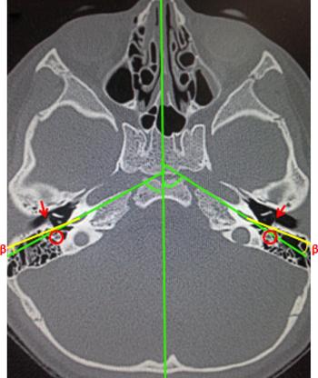
Axial CT scans were analysed in detail using the Philips Ingenuity 64-slice CT machine. A line was drawn along the mid-sagittal plane. A second line was drawn along the long axis of the basal turn of the cochlea. The angle that these two lines made with each other was called α (Figure 1). This angle was calculated on both sides. A third line was drawn along the proposed intra-operative axis of surgery, through the mastoid, facial recess and round window, running as near as possible to the posterior wall of the external auditory canal. The angle that this last line made with the second line was termed β (Figure 2). Two radiologists performed this calculation independently, and an average of both readings was taken. The values of α and β were then correlated with intra-operative difficulty in insertion of the electrode array.
Axial computed tomography scan demonstrating how angle α was measured.

Axial computed tomography scan demonstrating the measurement of angle β, which is the angle between the line of cochlear basal turn axis and the surgical axis (yellow lines). The red circles represent the facial nerve and the red arrows show the chorda tympani.

Results
A total of 107 patients were evaluated. Eleven patients were excluded because of cochleovestibular anomalies. Hence, our calculations were based on 96 patients (192 ears).
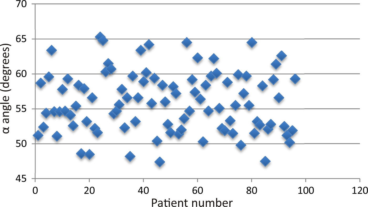
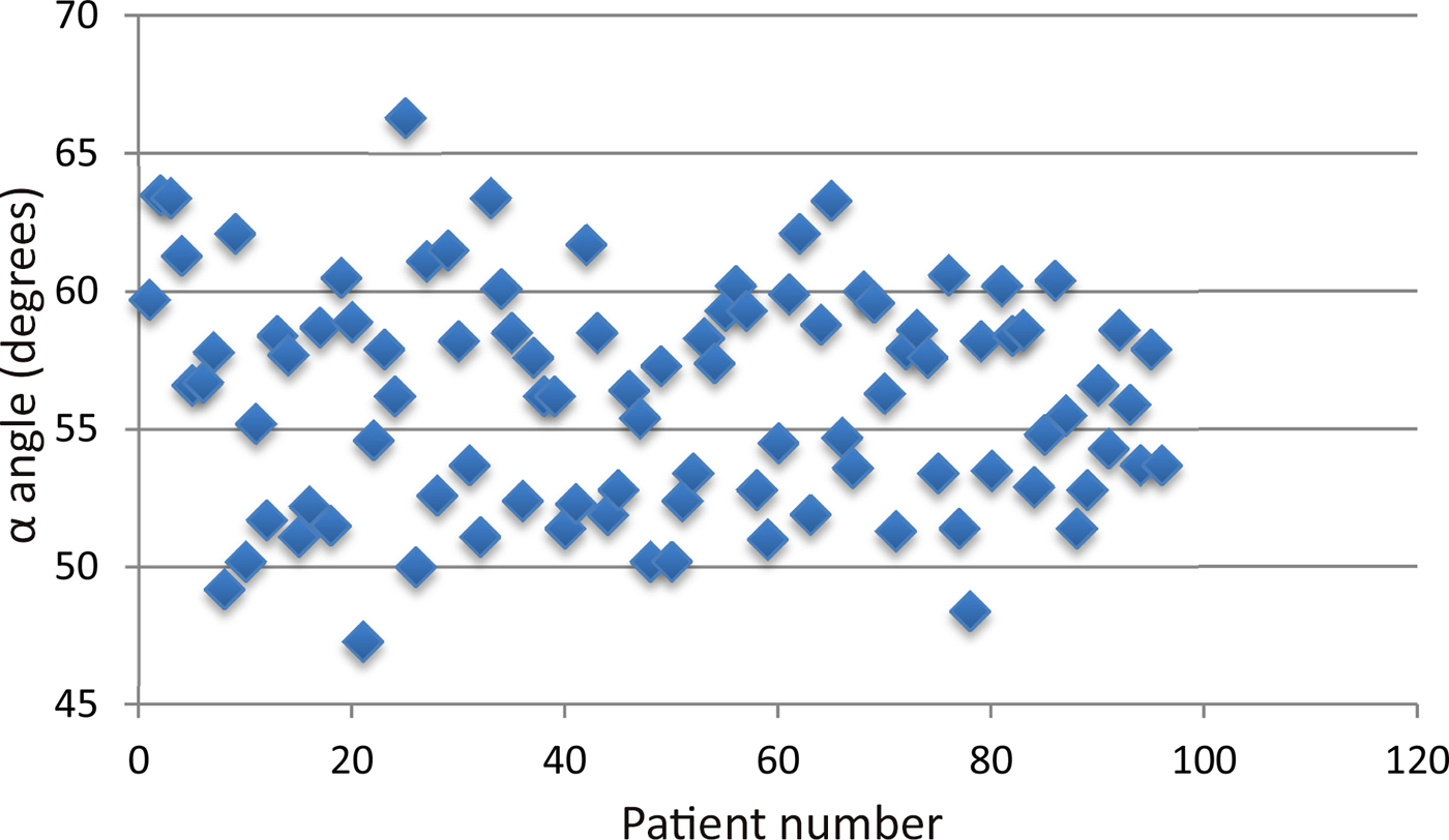
The average α angle was 55.95 degrees (range, 47.4–65.3 degrees) in the right ear (Figure 3) and was 56.19 degrees (range, 47.3–66.3 degrees) in the left ear (Figure 4), with an overall average of 56.07 degrees. In all seven patients who had an α angle of less than 50 degrees, the surgeon experienced difficulties in electrode insertion (Table I). However, there were four patients with an α angle of more than 50 degrees in whom the surgeon also experienced difficulties in electrode insertion. In all these patients, the β angle was more than 20 degrees (Table II).
Scatter diagram showing values of angle α in the right ear.

Scatter diagram showing values of angle α in the left ear.

Angle α values of patients in whom electrode insertion difficulties were experienced

*Indicates lowest of the two α values
Angle β values of operated ear in patients with angle α values over 50 with electrode insertion difficulties

*Indicates lowest of the two α values
Discussion
Most surgeons believe that cochlear rotation cannot be determined pre-operatively. Current data regarding cochlear orientation are limited. The few studies that have been conducted did not address the surgical implications of this orientation. Most of the studies focused on the change in cochlear orientation with age.
Spoor found a small but statistically significant difference in the orientation of the fetal and adult cochlea relative to the vestibule, when viewed on axial CT imaging.Reference Spoor1 The same author, in a more recent study using magnetic resonance imaging, confirmed that the labyrinth attains its adult size between 17 and 19 weeks of gestation.Reference Jeffery and Spoor2 This paper also stated that there may be a change in the orientation of the cochlea postnatally, although in their analysis this finding did not reach statistical significance. In addition, the authors observed statistically significant changes in semicircular canal orientation postnatally.Reference Jeffery and Spoor2
The only previous major study, by Lloyd et al. in 2010, discussed the change in cochlear orientation with age.Reference Lloyd, Kasbekar, Kenway, Prevost, Hockman and Beale3 In their study, the mean basal turn angle was 54.6 degrees (range, 46.8–63.8 degrees; standard deviation, 3.5). There was a statistically significant reduction in the angulation of the basal turn with increasing age (F = 10.1; p = 0.002). As per their observation, the three difficult cases had basal turn angles that were at the upper limit of the reference range. This mean basal turn angle is same as the α angle in our study, and the average value in our study of 56.07 degrees is similar.
However, the studies differ in regard to the surgical implication of this angulation. In our study, electrode insertion was found to be difficult in cases where the α angle was less than 50 degrees, which is the opposite of reports by Lloyd et al., in which difficulties were experienced in those cases with higher angles (66, 63 and 71 degrees).Reference Lloyd, Kasbekar, Kenway, Prevost, Hockman and Beale3 The study by Lloyd et al. reports a range of 46.8–63.8 degrees; however, confusingly, their discussion of difficult cases mentions angulations of 66 and 71 degrees in two of their patients and 64 degrees on the non-operative side in another patient. These numbers do not correlate. Furthermore, the difference in results could be because we attempted round window insertion in all our cases, whereas Lloyd et al. used a separate cochleostomy anterior to the round window niche.Reference Lloyd, Kasbekar, Kenway, Prevost, Hockman and Beale3
We will now consider the surgical implications of angle α. The lower the α angle, the more posteriorly rotated the cochlea; hence, the round window is more posterior and hidden from direct view through posterior tympanotomy when values of α are too low. In such circumstances, round window insertion would be difficult and in some cases not possible. If, however, the value of α is high, it means that the cochlea is anteriorly rotated, thereby making the round window easy to visualise through posterior tympanotomy and in a straight axis. In such cases, electrode insertion through the round window would be easy, because of good visualisation of the round window, direct axis to the cochlea and avoidance of a hook region.
Nevertheless, there are factors other than angle α that contribute to the surgery. For this reason, we calculated angle β. These other factors include the size and orientation of the facial recess, the position of the facial nerve and the chorda tympani, together with the orientation of the posterior canal wall of the external auditory canal. It is important that we understand the meaning and significance of angle β. If the angle of approach through the mastoid and posterior tympanotomy is the same as the angle of the basal turn relative to the sagittal plane, then passage of the implant electrode into the basal turn will be linear. However, with a more acutely angled basal turn, the cochlea will be more posteriorly rotated and the electrode will tend to abut against the medial wall of the basal turn as it passes through the cochleostomy, making insertion potentially more difficult, especially if round window insertion is being conducted.
Kennedy has demonstrated, using histological and endoscopic examination of the cochlea after electrode insertion, that there is often significant damage to the spiral ligament in the basal turn after insertion, and that passage of the electrode beyond the point at which resistance is felt can result in much more significant cochlear damage, including damage to the basilar membrane.Reference Kennedy4 A smooth linear approach into the basal turn is preferential to a more oblique approach. In cases with an obliquely angled basal turn, which is posteriorly located, it may be advantageous to consider an approach via the middle ear rather than through a posterior tympanotomy, or to consider a separate cochleostomy rather than round window insertion. The β angle indicates the extent of angulation between the surgical axis and axis of the basal turn of the cochlea. We cannot see inside the cochlea intra-operatively; however, pre-operatively, with help of β angle evaluation, we can decide whether to carry out electrode insertion via the round window or via a separate cochleostomy, to minimise intra-cochlear trauma.
• Cochlear implantation has become a standard treatment for severe to profound sensorineural hearing loss
• Transmastoid posterior tympanotomy is the standard approach for cochlear implantation
• Pre-operatively, high-resolution computed tomography was used to draw three lines, and two angles, α and β, were calculated
• In all seven patients with α angle less than 50 degrees and in four patients with β angle of more than 20 degrees, the surgeon had electrode insertion difficulties
• Calculation of cochlear orientation and its angle with the surgical axis (α and β) can aid surgical planning and cochleostomy site decisions
The position of the cochleostomy is critical for minimising damage to inner-ear structures. This is particularly true when we try to preserve residual hearing in patients undergoing cochlear implantation. The ideal position of the cochleostomy to prevent inner-ear damage is controversial, and recommendations in the literature vary from different positions in the otic capsule to round window insertion.Reference Li, Wang, Northrop, Merchant and Nadol5–Reference Balkany, Cohen and Gantz8 By calculating angle β, we will be in a better, though not ideal, position to define this site. However, increased precision and a bigger sample size would help us reach this goal.
Conclusion
Cochlear implantation surgery is not simply a matter of inserting electrodes inside the cochlea; instead, it aims to insert them in such a way that intra-cochlear trauma is minimised. Performing a cochleostomy at the right place, in the right manner, and conducting electrode insertion at the right angulation and speed, are crucial. Calculation of cochlear orientation and its angle with the surgical axis (α and β) will aid surgical planning, particularly with regard to the site of cochleostomy and preservation of residual hearing.
Competing interests
None declared.