1. Introduction
Middle Ordovician (Llanvirn, Darriwilian; Ogg, Ogg & Gradstein, Reference Ogg, Ogg and Gradstein2008, fig. 5.5) brachiopods were first recognized from western Thailand by Rong Jia-yu. Hagen & Kemper (Reference Hagen and Kemper1976, figs 2–4) provided a geological map of the Thong Pha Phum area and also indicated the location of many fossil localities, emphasizing that the Ordovician rocks consisted of a lower part dominated by limestones and an overlying part dominated by siliciclastics (Wolfart, Reference Wolfart2001a, fig. 1a, outcrop 7). Although Ordovician echinoderms have long been known from SE Asia (Indochina), early reports are still often taxonomically uncertain and the echinoderms rare, so any new occurrences are worth recording. For example, Noetling (Reference Noetling1890) described ‘Echinosphaerites kingi’ from near Mandalay, Myanmar (Burma), but Bather (Reference Bather and Reed1906, p. 6) already reported that ‘his description was quite inadequate and the type specimen cannot be traced’. Bather concluded that it was uncertain that the genus Echinosphaerites, even as then understood, occurred in Myanmar. Similarly, Lóczy (Reference Lóczy1897, Reference Lóczy1899a, b) described the first fossil echinoderms from China that included three isolated ‘cystoid’ plates from the Ordovician of Pu-piao, Yunnan Province. (See Stiller & Lin, Reference Stiller and Lin2015, for copies of the original illustrations and an English translation.) These plates can still only be referred to the superfamily Hemicosmitoida and probably the family Carycrinitidae, although Jaekel (Reference Jaekel1899, p. 313) named them as ‘Hemicosmites ? loczyi’. Reed (Reference Reed1906, Reference Reed1917) reported more extensive Ordovician faunas from the Northern Shan States, Myanmar and Yunnan Province, China, respectively. Bather (Reference Bather1918a, Reference Batherb, Reference Bather1919a, Reference Batherb, Reference Batherc, Reference Batherd) commented extensively on the latter paper. In both faunas the echinoderm fossils included numerous specimens preserving more or less complete thecae, but the echinoderm faunas have not been revised since Bather's comments. Sun (Reference Sun1936) described five additional taxa of Ordovician ‘cystoids’ from Kweichou, China, and later (Reference Sun1948) argued that several of these Ordovician taxa may have originated in China. More recently, Ordovician echinoderms and trilobites have been described from the same general region of Thailand as the present fauna by Wolfart (Reference Wolfart2001b, c). In addition, Paul et al. (Reference Paul, Donovan, Muir, Botting, Lin and Yuandong2016) have described a small early Ordovician fauna from Guizhou Province, South China.
Agematsu et al. (Reference Agematsu, Sashida, Salyapongse and Sardsud2006) described conodonts from the later Arenig to early Llanvirn (Dapingian to Darriwilian) in limestones from localities nearer to Thong Pha Phum than the new locality discussed herein, indicating that a facies change from limestone to siliciclastic beds occurs as one moves to the east in this area. Fortey (Reference Fortey1997) described later Ordovician trilobites from Satun Province, southern Thailand, similar to those present in the Pagoda Limestone of South China, and suggested a Caradoc age (Late Ordovician). The siliciclastic rocks of western Thailand from which our fauna was collected differ lithologically from the limestones that yielded Fortey's collection.
In 1997, Boucot and Tansathien found the roadcut locality discussed herein while carrying out a routine road reconnaissance along Highway 323 (Thong Pha Phum to Songkla Buri). Owing to the interruption of heavy rain, the initial collection was small, although clearly of ‘Middle Ordovician’ age. A second, far larger collection of large blocks of weathered, fossiliferous, calcareous mudstone was made in 2004. Both of these collections were initially prepared with a rock splitter in Boucot's Corvallis laboratory. The crumbly calcareous mudstone specimens were then ‘hardened’ with Alvar dissolved in acetone. Rong Jia-yu and Zhan Ren-bin in Nanjing then identified the brachiopods: Macrocoelia, Orthambonites, Porambonites, Saucrorthis and Taphrorthis, together indicating a Llanvirn (= Darriwilian) age. Trilobites are not present in the fauna, but the echinoderms are moderately diverse and form the subject of the present paper.
Our philosophy of open nomenclature follows Bengtson (Reference Bengtson1988). The specimens discussed herein are deposited in the Naturalis Biodiversity Center, Leiden, the Netherlands (prefix RGM, formerly Rijksmuseum van Geologie en Mineralogie).
2. Geological setting and locality
The specimens described below are from a locality at site Map Series 7018, Mapsheet Khao Bo Ngam (now Ban Mueang Song Tho) (4738I), UTM grid 479215E, 1639078N, Grid Zone 47P; latitude 14° 49′ 36″’ N, longitude 98° 48′ 24″’ E. The cut is located on the west side of the road, on the north side of the hill sloping down to the Valley of the Huai Chani, approximately 2 km south of Ban Mueng Song Tho and about 22 km ENE of the town of Thong Pha Phum.
Note that we had intended to provide a simple locality map, redrawn after the relevant Google Earth image. Although Thong Pha Phum and Highway 232 were located, this source indicated an inconsistency in the grid reference. Although Highway 323 passes through latitude 14° 49′ 36″’ N (see above), at that point Google Earth states the longitude to be 98° 39′ 59″’ E. The highway is in no way close to the stated longitude. The Google Earth coordinates define a point well to the east of the highway. We have been unable to clarify this inconsistency through the aid of our colleagues in Thailand.
The region is underlain by sedimentary rocks of the Cambro-Ordovician Pha Phum Group (Hagen & Kemper, Reference Hagen and Kemper1976; Bunopas, Reference Bunopas1981; Department of Mineral Resources, 1992). The upper part consists of siliciclastic rocks; the lower part is composed of limestones with some thin, argillaceous intercalations. Bedding at the fossil locality is steeply dipping (70–80°). The area is strongly folded. Most of the folds are oriented NNW–ESE, controlled by the Three Pagoda Fault Zone, itself a result of the Himalayan Orogeny.
Preliminary regional mapping shows the fossil locality in the Middle Ordovician Bo Ngam Formation of Siriphakdi, Salyapongs & Suteetorn (Reference Siriphakdi, Salyapongs and Suteetorn1985). These authors compiled the 1:250 000 geological map of the Tavoy Quandrangle and proposed the Song Tho Group for the limestone-dominated Ordovician rocks. They then subdivided the group into three divisions: in ascending order, the Unnamed Formation (O), the Bo Ngam Formation (O2) and the Kroeng Krawia Formation (O3). The Unnamed Formation consists of dark grey, wavy bedded limestones with abundant argillaceous films. The Middle Ordovician Bo Ngam Formation is composed of dark grey, well-bedded limestones with intercalated phyllitic shales containing cephalopods and crinoids in some places. The Upper Ordovician Kroeng Krawia Formation consists of limestones with argillaceous bands and thin-bedded shale. These Ordovician formations are overlain by the Silurian–Devonian Pong Sawae Formation, consisting of dark brown to black, well-bedded shale and siltstone containing tentaculitids and graptolites. The subdivision of the Ordovician by Siriphakdi, Salyapongs & Suteetorn (Reference Siriphakdi, Salyapongs and Suteetorn1985) was not accepted at that time because it could not be correlated with a subdivision of the Ordovician rocks in adjacent areas which employed the concept of the Thung Song and Thong Pha Phum Groups for the Ordovician sequences.
3. Columnals and crinoids
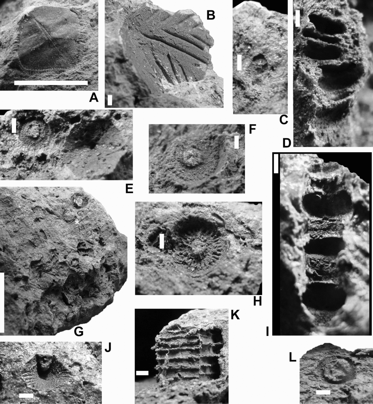
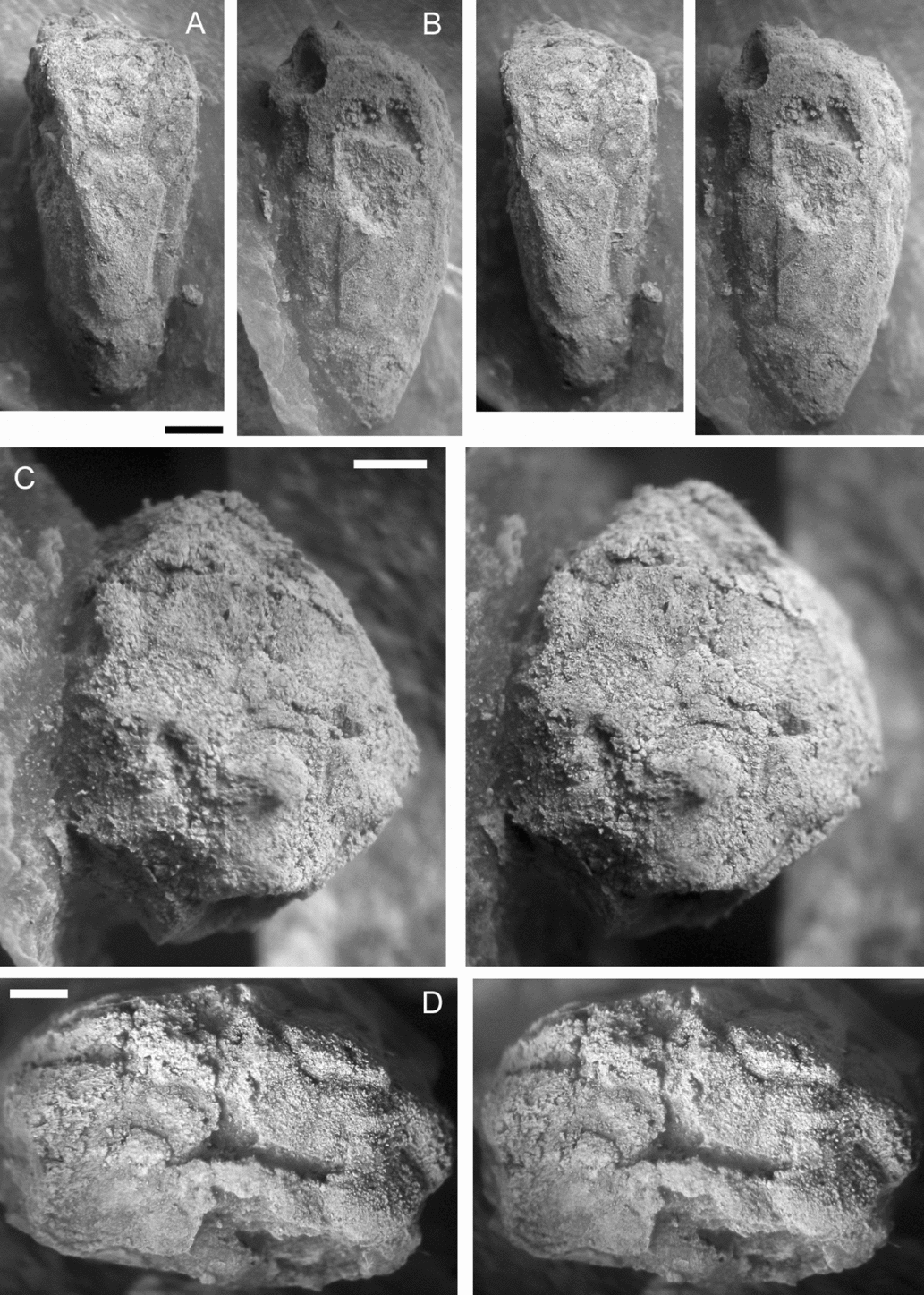
The columnal fauna of this rock collection is undoubtedly richer and more diverse than can be described herein, but the crude mouldic preservation in argillaceous matrix limits the available information. Shell hashes in these rocks are rich in small columnals (Fig. 1G), amongst other fragments, but these are both too fragile and too poorly preserved to be accurately determined in such a friable substrate; similarly, they are unworthy of detailed illustration. Further, what might appear to be a columnal in such a setting may instead be a broken fragment of a decalcified tube of an annelid or a mollusc. Larger columnals are identifiable (Figs 1C, E, F, HK, L, 2D, E), but rarely have an identifiable counterpart.
Pelmatozoan thecal plates (A, B, J) and columnals. Natural moulds. (A) RGM 792 530, tetragonal thecal plate bearing a superficial resemblance to a columnal of Colpodecrinus Sprinkle & Kolata Reference Sprinkle, Kolata and Sprinkle1982. Scale bar represents 10 mm. (B) RGM 792553b, ribbed thecal plate (see also Fig. 2C). (C) RGM 792 540, small Bystrowicrinus (col.) sp.; note the pentastellate lumen. (D) RGM 792 536, heteromorphic pluricolumnal (see also 2A). (E) RGM 792 538, hemicosmitoid columnal (left) and external mould of ‘Caryocrinites’ sp. thecal plate. (F) RGM 792 534, hemicosmitoid columnal. (G) RGM 792 543, general view of a shell hash. The specimen marked (*) is a columnal from the proxistele of a glyptocystitoid. Scale bar represents 10 mm. (H) RGM 792 541, hemicosmitoid columnal. (I) RGM 792 539, heteromorphic pluricolumnal (see also 2B). (J) RGM 792 550, radial plate (see also 2F). (K) RGM 792 558, pentameric pluricolumnal, probably caryocystitid. (L) RGM 792 547, glyptocystitoid proximal columnal. Specimens uncoated. Scale bars represent 1 mm unless stated otherwise.

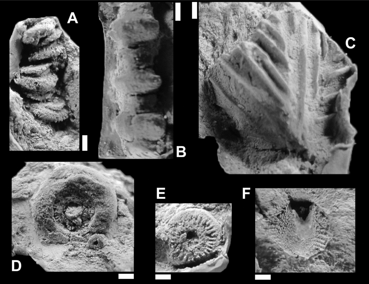
In a fauna in which thecal plates of ‘cystoids’ are common (see below), it is not unexpected that identifiable columnals are dominantly cystidean in origin and those that may have been derived from crinoids are a minor component, if that. Quite common and easily recognized are the columnals of hemicosmitoids. These have a circular outline and a central lumen with three-fold symmetry, commonly of moderate size (compare Figs 1E, F, H, 2D, E with Paul et al. Reference Paul, Donovan, Muir, Botting, Lin and Yuandong2016, fig. 9). A typical specimen, RGM 792 541 (Figs 1H, 2E), has a central lumen of rounded triangular outline; a circular areola; a marginal, radial symplectial articulation; and a concave epifacet.
Pelmatozoan thecal plates (C, F) and columnals. Latex casts from natural moulds. (A) RGM 792 536, heteromorphic pluricolumnal (see also Fig. 1D). (B) RGM 792 539, heteromorphic pluricolumnal (see also Fig. 1I). (C) RGM 792 553b, ribbed thecal plate (see also Fig. 1B). (D) RGM 792 561, hemicosmitoid nodal bearing a rounded epifacet and with internodals more centrally. (E) RGM 792 541, hemicosmitoid columnal (see also 1H). (F) RGM 792 550, radial plate (see also 1J). Specimens coated with ammonium chloride. Scale bars represent 1 mm.

The columnals of glyptocystitoid dichoporites are less common. The column is xenomorphic, with a conical proxistele formed from circular columnals with a broad axial canal and synarthrial articulation, and a slender, homeomorphic dististele of bead-like columnals with a narrow axial canal (Paul, Reference Paul1968a, Reference Paul1984). Two columnals from the proxistele are figured (e.g. 1G, L). Plates from the dististele may be among the small, indeterminate components in shell hashes.
Caryocystitids ‘have a meric, non-tapering column with a broad axial canal’ (Donovan, Reference Donovan1986, p. 19). One specimen, RGM 792 558 (Fig. 1K), is a pentameric pluricolumnal that fits this description (e.g. cf. Kesling, Reference Kesling and Moore1968, fig. 132.1a) and is determined to be a caryocystitid. Figure 1K shows that the meres of the pluricolumnal are not arranged perfectly into columnals and show longitudinal offset along the meric suture (cf. Donovan, Reference Donovan1986, text-fig. 5C).
The remaining columnals, tentatively regarded as crinoids (mainly because they are not identifiable as ‘cystoids’), are limited in the information that they can provide. The most distinctive, RGM 792 540 (Fig. 1C), is a Bystrowicrinus (col.) sp., a form of crinoid well known from the Ordovician, but rarer thereafter (e.g. Donovan & Keighley, Reference Donovan and Keighley2016). Columnals of comparable size and morphology were described as Bystrowicrinus? (col.) sp. A from the Hirnantian (uppermost Ordovician) of north Wales by Donovan (Reference Donovan1995, pp. 131–2, text-fig. 43H, I), with the ‘rays’ of the pentastellate lumen corresponding to the sides of the pentagonal columnal. A broadly similar columnal is figured as Digiticrinus sp. cf. D. bellus Stukalina, Reference Stukalina1980, by Wolfart (Reference Wolfart2001c, p. 221, pl. 13, figs 10, 11).
Pluricolumnals are rare (Figs 1D, I, 2A, B) and commonly have poorly preserved articular facets, so the gross features of the column remain unknown. However, they are at least reminiscent of the mesistele–dististele of primitive disparids such as Cincinnaticrinus Warn & Strimple, Reference Warn and Strimple1977. Three thecal plates are figured, two of which may be crinoidal. RGM 792 530 (Fig. 1A) is the exception and is probably cystidean, but is included as a curiosity. It is a tetragonal thecal plate, which bears a superficial resemblance to a columnal of Colpodecrinus Sprinkle & Kolata, Reference Sprinkle, Kolata and Sprinkle1982 (fig. 57F, pl. 22, figs 13, 14; see also Donovan, Reference Donovan1983). RGM 792 553a, b (part and counterpart) (Figs 1B, 2C) is large and bilaterally symmetrical, with a sculpture of regularly arrayed, branching ridges; it is likely to be crinoidal. The most distinctive of all is RGM 792 550 (Figs 1J, 2F), a polygonal radial plate with a simple ridged sculpture. This is crinoidal and not, for example, from a hemicosmitoid such as Caryocrinites (Sprinkle, Reference Sprinkle1975).
4. Systematic palaeontology
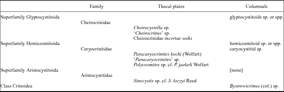
Remarks. Remains of ‘cystoids’ are not uncommon among our material. They represent at least five species-level taxa and can be attributed to the dichoporite superfamilies Glyptocystitoida and Hemicosmitoida, and the diploporite superfamily Aristocystitoida. Preservation is variable, but at least some of the hemicosmitoids are represented by complete thecae.
Superfamily GLYPTOCYSTITOIDA Bather, Reference Bather1899
Diagnosis. A dichoporite superfamily with well-developed, xenomorphic stem consisting of a rapidly tapering proximal portion composed of alternating inner and outer annular columnals, and a distal portion composed of alternating longer and shorter cylindrical columnals; theca composed of four basals, five infra-laterals, five laterals, four, five, six or ten radials, and seven orals (after Paul & Rozhnov, Reference Paul and Rozhnov2016, p. 260).
Remarks. Although genera, such as Macrocystella, Callaway, Reference Callaway1877, and Amecystis Ulrich & Kirk, Reference Ulrich and Kirk1921, which lack respiratory structures, and Cuniculocystis Sprinkle & Wahlman, Reference Sprinkle and Wahlman1994, which bears epispires, are included within the superfamily, most glyptocystitoid genera bear pectinirhombs. Pectinirhombs are rhombic respiratory structures developed in two adjacent thecal plates and defined by the presence of canals called dichopores that open externally in slits (Paul, Reference Paul1968b). The slits may be continuous along the entire length of the canal (conjunct pectinirhombs) or two separate slits may be present, one at each end of the canal (disjunct pectinirhombs). The theca may bear from one to over twenty pectinirhombs. As a result, when it disarticulates, only some of the isolated plates bear the characteristic half-rhombs. Nevertheless, isolated plates that bear half pectinirhombs can be assigned to the superfamily with confidence, although assigning isolated plates to families and genera is more speculative. Currently, eight families with Ordovician representatives are included within the superfamily, but only one of them occurs among the present material. The family Cheirocrinidae is represented by isolated plates and incomplete, partly articulated thecae.
Family Cheirocrinidae Jaekel, Reference Jaekel1899
Diagnosis. Glyptocystitida with six or more pectinirhombs; with a cylindrical theca having six radials; a large lateral periproct surrounded by five thecal plates and covered with a flexible plated integument; five, usually short, ambulacra with 5–15 brachioles each, rarely extending into clefts in the radial plates (modified from Paul, Reference Paul1984, pp. 97–8).
Remarks. Wolfart (Reference Wolfart2001c, p. 193, fig. 1, pl. 9, figs 1–3, pl. 12, figs 7, 8) described and illustrated isolated plates from the Thong Pha Phum Formation near Thong Pha Phum, which he referred to as Echinoencrinites sp. (family Echinoencrinitidae). However, all genera in the Echinoencrinitidae have disjunct pectinirhombs with very short oval slits (see e.g. Paul, Reference Paul1968b, pl. 135, figs 7, 8, 10), whereas the pectinirhombs Wolfart illustrated have conjunct slits, as do two of the plates described below. Conjunct pectinirhombs only occur in the families Cheirocrinidae and Pleurocystitidae. In the latter family and in the cheirocrinid genus Homocystites Barrande, Reference Barrande1887, the pectinirhombs are composed of advanced, confluent dichopores, whereas the present plates have discrete dichopores (see Paul, Reference Paul1968b, p. 703). Paul (Reference Paul1972a, p. 33) assigned all species of Cheirocrinidae with conjunct pectinirhombs composed of discrete dichopores to the genus Cheirocystella. Thus, some of the isolated plates from Thong Pha Phum can be assigned to a genus, even if it is uncertain that Cheirocystella is a monophyletic taxon. Among the material described herein is one plate with a disjunct pectinirhomb composed of discrete dichopores, which can only be attributed to the nominal genus ‘Cheirocrinus’ s.l. For the terminology describing isolated glyptocystitid plates, see Paul (Reference Paul1984, fig. 56).
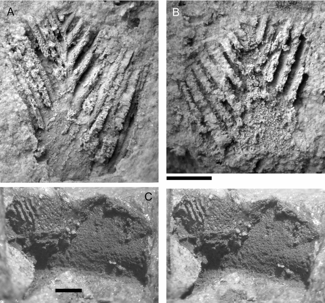
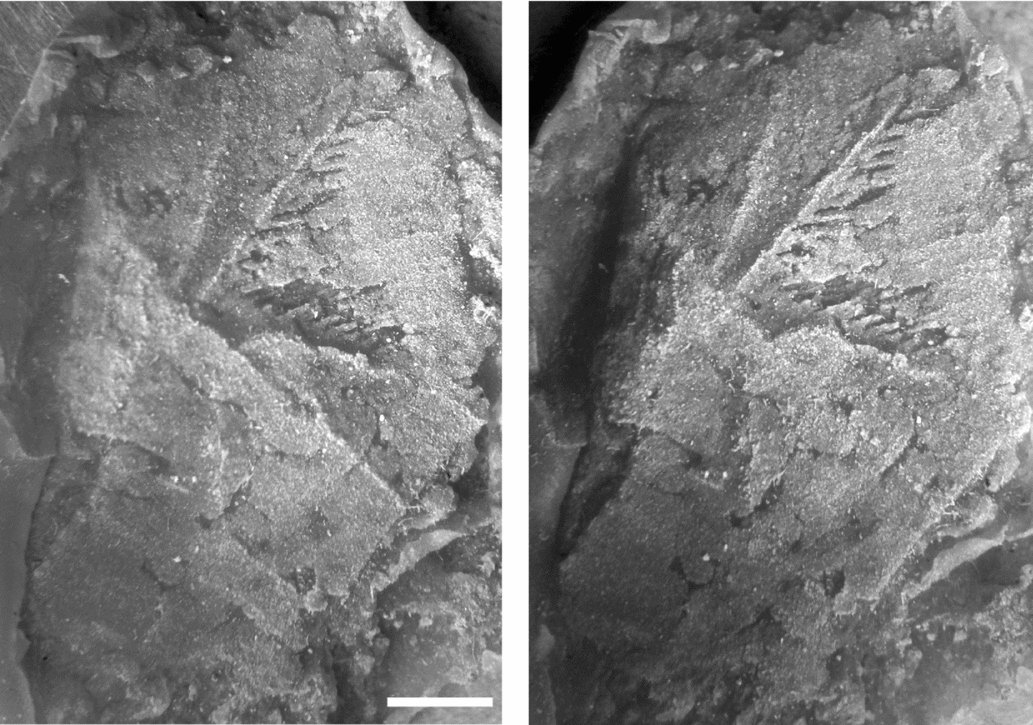
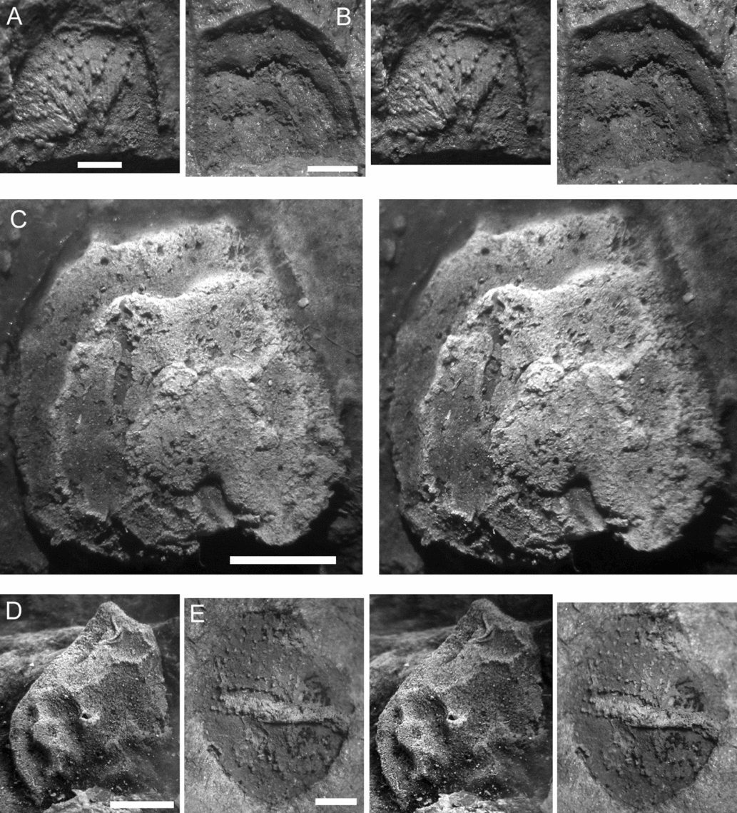
Cheirocystella sp. (A, B) RGM 702905a, b, part and counterpart natural moulds of an incomplete, isolated plate showing parts of two conjunct pectinirhombs. (C) RGM 702508a, stereophotos of a second, strongly folded radial(?) plate showing a small conjunct pectinirhomb. Scale bars = 1 mm, upper refers to both A and B, which were whitened with ammonium chloride.

Genus Cheirocystella Paul, Reference Paul1972a
Diagnosis. A genus of Cheirocrinidae with all plate circlets closed; short ambulacra confined to the flat oral surface; many (15–20 or more) conjunct pectinirhombs with discrete dichopores (modified from Paul, Reference Paul1972a, p. 33).
Remarks. As with all Glyptocystitoida, the theca in the Cheirocrinidae is composed of five circlets of plates called basals (B, plural BB), infra-laterals (IL, ILL), laterals (L, LL), radials (R, RR) and orals (O, OO). When all the plates in an individual circlet are in contact the circlet is described as closed; alternatively, if plates from the circlet below and above meet at a common suture, the circlet in between is described as open. Both arrangements are known within the Cheirocrinidae, but commonly it requires a complete theca to establish the difference unequivocally. To date, all species of Cheirocystella where the plate arrangement is known have all plate circlets closed, but isolated plates bearing conjunct pectinirhombs with discrete dichopores occur at localities from which no complete theca has been found. Thus, it is not certain that all species currently attributed to Cheirocystella do have all plate circlets closed. Previously described species of Cheirocystella come from the Lower to Middle Ordovician (Tremadoc to basal Darriwilian [Kunda]; Paul, Reference Paul1972a, p. 34). The present specimen extends the range of the genus into the upper Darriwilian.
Cheirocystella sp.
Figure 3
?Reference Reed1917 Echinoencrinites sp.; Reed, p. 19, pl. 4, fig. 7.
Reference Wolfart2001c Echinoencrinites sp. aff. E. senckenbergii (von Meyer, Reference Meyer1826); Wolfart, pp. 185, 186, 193, fig. 1b, c; pl. 9, figs 1–3; pl. 12, figs 7, 8 [non pl. 12, fig. 6 = a caryocrinitid plate].
Material. Part and counterpart of a single partially complete, isolated plate (RGM 792509a, b), plus an external mould of a second damaged radial? plate (RGM 702508a) (Fig. 3).
Description. The incomplete plate is 4.1 mm by 3.6 mm as preserved, and shows two half demi-rhombs with seven and six conjunct dichopores (Fig. 3A, B). The largest dichopore reaches almost to the apparent base of the plate and is 3.5 mm long. Slit spacing is one slit per 0.30 mm in the larger demi-rhomb and 0.31 mm in the smaller, both measured at the plate suture. The longest three dichopores in the larger demi-rhomb are slightly wider at the end furthest from the suture. The plate surface between the demi-rhombs lacks any ridges or tubercles and appears to be smooth. The second, radial? plate (Fig. 3C) is at least 6 mm high by 3 mm wide and shows a single conjunct half-rhomb with seven dichopores preserved. The longest dichopore reaches 1.3 mm, and slit spacing averages one slit per 0.41 mm. The plate is ornamented with radiating ridges or folds, in between which is a finely granular or pitted sculpture.
Comments. Wolfart (Reference Wolfart2001c, fig. 1b, c) illustrated the internal surfaces of two plates with two or more conjunct half-rhombs. As illustrated, the dichopores are not always perpendicular to the plate sutures, so interpretation of the drawings is not easy. However, it seems that both Wolfart's and our collections contain isolated plates of cheirocrinids with conjunct pectinirhombs. The shape of one plate is suggestive of a basal plate, although the stem facet cannot be seen. If it is a basal, then it is likely to be part of plate B2, which is the only basal plate to bear part of more than one pectinirhomb. The other plate appears to be a radial. Slit spacing is relatively coarse for cheirocrinids, but this is consistent with previous measurements of other species of Cheirocystella (Paul, Reference Paul1968b, table 1). The plates confirm the presence of the genus Cheirocystella, but otherwise little else can be said about them.
A comparison of the systematic diversity of primitive echinoderms determined from the thecal plates and columnals of the Darriwilian (Middle Ordovician) Bo Ngam Formation(?), Bang Mueang Song Tho, western Thailand.

Genus Cheirocrinus Eichwald, Reference Eichwald1856
‘Cheirocrinus’ sp.
Figure 4
Material. RGM 792507a, b, part and counterpart of a single, isolated thecal plate bearing a disjunct half-pectinirhomb with discrete dichopores (Fig. 4).
‘Cheirocrinus’ sp., RGM 792507, stereophotos of a latex cast of a large isolated plate showing a disjunct pectinirhomb. Thong Pha Phum. Scale bar = 2 mm. Whitened with ammonium chloride.

Description. The plate is incomplete, but at least 14 mm by 10 mm. The half-rhomb is 6 mm long (parallel to dichopores) by >7 mm wide, with at least 25 (estimated 30) dichopores, but the extreme edges of the rhomb are obscured. Slit spacing is one slit per 0.23 mm, and the slits reach 2.1 mm long by 0.15 mm wide in the centre of the half-rhomb. The plate is slightly folded about axial ridges (that is, ridges running from the plate centre to the middle of plate edges) and has one or two accessory ridges as well as radial ridges running to the corners of the plate on either side of the half-rhomb. The axial ridges suggest the complete plate was pentagonal in outline and, therefore, possibly truly from a species of Cheirocrinus sensu stricto. The plate inter-areas appear to be smooth (Fig. 4, below), as do all the ridges.
Remarks. If this plate is pentagonal, then either the infra-lateral or lateral circlet was open and it is most probably plate L2, which is the only pentagonal thecal plate to bear a single half-rhomb. Paul et al. (Reference Paul, Donovan, Muir, Botting, Lin and Yuandong2016, fig. 3, p. 146) illustrated a similar, pentagonal cheirocrinid plate from the Floian of Tonggao, South China.
Cheirocrinidae incertae sedis
Figure 5
Material. One partial theca with associated proximal stem, one group of associated plates and three other isolated plates (Fig. 5).
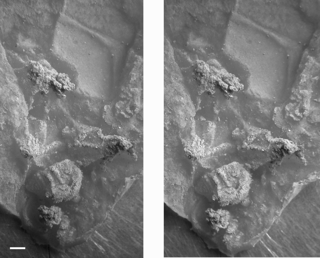
Cheirocrinidae incertae sedis, RGM 792512, stereophotos of a latex cast of several associated conical plates with strong radial ridges. The plate at the top right is a periproct border plate (probably IL4) seen from the interior. Scale bar = 1 mm. Whitened with ammonium chloride.

Description. The most complete specimen (RGM 792512; Fig. 5) is a small slab with moulds of a few proximal columnals and seven associated theca plates, six seen as external moulds and the seventh, a periproct border plate, seen as an internal mould (Fig. 5, above right). The stem is not well preserved (Fig. 5, below), but shows a wide lumen and is 2 mm in diameter adjacent to the theca. One external surface of an outer proximal columnal shows it had a rounded external flange ornamented with fine, vertical ridges. The associated plates appear to be one basal, two infra-laterals, two laterals and the lower part of a radial, all preserved showing the external surfaces. They are slightly disarticulated and measure no more than 12 mm from basal to radial. Even though some of the radial plate is probably obscured, the theca was small. All the plates are strongly vaulted (Fig. 5, centre), with strongly developed axial ridges originating in prominent plate umbones, plus a single accessory ridge on either side of some of the axial ridges. The largest plate is the one showing the internal surface and margin of the periproct, which is 6.3 mm high by 4.2 mm wide. It also appears to be flat and without ridges, so the strong ridges on the external surface of the other plates must be solid, rather than axes about which the plate is folded. The plates seen in external view all appear to be smaller, but only the centres of most plates can be seen. There is no sign of any pectinirhomb on exposed plates.
Other isolated plates include external moulds of two with reticulate ornament as well as part and counterpart of a tightly folded plate with axial and accessory ridges, but only finely granular ornament in between them. One of the more highly ornamented plates is probably a radial, 10 mm high by at least 6.7 mm wide. The latex of the external mould shows at least four axial and one radial ridge. The most prominent axial ridge divides as it approaches the plate edge, which is unusual. The plate inter-areas are covered with fine accessory ridges, themselves crossed by slightly weaker, concentric growth lines. A smaller plate is only 3.0 mm by 1.5 mm and also has many accessory ridges.
Remarks. Even though all these specimens lack traces of pectinirhombs, sufficient material is preserved of the partial theca to be certain of the family assignment. However, the isolated plates could belong to species of pleurocystitids. There are too few of them to be certain of their taxonomic affinities.
Superfamily Hemicosmitoida Jaekel, Reference Jaekel1918
Diagnosis. A dichoporite superfamily with well-developed heteromorphic stem; theca composed of three or four basals, six infra-laterals, eight or nine laterals all of which bear cryptorhombs, and additional plates in the oral region; with three or four primary ambulacra (after Lanc, McDermott & Paul, Reference Lanc, McDermott and Paul2015, p. 4).
Remarks. Hemicosmitoids, like glyptocystitoids, are characterized by a theca composed of a regular arrangement of plates and internal respiratory canals (dichopores) arranged in rhombs, but the details differ. At present the same terms are used to describe the plate circlets that bear rhombs, but it is by no means certain that plate circlets with the same name are homologous in the two superfamilies. In addition, caryocrinitids typically have a tegmen-like cover to the oral surface. It is most unlikely that this structure is homologous with any of the types of tegmina developed in crinoids. Herein we retain the terms for plate circlets as well as the terms ‘tegmen’ and ‘tegminal’ to facilitate comparisons with previous descriptions. There is a greater variation in the plate arrangements within the Hemicosmitoida, and the dichopores open in pores, not slits. Furthermore, typically all the pores in one half-rhomb are simple, whereas all the pores in the other half-rhomb are clusters of small pores, called compound pores (Paul, Reference Paul1968b, text-fig. 20) or sieve pores (Bockelie Reference Bockelie1979, pp. 371–2, figs 5–7). Bockelie (Reference Bockelie1979, pp. 372–3) showed that in hemicosmitids there are up to five systems of pore-rhombs, three of which produce currents parallel to the oral–aboral axis of the theca (systems 1–3) and two currents at right angles to the thecal axis (systems 4 and 5). This pattern is also present in caryocrinitids, except that system 3 is absent. Cryptorhombs are usually developed across all B:IL and IL:L sutures (systems 1 and 2). All pores in basal and lateral plates are sieve pores, which seawater entered (Paul, Reference Paul1968b, pp. 717, 719). All pores in systems 1 and 2 in infra-lateral plates are simple and acted as exits. In addition, cryptorhombs often occur across IL:IL and L:L sutures (Bockelie's systems 4 and 5), but may be less well-developed or even absent. Often they consist of just one or two dichopores, rather than a fully developed rhomb. The pattern of entrances and exits is also less systematically developed, and Bockelie (Reference Bockelie1979, p. 374) recorded one anomalous example in hemicosmitids. The pattern of cryptorhombs in caryocrinitids is identical to that of hemicosmitids except that there are no L:R rhombs (Bockelie's system 3) since caryocrinitids lack radial plates.
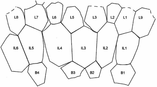
Currently, three families are recognized within the Hemicosmitoida, the Hemicosmitidae, Caryocrinitidae and Thomacystidae (see Paul, Reference Paul1984, fig. 57, p. 84). The last is based on the unique genus and species Thomacystis tuberculata Paul, Reference Paul1969, which is characterized by having three basal, six infra-lateral and eight lateral plates, four ambulacra developed on the oral surface, and the periproct between two lateral and two ‘adoral’ plates. The Hemicosmitidae may be most easily separated from the Caryocrinitidae by the position of the periproct, which lies between two infra-laterals, or more commonly at the junction between two infra-laterals and a lateral plate in Hemicosmites, or between two lateral plates in Corylocrinus (see Jaekel, Reference Jaekel1899, fig. 66, p. 296). Corylocrinus is also unique among the Hemicosmitidae in having only eight, not nine, lateral plates as do all genera in the Caryocrinitidae and Thomacystis. Herein, numbering of lateral plates follows Paul (Reference Paul1984, figs 87–9) in which it is assumed plate L4 of Hemicosmites is missing in all genera with only eight laterals. In the Caryocrinitidae the periproct opens above the lateral plate circlet, on the tegmen-like oral surface and sometimes not in contact with any lateral plate (e.g. see Frest, Reference Frest1975, figs 4a, 5, 8; Chauvel & Le Menn, Reference Chauvel and Le Menn1979, figs 2e, f, 3a–e, h, i; Figs 6–8 herein). In addition, many species of the Hemicosmitidae have a circlet of nine, rhomb-bearing, radial plates above the lateral circlet and below the oral surface (see Bockelie, Reference Bockelie1979). Several more or less complete thecae occur among the present material. None has a lateral periproct and all have only eight lateral plates, indicating that they belong in the family Caryocrinitidae.
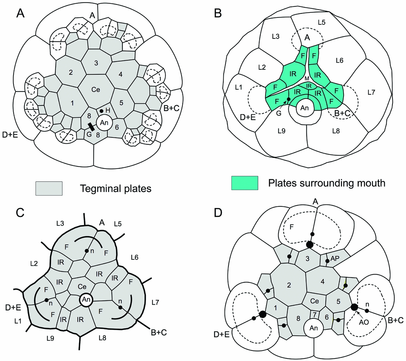
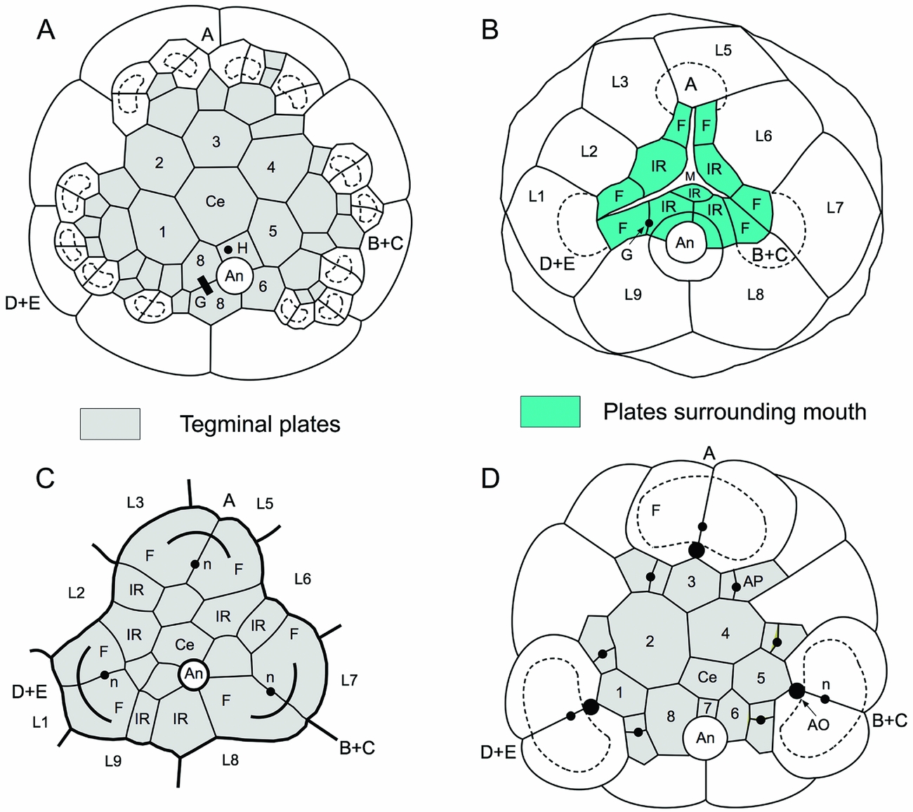
Oral areas of the Caryocrinitidae. (A) Caryocrinites sensu stricto (= C. ornatus Say, Reference Say1825, type species of Caryocrinites, redrawn after Frest, Reference Frest1975, fig. 3a). (B) Paracaryocrinites Chen & Yao, Reference Chen and Yao1993 (= an unnamed species, after Jaekel, Reference Jaekel1899, pl. 18, fig. 4). (C) Juglandocrinus von Koenen, Reference Koenen1886 (= J. sdzuyi (Chauvel & Le Menn, Reference Chauvel and Le Menn1979), after Chauvel & Le Menn, Reference Chauvel and Le Menn1979, fig. 2e, p. 554). (D) Stribalocystites Miller, Reference Miller1891 (= S. tribrachiatus Frest, Reference Frest1975, after Frest, Reference Frest1975, fig. 15, p. 103). Key: A, B + C, D + E = ambulacra; An = anus; AO = ambulacral orifice; AP = accessory tegminal plates; Ce = central plate of tegmen; F = facetal plates; G = gonopore; H = hydropore; IR = inter-radial plates; L1–L9 = lateral plates; M = mouth; n = nerve canal; 1–8 = peri-central plates sensu Frest (Reference Frest1975).
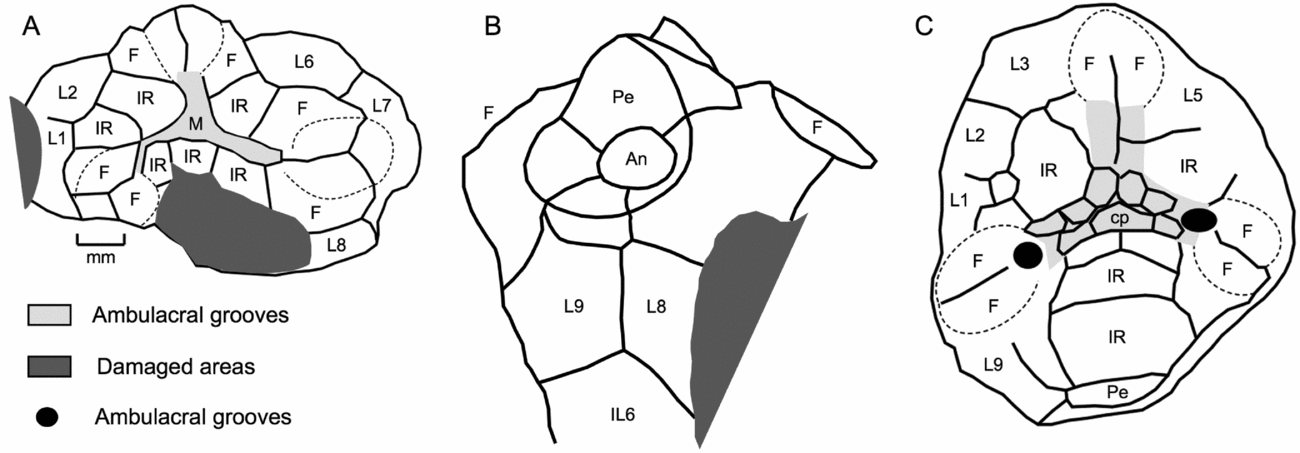
Camera lucida drawings of oral areas in Paracaryocrinites kochi (Reference WolfartWolfart, 2001c). (A) RGM 792508c, oral view showing three food grooves radiating from mouth (M). (B, C) RGM 792508b, posterior lateral view and oral surface, respectively. Note obvious ambulacral grooves, each ending in a single arm facet supported by two facetal plates (F) and with two ambulacral orifices (black) in (C). Darker shading indicates missing plates or damage. Key: An = anus; cp = ambulacral cover plates; IL6 = infra-lateral plate; IR = inter-radial plates; L1–L9 = lateral plates; Pe = periproct, which surrounds the anal opening. Scale in (A) applies to (B) and (C).

Thecal plate diagram of Paracaryocrinites kochi (Reference WolfartWolfart, 2001c). Note that the infra-lateral plates (IL1–IL6) are very elongate and may reach twice as high as wide (e.g. IL2). Key: B1–B4 = basal plates; L1–L9 = lateral plates. Dashed lines indicate inferred plate outlines.

Family Caryocrinitidae Bernard, Reference Bernard1895
Diagnosis. A family of Hemicosmitoida with four basals, six infra-laterals, eight laterals and a periproct on the oral surface; with three main ambulacra each with one to five arms (Lanc, McDermott & Paul, Reference Lanc, McDermott and Paul2015, p. 5).
Remarks. Kesling (Reference Kesling and Moore1968, p. S222) assigned five genera to the Caryocrinitidae, namely Caryocrinites Say, Reference Say1825; Juglandocrinus von Koenen, Reference Koenen1886; Stribalocystites S. A. Miller, Reference Miller1891; Ptychocosmites Jaekel, Reference Jaekel1918; and Oocystis Dreyfus, Reference Dreyfus1939. Since then, Chen & Yao (Reference Chen and Yao1993) have added the genus Paracaryocrinites. Of these, Ptychocosmites is still only known from isolated thecal plates, and its validity as a separate genus is uncertain. Paul (Reference Paul1984, p. 144) recommended referring all Ordovician caryocrinitid species to ‘Caryocrinites’ s.l. because none of the other genera could be characterized adequately. However, Lanc, McDermott & Paul (Reference Lanc, McDermott and Paul2015) reviewed all the available information about oral surfaces in the Caryocrinitidae, including some details overlooked by Paul (Reference Paul1984). Their conclusions as to the valid genera were as follows:
(1) Caryocrinites, type species C. ornatus Say, Reference Say1825, by monotypy (Fig. 6A), is characterized by a large tegmen that hides the plates surrounding the mouth and three ambulacra, all of which branch. Lanc, McDermott & Paul (Reference Lanc, McDermott and Paul2015, p. 6) attributed the Ordovician species C. septentrionalis Regnéll, Reference Regnéll1945, and C. cryptoporus Paul, Reference Paul1984, to Caryocrinites s.s., and added ‘Hemicosmites’ rugatus Salter, Reference Salter1866. All three species come from the Katian. Caryocrinites sensu stricto ranges from Upper Ordovician to Upper Silurian or possibly lowermost Devonian.
(2) Juglandocrinus, type species J. crassus von Koenen, Reference Koenen1886, by original designation (Fig. 6C), has an oral area with a single, relatively large ambulacral facet in each of the three ambulacra, but no trace of ambulacral grooves (Fig. 6C). Above the lateral plate circlet, Juglandocrinus has a circlet of 12 plates arranged in pairs. Each ambulacral facet is developed on a pair of adradial plates, and in between are two interradial plates of which the right-hand one (as viewed from the mouth) is usually the smaller. In addition, there are several smaller tegminal plates within this circlet covering the mouth. The periproct is not in contact with any lateral plate, but may be bordered by the two posterior interradial plates of the next adoral circlet. Lanc, McDermott & Paul (Reference Lanc, McDermott and Paul2015, p. 6) attributed ‘Caryocrinites’ europaeus (Quenstedt, Reference Quenstedt1874), ‘Corylocrinus’ elongatus Jaekel, Reference Jaekel1899, ‘Caryocrinites’ turbo Bather, Reference Bather and Reed1906, and ‘Caryocrinites’ sdzuyi Chauvel & Le Menn, Reference Chauvel and Le Menn1979, to Juglandocrinus. They also pointed out that Chauvel & Le Menn (Reference Chauvel and Le Menn1979, p. 559) regarded Oocystis vulgaris Dreyfus, Reference Dreyfus1939, type species of Oocystis, as a junior synonym of ‘Corylocrinus’ elongatus Jaekel, Reference Jaekel1899, as did Renard (Reference Renard1968, p. 67). Thus, Oocystis is also probably a junior synonym of Juglandocrinus.
(3) Paracaryocrinites, type species, P. subglobosus Chen & Yao, Reference Chen and Yao1993, has three obvious ambulacral grooves, each of which terminates in a single ambulacral facet developed on a pair of adradial facetal plates (Fig. 6B). In between are one or two interradial plates, except in the posterior interradius where there may be three or four. The periproct is on the oral surface and surrounded by four plates, where the number is known (Fig. 7B). Thus, Paracaryocrinites has an ambulacral system very similar to that of Hemicosmites, but a periproct on the oral surface, not laterally, and eight, not nine, lateral plates. Lanc, McDermott & Paul (Reference Lanc, McDermott and Paul2015, p. 7) attributed ‘Caryocrinites’ kochi Wolfart, Reference Wolfart2001c, and an as yet unnamed species described and illustrated by Jaekel (Reference Jaekel1899) as an aberrant specimen of Hemicosmites pyriformis von Buch, Reference Buch1840 (Fig. 6B herein), to Paracaryocrinites.
(4) Stribalocystites, type species S. tumidus S. A. Miller, Reference Miller1891, by original designation, has an oral area covered by a tegmen, like Caryocrinites s.s., but only a single facet in each ambulacrum (Fig. 6D). Indeed, Frest (Reference Frest1975) regarded Caryocrinites and Stribalocystites as synonymous, but Lanc, McDermott & Paul (Reference Lanc, McDermott and Paul2015, p. 7) thought it useful to distinguish Silurian species with a prominent tegmen-like structure and only a single facet per ambulacrum in the genus Stribalocystites. They also listed several points by which Juglandocrinus could be distinguished from Stribalocystites.
(5) Finally, Polycosmites, type species P. bohemicus Jaekel, Reference Jaekel1918, by monotypy, was described on the basis of an isolated plate and, until recently, was known only from the original specimen. Jaekel (Reference Jaekel1918, p. 98) attributed P. bohemicus to a monotypic family, the Polycosmitidae. However, there are reasons to believe that Polycosmites also belongs in the family Carycrinitidae (see below). Paul et al. (Reference Paul, Donovan, Muir, Botting, Lin and Yuandong2016, fig. 4) described and figured an additional plate of Polycosmites from the Floian of south China.
In summary, Ordovician species of caryocrinitids may be assigned to the genera Caryocrinites s.s., Juglandocrinus, Paracaryocrinites and Polycosmites. Silurian species are attributable to Caryocrinites s.s. and Stribalocystites. Species whose oral areas are unknown are best attributed to ‘Caryocrinites’ s.l. Among the material described below are a few examples in which at least some details of the oral areas can be determined. These show three ambulacral grooves terminating in a single brachiole facet, as in Hemicosmites, but a periproct above the lateral circlet. They are attributed to Paracaryocrinites Chen & Yao.
Genus Paracaryocrinites Chen & Yao, Reference Chen and Yao1993
Diagnosis. A genus of Caryocrinitidae with three exposed ambulacra on the oral surface, each terminating in a single arm facet, and the periproct above the lateral plate circlet in contact with L8 and L9.
Type species. Paracaryocrinites subglobosus Chen & Yao, Reference Chen and Yao1993, by original designation.
Stratigraphic and geographic range. Currently only known for certain from the ‘late Ordovician’ of Yunnan, China (Chen & Yao, Reference Chen and Yao1993), the Llanvirn (Darriwilian) of Thailand (Wolfart, Reference Wolfart2001c, and herein) and an unknown horizon in the Baltic area (Estonia or St Petersburg; Jaekel, Reference Jaekel1899).
Paracaryocrinites kochi (Wolfart, Reference Wolfart2001c)
Figures 7A–C, 8–10
Reference Wolfart2001c ‘Caryocrinites’ kochi n. sp., Wolfart, pp. 197, 199, fig. 2, pl. 9, figs 4, 6, 8.
Diagnosis. A species of Paracaryocrinites with elongate conical theca and the height of infra-lateral plates reaching twice or more their width.
Material. About ten thecae or partial thecae (including RGM 792473–792478), preserved as natural moulds, plus a large number of isolated thecal plates (including RGM 792465–792472; 792494–792505).
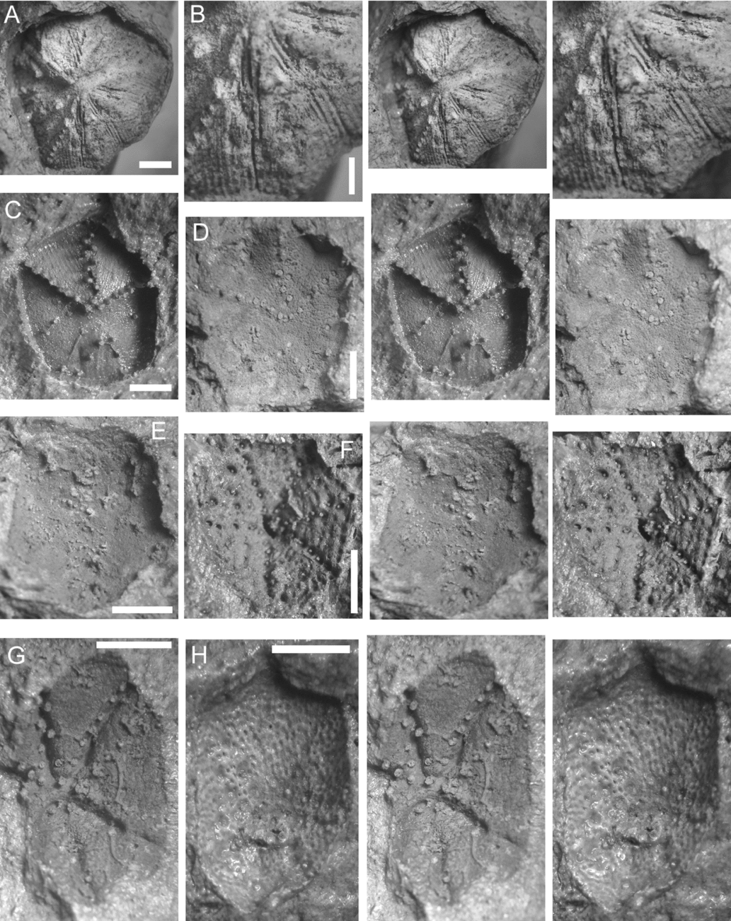
Description. Theca typically conical or elongate conical (Fig. 9A, B), but of variable shape. Small specimens are more globular (e.g. Fig. 10C), larger ones cylindrical, pyriform or elongate conical (Fig. 10), reaching up to 20 mm high by 13 mm maximum diameter. Composed of four basals, six elongate infra-laterals, eight laterals and at least 10 or 11 ‘orals’ (Fig. 8). Three ambulacra on the flat oral surface, each terminating in a single, relatively large facet (Figs 7B, D, 9C, D). Periproct above the lateral circlet on oral surface, supposed hydropore between the subcentral mouth and periproct (Figs 7C, 9B). Attached by a stem with circular columnals bearing radial crenellae.
Stereophotos of latex casts of thecae of Paracaryocrinites kochi (Reference WolfartWolfart, 2001c). (A–C) RGM 792508b. (A) Anterior lateral view centred on plate IL4 to show elongate infra-lateral plates (cf. Fig. 8). (B) Right lateral view to show conical thecal shape and large periproct (upper left) with prominent cover. (C) Oral surface with periproct below to show three radiating food grooves with central cover plates preserved and each ending in a single, relatively large, ambulacral facet (cf. Fig. 6B). The prominent tubercle over the periproct may be the position of the hydropore. (D) RGM 792508c, oral surface of a theca crushed in an anterior–posterior direction showing the three food grooves ending in single large facets. Black scale bar = 2 mm (A, B); white scale bars = 1 mm (C, D). All figures whitened with ammonium chloride.

Plates arranged in the standard pattern for a species of Paracaryocrinites (Fig. 8), relatively tall, especially infra-laterals where the height-to-width ratio can exceed 2.0. With variable external ornament, some finely granular (Fig. 10A), others with thick, smoother plates with deep sutures (Fig. 10B, D, G, I). On the largest example (and others), plates are very thick with deep sutures (Fig. 10G). Basals 2 and 3 may have a vertical, median ridge.
Stereophotos of Paracaryocrinites to show variation in thecal shape and ornament. (A) RGM 792474, ovoid theca with granular ornament and shallow sutures. (B) RGM 792476, cylindrical theca with smooth plates separated by deeper sutures. (C) RGM 792475, small, conical theca. (D, F, H) RGM 792477. (D, F) Part and counterpart of pyriform theca with deeper sutures. (H) Part of the oral surface showing one arm facet and uncovered food groove. (E) RGM 792478, small globular theca. (G) RGM 792465a, part of the largest theca to show extremely thick plates with very deep sutures. (I) RGM 792473, cylindrical theca with deep sutures and one arm facet. Scale bars = 2 mm. All latex moulds whitened with ammonium chloride.

Cryptorhombs present across all B:IL and IL:L sutures, and usually fully developed, uncommon isolated dichopores across IL:IL and more rarely L:L sutures. As far as is known, all pores in BB and LL are sieve pores, whereas all pores in Bockelie's (Reference Bockelie1979) systems 1 and 2 within ILL plates are simple. The pattern of sieve and simple pores in cryptorhombs of Bockelie's series 4 and 5 is not well enough known to make any generalizations.
Oral surface preserved in part on several examples, and known from both internal and external moulds. The mouth appears to have been surrounded by single plates in the AB and EA interrays, and additional inter-radial plates in the CD inter-ray, plus the three pairs of adradial facetal plates (Figs 7A, C, 9C, D). Three food grooves radiated out from the mouth and terminated in a single, relatively large, elongate facet developed on a pair of facetal plates. The food grooves were covered with a single series of small, alternating cover plates, of which the posterior is the largest (Fig. 9C). The ambulacral structures that attached to the facets have not been preserved, but were presumably arms as in Caryocrinites s.s. (Sprinkle, Reference Sprinkle1975; Lanc, McDermott & Paul, Reference Lanc, McDermott and Paul2015). Subcentrally in the facets is a nerve canal. Facets are separated by a single large, inter-radial plate, except in the posterior inter-radius, where there may be two or three (Figs 7B, D). Small, additional plates may also occur on the oral surface.
Periproct with a large, prominent aperture surrounded by four plates enclosing an oval anal opening 1.0 mm across in RGM 792508b (Figs 7C, 9B). Cover plates missing. Apparently much less conspicuous in other examples.
Gonopore not detected.
A small structure between the mouth and anus may be the hydropore, by analogy with Frest's (Reference Frest1975) interpretation of the position of the hydropore in Silurian species of Caryocrinites.
Remarks. Among other species of caryocrinitids, only Juglandocrinus elongatus (Jaekel, Reference Jaekel1899) has such an elongate theca. However, J. elongatus is much larger (70 mm high), spindle-shaped and lacks exposed ambulacral grooves on the oral surface. Furthermore, plates of both the basal and lateral circlets are taller than those of the infra-lateral circlet (see Jaekel, Reference Jaekel1899, pl. 17, fig. 4). In contrast, Paracaryocrinites kochi is sometimes widest at the oral surface, and the infra-lateral plates are distinctly taller than either the basals or laterals (Figs 8, 9A). Indeed, no other described caryocrinitid has such elongate infra-lateral plates.
The oral area is not well exposed in most examples, but we believe sufficient details can be seen to confirm this species belongs to Paracaryocrinites Chen & Yao. Thecal shape and plate ornament vary considerably in our material (Figs 9A, B, 10), and it is not certain that all specimens belong to a single species. Isolated plates show even more variation (see below). Wolfart (Reference Wolfart2001c) described two species based on internal moulds, of which the second, ‘Caryocrinites’ jaekeli, was characterized by unusual cryptorhombs with intermediate pores. His description and illustrations (especially pl. 10, figs 2, 3, and fig. 5k, l, therein) suggest the genus Polycosmites, which was hitherto only known from an isolated plate from Bohemia (Jaekel, Reference Jaekel1918), but has recently been reported from the Lower Ordovician (Floian) of China (Paul et al. Reference Paul, Donovan, Muir, Botting, Lin and Yuandong2016). None of our articulated material bears these unusual cryptorhombs, but we do have isolated plates with intermediate pores (described below).
‘Paracaryocrinites’ sp.
Figure 11
Reference Wolfart2001c ‘Caryocrinites’ sp. aff. ‘C.’ rugatus (Forbes, Reference Forbes1848); Wolfart, p. 204, fig. 4, pl. 10, fig. 4.
Reference Wolfart2001c ‘Caryocrinites’ sp.; Wolfart, fig. 5a–j (non-fig. 5k, l = Polycosmites sp. of Jaekel, Reference Jaekel1918).
Material. Many isolated thecal plates preserved as both internal and external moulds.
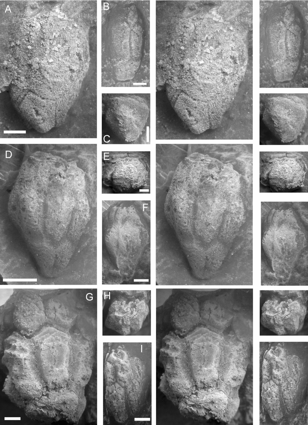
Description. Isolated thecal plates are common and range up to 10 mm by 12 mm (Fig. 11A). Plate type (basal, infra-lateral, lateral) and orientation (adoral–aboral axis) can be determined from plate outline (see Fig. 8) and arrangement of cryptorhombs. Basals may show the outline of the stem facet, are pentagonal (B2, B3) or hexagonal (B1, B4) and only have cryptorhombs developed across the upper (adoral) two or three sutures. The growth centre of basals is often close to the lower margin. Similarly, laterals are quadrilateral (L2, L5) or pentagonal (all others) and only have complete cryptorhombs across their lower one or two sutures. Growth centres of lateral plates are usually very close to the upper margins. Infralaterals may be pentagonal (IL1, IL5; Fig. 11C–E), hexagonal (IL3, IL6; Fig. 11G, H) or heptagonal (IL2, IL4; Fig. 11A, F). They tend to have complete cryptorhombs across both their upper and lower sutures (series 1 and 2 of Bockelie, Reference Bockelie1979) and may have incomplete cryptorhombs consisting of a few isolated dichopores across their lateral (IL:IL) sutures (Bockelie's series 4; Fig. 11C, D).
Stereophotos of natural moulds of plates of ‘Paracaryocrinites’ sp. (A, B) RGM 792466. (A) Complete internal mould of IL2 or IL4 showing fully developed cryptorhombs across all sutures. (B) Detail of two cryptorhombs to show that the dichopores penetrated well into the thecal cavity (above). (C) RGM 792470, internal mould of IL1 or IL5 showing two complete cryptorhombs above and one below, plus casts of two perpendicular canals on the left and another on the right. (D) RGM 702472, external mould of IL1 or IL5 with a very similar arrangement of cryptorhombs. Note a single sieve pore (left centre) and the facet-like surfaces to the plate above. Simple pores of the upper cryptorhombs opened in shallow channels on the plate surface. A few simple pores of the lower cryptorhomb can be seen below. (E) RGM 792469, a smooth, tumid lateral plate with obvious sieve pores. Casts of sieve canals can be seen coalescing in two examples (below). (F) RGM 792467, a vaulted and highly ornamented heptagonal plate (IL2 or IL4) with simple cryptorhomb pores in the crests of ridges running to the corners of the plate. (G) RGM 792468, a similar vaulted hexagonal plate (IL3 or IL6) with simple cryptorhomb pores beside strong radial ridges. (H) RGM 792471, a granular hexagonal plate (IL3 or IL6) with inconspicuous cryptorhomb pores. Orientation of (E) uncertain; all others orientated with the oral surface above. Scale bars = 2 mm except for (B), where the scale bar = 1 mm.

Cryptorhombs are most easily seen on internal moulds and may occur across all sutures (e.g. Fig. 11A), or only across the upper and lower sutures (Fig. 11C, D). So far it has not been possible to determine whether this correlates with other characters and so its taxonomic significance is uncertain. Cryptorhomb pores of Bockelie's series 1 and 2 are sieve pores in basal and lateral plates (e.g. Fig. 11E), but simple pores in infra-lateral plates (Fig. 11D, F, G).
Internal moulds commonly preserve casts of the perpendicular canals of the dichopores (Fig. 11C, E) and invariably traces of the tangential canals (Fig. 11A–C). Plates are usually thickened internally in the areas between the cryptorhombs during growth (e.g. Fig. 11A). This enables the number and completeness of the rhombs to be determined.
External moulds reveal the plate ornament, which varies from smooth (Fig. 11D, E) to granular (Fig. 11H). In addition, some isolated plates have strongly developed radial ridges with cryptorhomb pores within or beside them (Fig. 11F, G). So far we have found no complete or partial thecae with this ornament.
Remarks. Little can be said about these isolated thecal plates other than that they clearly bear cryptorhombs and therefore originate from hemicosmitoid cystoids sensu lato. The range of external ornament is quite broad, but there is insufficient evidence to be sure that more than one species is represented among the material. A few plates had cryptorhombs across all sutures, as Dreyfus (Reference Dreyfus1939) described for Oocystis, but most have incomplete rhombs across intra-circlet sutures or even no dichopores at all across these sutures. This may reflect the small size of the specimens. One would expect the respiratory requirements to increase with growth and so intra-circlet rhombs may well develop later in life. At present it seems best to group all these plates together as ‘Paracaryocrinites’ sp.
Although the name has been widely quoted in the literature (see Paul, Reference Paul1984, p. 148; Lanc, McDermott & Paul, Reference Lanc, McDermott and Paul2015), Forbes (Reference Forbes1848) never used the name Hemicosmites rugatus. However, ‘Hemicosmites rugatus Forbes’ was listed as a nomen nudum by Phillips & Salter (Reference Phillips and Salter1848, p. 302).
Genus Polycosmites Jaekel, Reference Jaekel1918
Figure 12
Diagnosis. A genus of Caryocrinitidae with multidisjunct cryptorhombs with a complex arrangement of perpendicular canals forming a rhomb-in-rhomb structure. Primary thecal plates arranged as in the Caryocrinitidae. Oral area unknown.
Remarks. Jaekel (Reference Jaekel1918, p. 98) associated his two monotypic new families, Polycosmitidae and Stichocystidae, in a suborder because he thought both families lacked calcified tangential canals connecting the abundant perpendicular canals that formed the complex rhombs. Kesling (Reference Kesling and Moore1968, p. S227) accepted Jaekel's views and formally erected a superfamily Polycosmitida. However, Bockelie (Reference Bockelie1981) thoroughly redescribed Stichocystis on the basis of more extensive and better-preserved material. He showed that Stichocystis had multiple tangential canals within delicate external, raised ridges that were seldom preserved undamaged, and that the rhombs were a specialized form of humatirhomb. Bockelie also showed that all other morphological features of Stichocystis, especially the plating around the oral area and the relative positions of the peristome, periproct and gonopore, were typical of caryocystitid fistuliporites. He therefore abandoned the family Stichocystidae and transferred Stichocystis to the family Caryocystitidae.
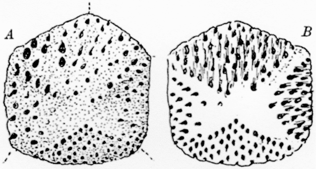
Hitherto, Polycosmites was only known from an isolated, pentagonal, infra-lateral plate from Vosek in Bohemia, illustrated by Jaekel (Reference Jaekel1918, fig, 90, p. 98, reproduced herein as Fig. 12). This showed multiple pores over both external and internal surfaces. The pores are more obviously arranged in rhombs on the internal surface, which also shows traces of the connecting internal tangential canals. The complex arrangement of pores in Polycosmites is analogous to the arrangement of slits in multidisjunct pectinirhombs and we propose to refer to the rhombs in Polycosmites as multidisjunct cryptorhombs.
Polycosmites bohemicus Jaekel, Reference Jaekel1918. Copy of the original diagram (Jaekel, Reference Jaekel1918, fig. 90).

Although Polycosmites was only known from isolated plates, Jaekel (Reference Jaekel1918, p. 98) suggested that it had a plate arrangement similar to that of hemicosmitoids. Recently, Wolfart (Reference Wolfart2001c, pl. 10, figs 2, 3) illustrated the holotype of his new species ‘Caryocrinites’ jaekeli, which has multidisjunct cryptorhombs, and a theca with four basals, six infra-laterals and eight laterals, as in Caryocrinites. Thus, it seems reasonable to abandon the family Polycosmitidae and transfer the type genus, Polycosmites, to the Caryocrinitidae. Wolfart had only internal moulds, and the plating around the oral area of Polycosmites jaekeli remains unknown.
Among the material we have are one matching internal and external mould, plus two other internal moulds of isolated plates showing multidisjunct cryptorhombs. The matching external mould has a distinctive surface ornament and we have three other external moulds of isolated plates with similar ornament that may belong to this species. Paul et al. (Reference Paul, Donovan, Muir, Botting, Lin and Yuandong2016, p. 148, fig. 4) described and illustrated a single, probable lateral plate of Polycosmites from the early Ordovician (Floian) of Tonggau, Yunnan Province, China.
Polycosmites sp. cf. P. jaekeli Wolfart, Reference Wolfart2001c
Figure 13
?Reference Wolfart2001c ‘Caryocrinites’ jaekeli n. sp., Wolfart; pp. 200, 201, fig. 3, pl. 10, figs 1–3; pl. 11, figs 1–3.
Reference Wolfart2001c ‘Caryocrinites’ (pars); Wolfart, fig. 5k, l (non- fig. 5a–j = Paracaryocrinites sp.).
Material. Natural internal and external moulds of six complete or partial isolated plates showing the characteristic rhomb structure or surface ornament (RGM 792486–792493) (Fig. 13).
Stereophotos of Polycosmites sp. (A-C) RGM 792490a, b, part of an isolated thecal plate. (A) Internal mould to show multiple perpendicular canals in three half-rhombs. (B) External mould to show irregular thecal ornament and inconspicuous pores. (C) Latex of the external mould. (D) RGM 792492, latex of an external mould of another partial plate with similar irregular thecal ornament. (E) RGM 792488, internal mould showing multiple perpendicular canals arranged in a rhomb-in-rhomb manner. Scale bars = 2 mm. (C) and (D) whitened with ammonium chloride.

Description. The most complete plate (Fig. 13E) is elongate, pentagonal in outline, 8.0 mm long by about 6 mm wide and has two half multidisjunct cryptorhombs confined to one end of the plate, but with evidence of other dichopores at the other end, too. It must be an infra-lateral plate, probably IL1 or IL5. Both the multidisjunct cryptorhombs show at least four parallel rows of pores, with up to nine in the longest row. The internal mould of the matching pair (Fig. 13A) shows three adjacent half multidisjunct cryptorhombs all of which have two rows of pores. The preserved part of the external mould is 6.3 mm wide and shows a distinctive irregular surface ornament (Fig. 13B, C). The third internal mould is rounded pentagonal in outline, 9.5 mm high by 8.3 mm wide, and bears three half multidisjunct cryptorhombs which have additional pores within the rhombs, but preservation is too poor to make out the details. In addition, two further external moulds show irregular surface ornament, which resembles that of the matching internal and external moulds, and are included here. One has irregular pitted ornament (Fig. 13D) and may be a lateral plate, but it is incompletely preserved.
Remarks. If Wolfart's ‘Caryocrinites’ jaekeli does possess multidisjunct cryptorhombs then the plate arrangement of Polycosmites is established and does not differ from that of other caryocrinitid genera, at least as far as the lowest three plate circlets are concerned. It is also a significant discovery, as hitherto the genus was only known from Bohemia and China. There is a potential taxonomic problem here as Sun (Reference Sun1936, p. 481, pl. 2, fig. 3) described and illustrated a cystoid as Hemicosmites jaekeli. Sun's description stated that this cystoid had nine lateral plates. However, he also stated the species was based on a single, incomplete natural external mould, raising doubts as to whether or not he directly observed the nine lateral plates. More importantly, his illustration shows two heptagonal infra-lateral plates separated by a hexagonal infra-lateral. These can only be IL2, 3 and 4 (see Fig. 8). However, in Hemicosmites all three of these infra-lateral plates are heptagonal in outline (see Bockelie, Reference Bockelie1979, text-fig. 4a, p. 371). This is because in Hemicosmites IL3 lies directly below L4, which is the lateral plate that is present in the Hemicosmitidae, but missing in the Caryocrinitidae. Thus, Sun's species belongs to ‘Caryocrinites’ s.l. and not to Hemicosmites, and becomes a senior synonym of ‘Caryocrinites’ jaekeli Wolfart, Reference Wolfart2001c. Sun's material all came from the Middle Ordovician of Kweichou (China) and his fauna shows considerable resemblance to that described from Thong Pha Phum. For example, both areas have yielded species of Sinocystis, ‘Caryocrinites’ s.l., Heliocrinites and Stichocystis. Sun's illustration is remarkably similar in general outline to Wolfart's specimens of his ‘C.’ jaekeli and it is distinctly possible that they are the same taxon. Unfortunately, Sun made no mention of the rhombs and only discussed the plating of the oral area in the most general terms. Thus, it is unwise to accept this synonymy at present.
Superfamily Aristocystitoida Neumayr, Reference Neumayr1889
Diagnosis. Directly attached diploporites with elongate oral area surrounded by at least eight plates (four inter-radial peri-orals [POO] and four radial circum-orals [COO]), and covered by a double biseries of cover plates; with two to five ambulacra bearing one to four brachioles each; with a large hydropore shared by plates PO1 and PO6, and often a spout-like gonopore usually within a single plate near the periproct; with thecal plates densely covered with diplopores that are commonly sealed externally by a thin epistereom and sometimes extended into spine-like projections.
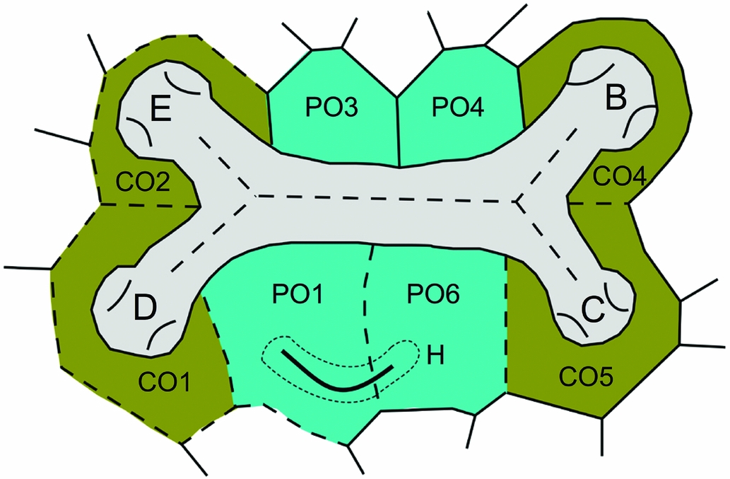
Remarks. This diagnosis is significantly different from preceding diagnoses of this superfamily (e.g. Kesling, Reference Kesling and Moore1968, p. S250), largely because the arrangement of plates in the oral area was not previously fully appreciated, but also because genera that would now be included in the family Holocystitidae were previously included within the Aristocystitidae. All aristocystitid genera where the oral plating and ambulacra are known have a mouth elongated at right angles to the oro-anal axis, and surrounded by eight plates interpreted by Paul (Reference Paul2017) as four central, peri-oral plates (PO1, PO2, PO4 and PO5) and four distal circum-oral plates (CO1, CO2, CO4 and CO5) (Fig. 14). In Aristocystites, only two, weakly developed, ambulacral facets occur, one at each end of the elongate mouth (see e.g. Parsley Reference Parsley1990, p. 284, fig. 6.6). Paul (Reference Paul2017, fig. 7.1) interpreted the two ambulacra as B and D on the grounds that the facets were developed on plates CO1 and CO4. In Calix Rouault, Reference Rouault1851, the ambulacra divide at each end and give rise to four ambulacral facets, one on each of the COO plates. In the type species, Calix sedgwicki Rouault, Reference Rouault1851, each facet gives rise to four long brachioles (see Chauvel, Reference Chauvel1977, pl. 1, fig. 6). Although numerous species of Calix have been described, as far as we are aware in no other species is the oral area known. Even so, we are inclined to restrict the genus Calix to include only those species which have what Chauvel (Reference Chauvel1977, p. 314) called ‘bouquets of brachioles’. The only other genus to share such ‘bouquets of brachioles’ is Oretanocalix Guttiérrez-Marco, Reference Gutiérrez-Marco2000, which differs from Calix s.s. in having all five ambulacra developed (Guttiérrez-Marco Reference Gutiérrez-Marco2000, pp. 83, 84, fig. 1e). Photographs kindly supplied by Guttiérrez-Marco suggest that Oretanocalix has an extra circum-oral plate (CO3) bearing the extra ambulacral facets in ambulacrum A. Sinocystis Reed, Reference Reed1917 (Fig. 14), Lepidocalix Termier & Termier, Reference Termier and Termier1950a, Glaphocystis Chauvel, Reference Chauvel1966, and the cystoid described by Prokop (Reference Prokop1965) as Hippocystis sculptus (Barrande, Reference Barrande1887) all have four ambulacral facets, from each of which it appears only a single brachiole arose. However, Guttiérrez-Marco (Reference Gutiérrez-Marco2000, p. 84, fig. 1h) illustrated the oral surface of ‘Calix’ inornata Meléndez, Reference Meléndez1958, with each of the four ambulacra divided to give a total of eight ambulacral facets. In addition, Chauvel (Reference Chauvel1978, p. 39, fig. 5b, pl. 3, fig. 1) has illustrated, under the name Phlyctocystis sp., another aristocystitid cystoid with four divided ambulacra, giving a total of eight facets; yet, in Phlyctocystis Chauvel, Reference Chauvel1966, the ambulacral branches are much longer. Without examining the specimens, we are uncertain whether or not these two aristocystitids with eight facets could usefully be combined in a single genus. If so, Phlyctocystis Chauvel, Reference Chauvel1966, would be a suitable name for it. As the name implies, ‘Calix’ inornata has a smooth external surface, whereas Phlyctocystis has many tumid plates with deep sutures that are often ornamented with tubercles. Finally, the unique Silurian genus Triamara Tillman, Reference Tillman1967, has three ambulacral grooves, each of which ends in a double facet. It also has a much smaller mouth, almost certainly surrounded by only the four peri-oral plates. The oral area is unknown in other aristocystitid genera.
Ambulacra and oral plating in Sinocystis Reed, Reference Reed1917. In aristocystitids the mouth (not shown) lies within a broad food groove, which extends laterally left and right. The mouth is surrounded by four, central, peri-oral plates (PO1, PO3, PO4 and PO6), plus four more distal circum-oral plates (CO1, CO2, CO4 and CO5). In Sinocystis both lateral food grooves divide once to give four large facets, each of which lies within a single circum-oral plate and apparently gave rise to a single ambulacral appendage. The hydropore (H) lies across the PO1:PO6 suture. Key: B, C, D and E = ambulacra B–E. Redrawn from Reference BatherBather (1918b, fig. 9, p. 535).

As currently defined, the superfamily Aristocystitoida contains one family, the Aristocystitidae. Termier & Termier (Reference Termier and Termier1950b) recognized a family Calixidae (sic), and Chauvel (Reference Chauvel1966) the subfamilies Hippocystinae and Calicinae, based on the form of the diplopores and the number of ambulacral facets. These subfamilies have not been generally accepted, although Makhlouf et al. (in press) have recognized the Calicinae.
Family Aristocystitidae Neumayr, Reference Neumayr1889
Diagnosis. As for the superfamily.
Remarks. Aristocystitids are characterized by a mouth set within a wide ambulacral tract composed of two ambulacral grooves, which extend from the mouth, may or may not divide, and terminate in large ambulacral facets. Paul (Reference Paul2017, fig. 7) has interpreted the plating around the mouth in several genera with two or four ambulacra (Aristocystites Barrande, Reference Barrande1887; Calix Rouault, Reference Rouault1851; Glaphocystis Chauvel, Reference Chauvel1966; Lepidocalix Termier & Termier, Reference Termier and Termier1950a; Sinocystis Reed, Reference Reed1917) as consisting of four radial ‘circum-oral’ plates bearing the ambulacral facets and four inter-radial ‘peri-oral’ plates. This pattern of oral plating also occurs in ‘Hippocystis sculptus’ as described by Prokop (Reference Prokop1965), although Hippocystis Bather is a junior synonym of Aristocystites Barrande, Reference Barrande1887. The material described by Prokop needs a new generic name (Paul, research in progress). Makhlouf et al. (in press) have accepted this interpretation of the oral plating in Lepidocalix Termier & Termier.
The ambulacral structures that arose from the facets are poorly known. However, Chauvel (Reference Chauvel1977, figs a, b, pl. 1, figs 1, 5, 6) illustrated the oral area and ambulacral structures in Calyx sedgwicki Rouault, Reference Rouault1851, type species of Calix Rouault, Reference Rouault1851, in which four biserial brachioles arose from a single large ambulacral facet. It is quite likely that the details of the ambulacral system in aristocystitids may be used to characterize subfamilies, but the details are unknown in too many nominal genera to be sure how consistent they are. Nevertheless, Aristocystites Barrande, Reference Barrande1887, is characterized by two unbranched ambulacra ending in single facets, although Kesling (Reference Kesling and Moore1968, p. S251, fig. 148.1i) illustrated an abnormal example with three facets (copied from Jaekel, Reference Jaekel1899, pl. 3, fig. 11). Sinocystis Reed, Reference Reed1917, Lepidocystis Termier & Termier, Reference Termier and Termier1950a, Glaphocystis Chauvel, Reference Chauvel1966, and the ‘Hippocystis’ of Prokop (Reference Prokop1965) are characterized by two ambulacra that branch once, and end in single facets, giving a total of four. The facets appear to have given rise to a single brachiole each in Glaphocystis (see Chauvel, Reference Chauvel1966, pl. 4, fig. 1e, pl. 9, fig. 2). In the Silurian genus Triamara Tillman, Reference Tillman1967, the left ambulacrum branches once, but the right is unbranched, resulting in three food grooves, each of which terminates in a pair of ambulacral facets, giving a total of six. In Phlyctocystis Chauvel, Reference Chauvel1966, both ambulacra divide twice, giving a total of eight facets (see Chauvel, Reference Chauvel1978, fig. 5b). Finally, Gutiérrez-Marco (Reference Gutiérrez-Marco2000) has reinterpreted ‘Echinosphaerites’ murchisoni Verneuil & Barrande, Reference Verneuil and Barrande1855, as an aristocystitid and attributed it to a new genus Oretanocalix Gutiérrez-Marco, Reference Gutiérrez-Marco2000. Oretanocalix, which is probably the least derived aristocystitid genus, has five ambulacra, although ambulacrum A is reduced compared with the other four. It also appears to have the compound facets that gave rise to more than two brachioles that typify Calix s.s.
Finally, as currently understood (e.g. Gutiérrez-Marco & Colmenar, Reference Gutiérrez-Marco, Colmenar, Gutiérrez-Marco, Rabáno and Garcia-Bellido2011), the genus Calix includes species with distinctly different oral areas. The type species, C. sedgwicki, has both food grooves divided once, giving a total of four large ambulacral facets, but all four facets are compound and give rise to several brachioles; four in the one ambulacrum where the brachioles are preserved (Chauvel, Reference Chauvel1977, pl. 1, figs 1, 6). In ‘Calix’ inornata Meléndez, Reference Meléndez1958, the food grooves divide once, but each ends in a pair of facets, giving a total of eight, although each pair appears to be developed on a single circum-oral plate.
In all the other genera attributed to the Aristocystitidae, namely Baculocystites Barrande, Reference Barrande1887, Batalleria Chauvel & Meléndez, Reference Chauvel and Meléndez1978, Maghrebocystis Chauvel, Reference Chauvel1966, and Pachycalix Chauvel, Reference Chauvel1941, details of the oral area remain unknown. ‘Aristocystis’ sinica Sun, Reference Sun1936, was originally based on an internal mould. Later, Sun (Reference Sun1948, pl. 1, fig. 1b) illustrated a petaloid oral area with five ambulacra, thus demonstrating that his species is probably not an aristocystitid.
Material we have from Thailand has both tubercular and sunken diplopores, as well as inconspicuous ambulacral facets beneath which are numerous thecal canals. These are all features mentioned by Reed (Reference Reed1917) and Reference BatherBather (1918a, b) in describing Sinocystis from the Ordovician of Yunnan.
Genus Sinocystis Reed, Reference Reed1917
Diagnosis. A genus of Aristocystitidae with a large, globular theca, four ambulacral facets, thecal plates of several generations all bearing conspicuous diplopores which may open externally in tubercles or sunken peripores in the same individual.
Sinocystis sp. cf. Sinocystis loczyi Reed, Reference Reed1917
Figures 14, 15
Material. At least five partial thecae and several fragments composed of a few associated plates (Fig. 15).
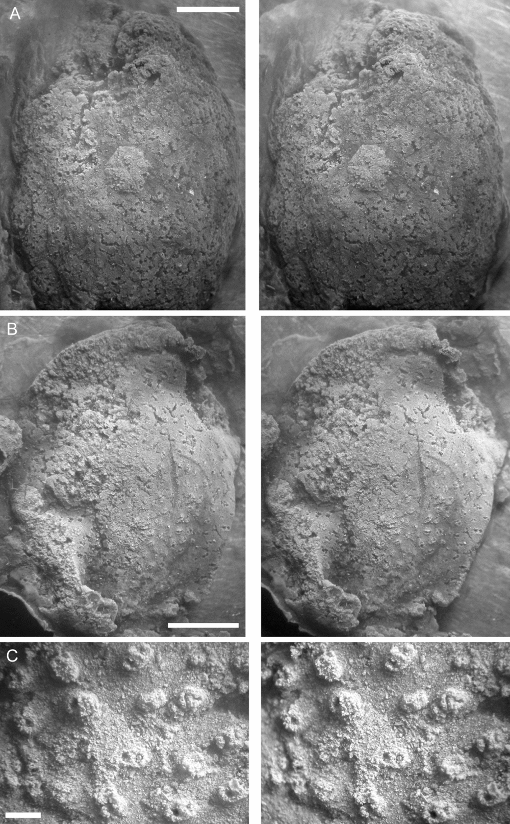
Stereophotos of latex casts of Sinocystis. (A) RGM 792461, an ovoid theca showing the peripropct (centre) and poorly preserved oral area (above). Note the sunken diplopores (below). (B, C) RGM 792462. (B) A crushed theca showing some diplopores in raised tubercles. (C) Detail of tubercular diplopores. Scale bars = 5 mm (A, B) and 0.5 mm (C). All whitened with ammonium chloride.

Description. Partial thecae reach at least 30 mm by 22 mm, and were originally ovoid and much larger. They are composed of numerous small plates, which reach about 7 mm in maximum dimension and almost all of which bear diplopores. The diplopores may be raised in distinct tubercles (Fig. 15B, C) or sunken into the theca (Fig. 15A). Both types are preserved in the same theca and the difference may be preservational to some extent. All the plates appear to have been densely penetrated by randomly arranged pore canals.
The oral area appears to be present in RGM 792461 (Fig. 15A, top), but is poorly preserved. Two eroded mounds to the right of an irregular, elongate slit probably represent two of the ambulacral facets at the right end of the ambulacral groove. However, no clear mouth nor articulation surfaces can be discerned. Below the supposed ambulacral groove is another narrower irregular slit, which is in the right position to represent the hydropore. The periproct can also be seen in RGM 792461 (Fig. 15A, centre). It lies about a third of the way down the theca, is 4 mm in diameter, hexagonal and covered by six triangular plates.
Remarks. Despite having several near-complete thecae that preserve the surface ornament to varying degrees, only one specimen reveals anything about the oral area, and the details are poorly preserved. If the mounds referred to above are indeed remains of ambulacral facets, then the diplopore canals are very densely packed below them. Equally, there is nothing in the associated irregular groove to confirm that it does represent an ambulacrum. Only the periproct is clearly seen, and the other structures are interpreted by comparison with better-preserved examples of Sinocystis from Yunnan.
5. Comments on Wolfart's (Reference Wolfart2001c) other pelmatozoan taxa
Stichocystis thailandica Wolfart, Reference Wolfart2001c. This is a genuine Stichocystis, but the illustrations (especially fig. 7a, b) are upside down. The mouth lies at the summit of the thecal elongation, which is not a stem-like aboral extension. Although this thecal shape is unknown in the type species (see Bockelie, Reference Bockelie1981), it is not uncommon in other caryocystitid cystoids.
Heliocrinites sp. aff. H. qualus Bather, Reference Bather and Reed1906. It is likely that this species should be attributed to the genus Lophotocystis Paul, Reference Paul1972b.
Gomphocystites? sp. indet. The illustrations are quite unlike Gomphocystites and I suspect this is an incompletely preserved cephalon of a lichid trilobite. Reed (Reference Reed1917, pl. 8, figs 8, 9) illustrated two species of Lichas (Metopolichas) from Yunnan, which resemble Wolfart's illustrations.
Codiacystis sp. aff. C. bohemicus (Barrande, Reference Barrande1887). This, too, is not an echinoderm, but probably a bryozoan.
Aristocystis [sic] sp. A of Paul, Reference Paul1973. This is probably an aristocystitid cystoid, but little else can be said about it. It does not resemble the material of Sinocystis that we have because the diplopores are too sparsely developed. Bather (Reference Bather and Reed1906, p. 8, pl. 2, figs. 1–5) described and illustrated ‘Aristocystis’ dagon from the Ordovician of Sedaw, ‘Burma’, which may well be a genuine Aristocystites. It is possible Wolfart's specimen may be another example.
Incertae sedis sp. C of Paul, Reference Paul1973. Wolfart's illustrations (2013c, pl. 13, figs 7, 8) appear to show an external mould. If this interpretation is correct, the surface bears circular pits with a raised centre excavated into the external surface. This does not resemble the diploporite ‘Incertae sedis sp. C’ of Paul, Reference Paul1973, which had diplopores developed in raised tubercles. It is uncertain that this is an echinoderm.
6. Discussion
The study area is part of the Sibumasu terrane, an early Palaeozoic palaeocontinent, which included Sumatra, Malaysia, West Thailand and Burma (Fortey & Cocks, Reference Fortey, Cocks, Hall and Holloway1998). During the Ordovician, this terrane was marginal to northern Gondwana and equatorial (Torsvik & Cocks, Reference Torsvik, Cocks, Harper and Servais2013). As recently as Paul's global analysis (1976, text-fig. 3, upper), there were no Ordovician echinoderm localities recorded from Thailand. Unfortunately, there is no change in the latest major review of the distribution of the Ordovician echinoderms (Lefebvre et al. 2013), in which the orientation of all reconstructions hides the Sibumasu terrane in the concealed hemisphere of the Ordovician globe (compare various figures therein with Torsvik & Cocks, Reference Torsvik, Cocks, Harper and Servais2013, fig. 2.13).
The Ordovician echinoderms of Thailand remain poorly known, yet it is apparent that disarticulated remains, at least, are locally common. For example, Fortey (Reference Fortey1997, p. 398) noted ‘massive pelmatozoan limestones’ in the lower part (Caradoc) of the Pa Kae Formation of southern Thailand. Similarly, Agematsu et al. (Reference Agematsu, Sashida, Salyapongse and Sardsud2006) recorded crinoid debris from all three sections that they measured in the Thong Pha Phum district of western Thailand. Other passing comments doubtless exist in the literature, demonstrating the presence of echinoderm debris, but have done little to progress its identification.
Wolfart (Reference Wolfart2001c) provided the first detailed analysis of some of the Middle – early Late Ordovician pelmatozoans of the region; the present contribution both expands on and revises this pioneering study. But these are only pinpricks in a broad region awaiting a detailed monographic analysis. Eleutherozoans and Lower Ordovician echinoderms remain unknown; the Late Ordovician has been inadequately sampled (Wolfart, Reference Wolfart2001c); and the relatively well-known echinoderms of the Middle Ordovician have been the subject of only two papers. As noted by one of our reviewers, ‘If this fauna had been found in the U.S, or the U.K., it might not have been worthy of description. Because it is from Thailand, it needs to be documented.’
The echinoderms described by Wolfart (Reference Wolfart2001c) from the Thong Pha Phum region closely resemble those described above. We have followed the methodology of Paul et al. (Reference Paul, Donovan, Muir, Botting, Lin and Yuandong2016) in examining thecae and thecal plates separately from columnals, while combining these data in examining the complete echinoderm fauna. This compilation (Table 1) provides broad agreement that glyptocystitoids and hemicosmitoids are present, although it is the details of the thecal plates that have enabled division into separate species, even if most are left in open nomenclature. Crinoid thecal plates(?) were identified in only the broadest sense, but the better-preserved crinoid columnals were identifiable to morphogenus, namely Bystrowicrinus (col.) sp. As recognized by Paul et al. (Reference Paul, Donovan, Muir, Botting, Lin and Yuandong2016, p. 154), the information provided by the thecae, thecal plates and columnals are essential components for determining the most complete estimate of generic diversity.
The mixture of rare thecae and common disarticulated ossicles is a common association of Ordovician echinoderm assemblages. The absence of trilobites, a coeval group which also has a multi-element calcareous skeleton that would provide a supporting biostratinomic signal, may be significant (Speyer, Reference Speyer and Donovan1991). The lack of even disarticulated fragments of trilobites from a fauna with abundant echinoderm ossicles is considered to indicate a true absence; that is, trilobites were not part of the Darriwilian fauna of Bang Mueang Song Tho. Further, the occurrence of rare articulated echinoderm endoskeletons – thecae, pluricolumnals – suggests minimal transport, and the common disarticulated ossicles are the subject of slow breakdown of unmineralized connecting tissues following death (Donovan, Reference Donovan and Donovan1991); that is, the echinoderm assemblage may be parautochthonous.
Not only are records of Ordovician echinoderms from SE Asia rare, but so are geographically intermediate faunas that might provide links with the better-known faunas of Europe and North America. Recently, however, a few intermediate Ordovician faunas have been recorded. Lefebvre, Ghobadipou & Nardin (Reference Lefebvre, Ghobadipou and Nardin2005) described two Ordovician echinoderms faunas from Iran, and Rozhnov, Minjin & Kushlina (Reference Rozhnov, Minjin and Kushlina2009) two fistuliporite genera from Mongolia, while Nardin, Göncüoğlu & Lefebvre (Reference Nardin, Göncüoğlu and Lefebvre2014) have recorded Ordovician diploporites (probably sphaeronitids) from Turkey. The new discoveries have greatly increased the ranges of some genera. The Thai cystoid fauna includes representatives of the families Cheirocrinidae, Caryocrinitidae, Caryocystitidae and Aristocystitidae. The affinities of the fauna are closest to those of other cystoid faunas from SE Asia (Bather, Reference Bather and Reed1906; Reed, Reference Reed1917; Sun, Reference Sun1936; Paul et al. Reference Paul, Donovan, Muir, Botting, Lin and Yuandong2016), but there is some evidence of links to other Ordovician faunas. The Cheirocrinidae are largely cosmopolitan (Paul, Reference Paul and Bassett1976). Even the single identified genus, Cheirocystella, is known from North America, Baltica and southern Europe. Among the caryocrinitids, Paracaryocrinites has been recorded from a single example in Baltica (Lanc, McDermott & Paul, Reference Lanc, McDermott and Paul2015), and Polycosmites was originally described from the Czech Republic (Jaekel, Reference Jaekel1918). Among the Caryocystitidae, ‘Heliocrinites’ is almost as cosmopolitan as the Cheirocrinidae, but Stichocystis is more restricted. Paul (Reference Paul and Bassett1976, p. 557) was cautious about accepting Sun's (1936) record of Stichocystis from China. The figure is a coarse halftone photograph in which it is difficult to be certain that the characteristic rows of pores are present. However, with the more recent reports of undoubted Stichocystis from Thailand (Wolfart, Reference Wolfart2001c) and Mongolia (Rozhnov, Minjin & Kushlina, Reference Rozhnov, Minjin and Kushlina2009), it is now known that the genus occurs quite widely in the Far East. Chauvel (in Chauvel & Le Menn, Reference Chauvel and Le Menn1979) also recorded Stichocystis from the ‘Ashgillian’ of Fombuena, Spain. Finally, Sinocystis is known from Iran (Gansser & Huber, Reference Gansser and Huber1962; Lefebvre, Ghobadipou & Nardin, Reference Lefebvre, Ghobadipou and Nardin2005) as well as SE Asia. These few additional records do not give a clear picture of the affinities of the Thai Ordovician cystoid fauna, except perhaps that so far it has little in common with the North American fauna and maybe more affinities with southern Europe than Baltica.
Acknowledgements
We thank Rong Jia-yu and Zhan Ren-bin for their identifications of the Middle Ordovician brachiopods. We devote this paper to the memory of the late Terry Frest, who was originally intended to identify the echinoderms documented herein. The considered comments of Professor Thomas E. Guensburg (Rock Valley College, Illinois) and a second, anonymous reviewer are gratefully acknowledged.