Anomalous aortic origin of a coronary artery that courses between the two great vessels is a rare congenital anomaly with increased risk of sudden cardiac death in children and young adults.Reference Basso, Maron, Corrado and Thiene1, Reference Taylor, Byers, Cheitlin and Virmani2 Marfan syndrome is a connective tissue disorder that carries a risk of sudden death that is due to progressive aortic root dilation and subsequent aortic dissection or rupture.Reference Pyeritz3 We present a case of two brothers with Marfan syndrome who were serendipitously found to have anomalous aortic origin of a coronary artery with an inter-arterial and intramural course. Even more uniquely, they have the opposite coronary anomaly: one with anomalous left coronary artery from the right sinus of Valsalva and the other with anomalous right coronary from the left sinus. To our knowledge, anomalous aortic origin of a coronary artery and Marfan syndrome together have been reported rarely,Reference Matsuda, Ichikawa, Iwai and Takahashi4, Reference Papadopoulos, Moyssakis and Votteas5 but siblings with anomalous coronary artery and Marfan syndrome have never been documented.
Case report
We report a case of two brothers, aged 7 and 9 years, respectively, of Haitian descent with Marfan syndrome who were referred to our institution for a second opinion regarding management options for the complex findings of anomalous aortic origin of a coronary artery and Marfan syndrome. The brothers were diagnosed with Marfan syndrome in 2002 after genetic testing showed the same mutation on the fibrillin-1 gene as their father who also has Marfan syndrome. Both were asymptomatic from a cardiovascular perspective and were exercise-restricted. At the time of diagnosis, the 7-year-old had an aortic root z score of greater than 3 and qualified for enrolment in a national Marfan trial sponsored by the National Institutes of Health comparing atenolol (selective beta-1 receptor antagonist) to losartan (angiotensin II receptor antagonist);Reference Lacro, Dietz and Wruck6 the 9-year-old boy did not meet the criteria for trial inclusion and was maintained on atenolol 50 milligrams daily.
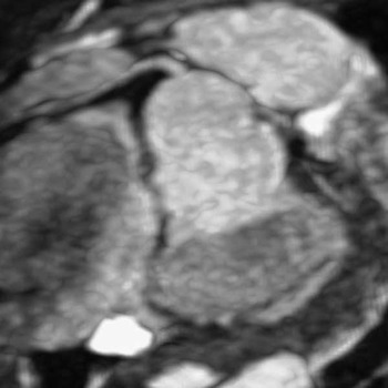
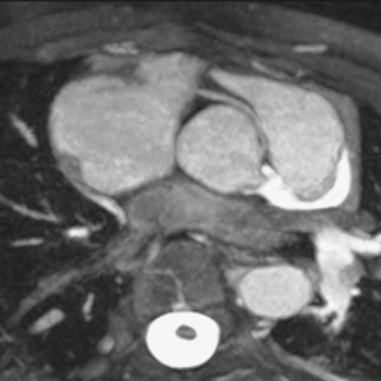
On physical examination, both boys had a Marfanoid body habitus: they were tall and thin and had arachnodactyly. The 9-year-old boy has bilateral lens subluxation. Cardiovascular examinations revealed a normal S1 and S2, and a systolic click but no murmurs. Electrocardiograms were normal. Both boys had recent echocardiograms. The 7-year-old boy had mild tricuspid and mitral valve prolapse without mitral insufficiency. Owing to height and weight gain, his aortic root at the sinuses of Valsalva now had a z score of 2.5, measuring 28 millimetres. The left coronary artery arose from the right sinus of Valsalva and coursed between the aorta and the pulmonary artery. The 9-year-old boy had mitral valve prolapse with mild mitral valve regurgitation. His aortic root at the sinuses of Valsalva measured 31 millimetres, or a z score of 1.6. The right coronary artery arose from the left sinus of Valsalva, coursing between the great vessels. Exercise stress tests with nuclear perfusion were normal without evidence of ischaemia or perfusion defects. The brothers also underwent cardiac magnetic resonance imaging to further delineate the coronary anatomy (Figs 1 and 2).
Magnetic resonance coronary imaging at a plane through the aorta at the level of the sinuses shows the anomalous left coronary artery originating immediately to the right of the inter-coronary commissure and coursing tangentially between the aorta and the pulmonary artery. The aortic root measures at the upper limits of normal.

Magnetic resonance coronary imaging at a plane through the aorta at the level of the sinuses shows the anomalous right coronary artery originating from the left sinus of Valsalva and coursing tangentially between the aorta and the pulmonary artery. The aortic root is mildly dilated.

Discussion
We report the first ever case of siblings with Marfan syndrome and anomalous aortic origin of a coronary artery. Marfan syndrome is known to have an autosomal dominant inheritance.Reference Judge and Dietz7 Despite there being no known inheritance pattern with anomalous aortic origin of a coronary artery, familial occurrences are reported.Reference Brothers, Stephens, Gaynor, Lorber, Vricella and Paridon8
Anomalous aortic origin of a coronary artery is a rare congenital anomaly with an estimated prevalence of 0.1–0.3%.Reference Davis, Cecchin, Jones and Portman9, Reference Yamanaka and Hobbs10 Sudden death most commonly occurs during or just after exercise in otherwise healthy children and young adults.Reference Basso, Maron, Corrado and Thiene1, Reference Taylor, Byers, Cheitlin and Virmani2 The mechanism of sudden death is thought to be related to one or more of the following: an acute angle of the coronary artery at the take-off from the aorta, presence of an ostial ridge, and/or a proximal intramural course that narrows as it courses between the great arteries or within the aortic wall. The increased cardiac output and aortic distension that occur during exercise more likely increase the risk of ischaemia. Most physicians agree that surgery is indicated if a patient with anomalous aortic origin of a coronary artery presents with signs and/or symptoms of myocardial ischaemia. Despite anomalous left coronary artery being less common than anomalous right, it appears to be a higher-risk lesion for sudden death; those with anomalous left coronary artery are usually recommended for surgical repair regardless of symptoms. Surgical repair for asymptomatic anomalous right coronary artery is more controversial.
These cases represent a unique management dilemma, especially for the boy with anomalous left coronary artery. Marfan syndrome is a connective tissue disorder with increased tissue friability, and therefore any surgery may confer a much higher risk of surgical complications. Thus, the risk-to-benefit ratio for operating to unroof the coronary artery in either boy is very unclear. To date, there is only one report of aortic root remodelling in an adolescent with Marfan syndrome and anomalous aortic origin of a coronary artery; however, the anomalous coronary artery was not intervened upon and only short-term results were reported.Reference Matsuda, Ichikawa, Iwai and Takahashi4
For these brothers we are advocating a non-surgical approach, even in the boy with anomalous left coronary artery. If we add the cumulative risk of performing open-heart surgery in an asymptomatic child with Marfan syndrome who does not need aortic root replacement, is restricted from exercise and is taking atenolol or losartan, we believe the non-surgical approach prevails over the risk of surgery. We cannot know the risk of the unroofing procedure in a child with a connective tissue disorder, and therefore we would expect it to be increased given their tissue friability. Certainly, if we are not going to operate, then aggressive medical management is necessary to prevent aortic root dilation; we do not know the risk of a dilated aorta and impingement on an already angulated anomalous coronary artery ostium.
For the reasons cited above, we recommended the following medical management: exercise restriction, regular echocardiographic evaluations to assess for aortic root dilation, periodic stress tests with nuclear perfusion to evaluate for ischaemia, and optimisation of medication to decrease progressive aortic root dilation. Certainly, if either boy becomes symptomatic or there is evidence of ischaemia, then surgery would be recommended. Further, if the aortic root dilates enough whereby aortic root replacement is needed, then an unroofing procedure may be warranted as part of this operation. This report shows the unique management dilemma of coexistent anomalous aortic origin of a coronary artery and Marfan syndrome and serves to remind us of the principal tenet of medicine: “first do no harm”.