Introduction
The black armoured catfish, Rhinelepis aspera, is an important commercial siluriform fish found in the São Francisco River, Brazil. The meat of this fish is firm, tasteful and almost completely lacks intramuscular bones and fats. Furthermore, it is the largest loricariid fish in the river basin reaching a body weight of up to 4 kg. The black armoured catfish also occurs in the Paraná River Basin. Despite being rare in the São Francisco River Basin, this catfish is still captured in its main tributary, the Paracatu River (Sato et al., Reference Sato, Fererich-Verani, Verani, Godinho and Sampaio1998). At the Paraná River Basin, gonadal maturation of R. aspera starts in October (Agostinho et al., Reference Agostinho, Barbieri, Verani and Agostinho1986) while spawning occurs between December and February (Suzuki et al., Reference Suzuki, Agostinho and Winemiller2000). However, despite the economic importance of this species, there are only a few studies on it (Agostinho et al., Reference Agostinho, Barbieri, Verani and Agostinho1986, Sato et al., Reference Sato, Fererich-Verani, Verani, Godinho and Sampaio1998, Suzuki et al., Reference Suzuki, Agostinho and Winemiller2000, Rizzo et al., Reference Rizzo, Sato, Barreto and Godinho2002) and none of them have studied embryonic and larval development.
The eggs of teleosts can be non-adhesive, weakly adhesive and strongly adhesive. Most migratory species have non-adhesive eggs, while sedentary species possess eggs with some degree of adhesiveness (Rizzo et al., Reference Rizzo, Sato, Barreto and Godinho2002). Freshwater neotropical teleosts that lack parental care usually have eggs that are non-adhesive, small, with a large perivitelline space, a high fecundity and fast embryonic development. On the other hand, species that display parental care usually have eggs that are adhesive, large, with a small perivitelline space, low fecundity and slow embryonic development (Sargent et al., Reference Sargent, Taylor and Gross1987; Gillooly et al., Reference Gillooly, Charnov, West, Savage and Brown2002; Goodwin et al., Reference Goodwin, Dulvy and Reynolds2002; Kolm & Ahnesjö, Reference Kolm and Ahnesjö2005). The oocytes of Siluriformes fish have a jelly coat that, in some species, might be associated with the adhesiveness of the eggs. A comparative ultrastructural study of the oocyte surface showed several different morphological patterns of micropyle, oocyte surface structure and eggs adhesiveness in the Siluriformes (Rizzo et al., Reference Rizzo, Sato, Barreto and Godinho2002).
The analysis of the first stages of the life cycle of teleost fish is essential in order to elucidate taxonomic and ecologic issues (Sanches et al., Reference Sanches, Nakatani and Bialetzki1999; Marques et al., Reference Marques, Nakaghi, Faustino, Ganeco and Senhorini2008). Furthermore, knowledge of embryogenesis is important in order to characterize the chronological and morphological events that occur during embryonic development. This understanding would make it possible to identify eggs and larvae collected in natural environments, thus allowing the identification of spawning sites that then can be included as priority areas for management and conservation (Nakatani et al., Reference Nakatani, Agostinho, Baugartnet, Bialetzki, Sanches, Makrakis and Pavanelli2001; Reynalte-Tataje et al., Reference Reynalte-Tataje, Zaniboni-Filho and Muelbert2001). Embryonic development is a complex process that has many applications including biotechnology experiments, genetic manipulation and as an indicator of environmental conditions (Ninhaus-Silveira et al., Reference Ninhaus-Silveira, Foresti and Azevedo2006). This process can also help in the evaluation of the effect of toxic substances on the aquatic fauna (Flores et al., Reference Flores, Araiza and Valle2002; Ninhaus-Silveira et al., Reference Ninhaus-Silveira, Foresti and Azevedo2006; Marques et al., Reference Marques, Nakaghi, Faustino, Ganeco and Senhorini2008).
Several events during larval development are important for pisciculture. In particular, the time of absorption of the yolk sac and the opening of the mouth and of the intestinal lumen, are indicators of the end of endogenous feeding and the need for exogenous food supplies by larvae (Godinho et al., Reference Godinho, Santos, Sato, Godinho and Godinho2003; Borçato et al., Reference Borçato, Bazzoli and Sato2004). The formation and differentiation of the gonads are also important events of the larval ontogenesis. The primordial germ cells (PGCs) that are important gamete precursors have been poorly studied even though they might have applications for biotechnology, including transgenic production, gene knockout studies and gene resources preservation through cell cryopreservation (Yoshizaki et al., Reference Yoshizaki, Takeuchi, Kobayashi, Ihara and Takeuchi2002). Studies on zebrafish demonstrated that after hatching, the PGCs are found between the yolk sac and the renal ducts. After the formation of the gas bladder, PGCs are located caudal to this structure and dorsal to the intestine (Braat et al., Reference Braat, Zandbergen, Van de Water, Goos and Zivkovic1999).
Because of the above and of the lack of studies about the biology of embryos and larvae of R. aspera, the objective of this study was to analyze oocyte morphology, embryogenesis and larval ontogenesis of this important species found in the São Francisco River Basin.
Material and methods
Fish
R. aspera specimens, 15 females with total length of 49.8 ± 6.0 cm and body weight of 1.5 ± 0.53 kg and 15 males with total length of 48.3 ± 6.0 cm and body weight of 1.34 ± 0.4 kg, were captured at the Paracatu River. They were kept at the Hydrobiology and Hatchery Station of Três Marias–CODEVASF (18°11′58″S, 45°15′07′W), in Minas Gerais, Brazil, in 200 m2 aquaculture tanks with a mean depth of 1 m and a fish density of 0.1–0.2 kg fish/m2. The fish were fed with commercial pelleted feed with 37% crude protein content, provided five days a week in a proportion of 1.5–2.0% of live weight per day.
Reproduction induction and embryogenesis
For the process of reproductive induction, five breeders from each sex were transferred to tanks at 26°C with constant water flow. Hypophysation was performed by coelomic injection of crude common carp (Cyprinus carpio) pituitary extract in a unique dose for males (2.5–3.0 mg/kg body weight) and two doses for females (0.5–1.0 mg/kg and 5.0–6.0 mg/kg body weight) applied with a 15 h interval. Oocyte and semen extrusion was performed manually and fertilization was carried out using the dry method. Eggs were then kept in funnel-type 20 l incubators with constant water flow at 24°C. Egg samples were collected every 10 min. The eggs were observed when fresh and photographed under a stereomicroscope for the observation of embryonic development until hatching.
Histology and histochemistry
For the histological and histochemical analyses ovary fragments of 10 females and recently extruded oocyte samples were fixed in Bouin's solution for 8 to 10 h. For analysis of the ovary fragments, females were killed by transverse section of the cervical medulla, in accordance with the ethics principles established by the Brazilian College for Animal Experimentation (COBEA). The samples were embedded in paraffin, cut in 4–5 μm sections, stained with haematoxylin–eosin, PAS (periodic acid–Schiff) and Alcian blue pH 2.5 and 0.5. The diameter of recently extruded oocytes was determined by measuring 25 oocytes from each female using a micrometric ocular attached to a light microscope (mean ± standard deviation). The thickness of the zona pellucida and the height of follicular cells were also measured in the ovary sections.
Scanning electron microscopy
For the analysis of oocyte surface by scanning electron microscopy, recently extruded oocytes were fixed in Karnovsky solution (2.5% glutaraldehyde and 2% paraformaldehyde in phosphate-buffered saline 0.1 M, pH 7.3) for 18–22 h at 4–8°C. The samples were fixed with 1% osmium tetroxide for 2 h at ambient temperature, followed by 1% tannic acid for 20 min and 1% osmium tetroxide for another 2 h. After fixation the oocytes were dehydrated using CO2, arranged in metallic stubs and metalized with gold for 1–2 min at 15 mV and examined using a scanning electron microscopy at 10–20 kV.
Larval ontogenesis and biometry
After hatching, 10 larvae were collected every 12 h for 7 days. Larvae were anaesthetized with MS222, fixed in Bouin's solution for 8–12 h and embedded in historesin. Histological sections of 3 μm were stained with toluidine blue–sodium borate and analyzed by light microscopy in order to follow larval development.
For the biometry study 10 larvae were directly fixed in 4% neutral formaldehyde and analyzed under the stereomicroscope. Based on the methodology used by Guimarães-Cruz et al. (Reference Guimarães-Cruz, Santos, Sato and Veloso-Júnior2009), the following parameters were measured: total length (TL), standard length (SL), pre anal distance (PAD), head length (HL), head height (HH), snout length (SNL), eye diameter (ED), yolk sac length (YSL), yolk sac height (YSH) and body height (BH).
Results
Females (80%) subjected to hypophysation responded positively to the treatment. Oocyte extrusion was performed 8 h and 30 min after the second hormone dose and the fertilization rate was 72.4 ± 8.8%.
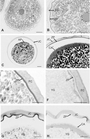
The vitellogenic oocytes of R. aspera are spherical, yellow and adhesive. They present elliptical yolk globules, cortical alveoli with small vesicles, prismatic follicular cells measuring 25.3 ± 7.8 μm of height and a zona pellucida thickness of 4.8 ± 1.0 μm (Fig. 1A and B). After extrusion, the oocytes reported a diameter of 1.36 ± 0.04 mm and had an evident perivitelline space, basophilic material at the animal pole and a conspicuous jelly coat covering the zona pellucida (Fig. 1C, D). Histochemical analysis of the vitellogenic oocytes and of the recently extruded oocytes showed a positive PAS reaction at the yolk globules and the zona pellucida, indicating the presence of neutral glycoproteins in these structures. Positive reactions to PAS and AB pH 2.5 were detected at the follicular cells and at the jelly coat. This indicates the presence of neutral glycoproteins and carboxylated acid glycoconjugates, forming mucosubstances (Fig. 1E–H). There was no positive reaction to AB pH 0.5 in the analysed structures.
Oocytes of R. aspera. (A) Oocyte before extrusion, HE; (B) detail of the oocyte before extrusion, HE; (C) oocyte after extrusion with perivitelline space, HE; (D) detail of the oocyte after extrusion with basophile material at the animal pole and a conspicuous jelly coat, HE; (E) positive PAS reaction in zona pellucida, follicular cells and yolk globules at the oocyte before extrusion; (F) positive AB pH 2.5 reaction in follicular cells and cortical alveoli at the oocyte before extrusion; (G) positive PAS reaction in zona pellucida, jelly coat and yolk globules at the oocyte after extrusion; and (H) positive AB pH 2.5 reaction in jelly coat at the oocyte after extrusion. Scale bars = 150 μm in A–D; 50 μm in E–H. AP, animal pole; CA, cortical alveoli; FC, follicular cells; JC, jelly coat; YG, yolk globules; ZP, zona pellucida.

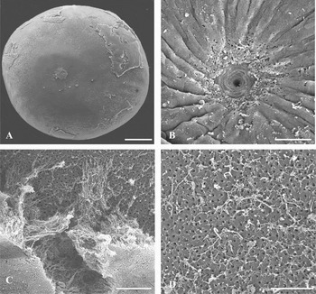
Ultrastructural analysis using scanning electron microscopy showed a thick jelly coat that was irregularly arranged over the surface of the oocytes and the presence of a micropyle disc at the animal pole. The animal pole presented a micropylar disc surrounding the micropyle vestibule with mucous aspect but with no evidence of filaments associated with the mucous material (Fig. 2).
R. aspera oocyte surface under scanning electron microscopy. (A) Oocyte with irregularly distributed jelly coat and micropyle, ×64; (B) adhesive disc surrounding the micropyle, 3387×; (C) detail of the jelly coat over the zona radiata, ×890; and (D) detail of the porous zona radiata, ×4250. Scale bars = 200 μm in A; 20 μm in B and 5 μm in C–D.

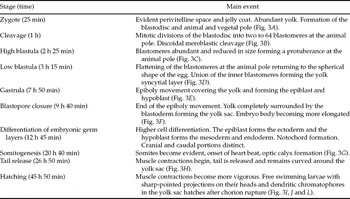
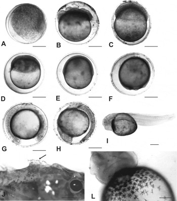
The embryonic development of R. aspera was completed 45 h and 50 min after fertilization at 24°C. The main events of the embryogenesis and their respective times are found in Table 1 and Fig. 3.
Main events of the embryonic development of R. aspera and their respective times after fertilization at 24°C.

Stages of embryonic development of R. aspera. (A) Early fertilized egg; (B) eight blastomeres stage; (C) high blastula; (D) low blastula; (E) gastrula; (F) blastopore closure; (G) somites development; (H) tail release; (I) hatching; (J) lateral view of the head showing dorsal sharp-pointed projections (arrow); and (L) ventral view showing dendritic chromatophores in the yolk sac. Scale bars = 400 μm in A–J; 200 μm in L.

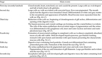
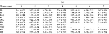
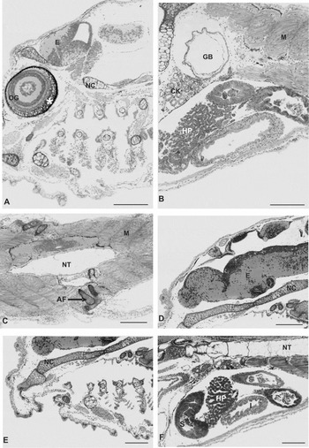
Recently hatched larvae presented spine shaped projections on their heads and numerous dendritic chromatophores in the yolk sac (Fig. 3). Fin rays were formed in the following sequence: caudal, pectoral, dorsal, anal and pelvic. The main morphologic features of larval ontogenesis and their respective times after hatching until the seventh day are shown in Table 2 and Figs 4, 5 and 6. Biometric parameters showed a gradual increase from the first to the seventh day, with the exception of yolk sac length and yolk sac height, which gradually reduced from the first to the fifth day, when the structure was completely absorbed (Table 3).
Main morphologic events occurring during the development of R. aspera larvae from hatching to the seventh day.

Body measurement (in mm) of R. aspera larvae from the first to the seventh day after hatching.

TL = total length, SL = standard length, PAD = pre anal distance, HL = head length, SNL = snout length, ED = eye diameter, YSL = yolk sac length, YSH = yolk sac height, HH = head height, BH = body height.
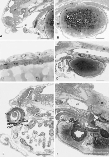
Stages of larval ontogenesis of R. aspera. (A) Second day after hatching, cranial portion; (B) second day after hatching, medial portion; (C) second day after hatching, detail of the yolk syncytial layer; (D) third day after hatching, medial portion; (E) cranial portion on the third day after hatching; (F) digestive system on the third day after hatching. B, barbells; CK, cranial kidney; DT, digestive tract; E, encephalon; G, ganglionar cell; GA, gill arches; H, heart; HP, hepato-pancreas; IN, inner nuclear layer; IP, inner plexiform layer; M, miomeres; MO, mouth; NC, neurocranium chondrogenesis; NT, notochord; OG, ocular globe; OP, outer plexiform layer; ON, outer nuclear layer; YS, yolk sac; YSL, yolk syncytial layer; *, pigmented retina. Scale bars = 200 μm in A, B, D, E and F; 20 μm in C.

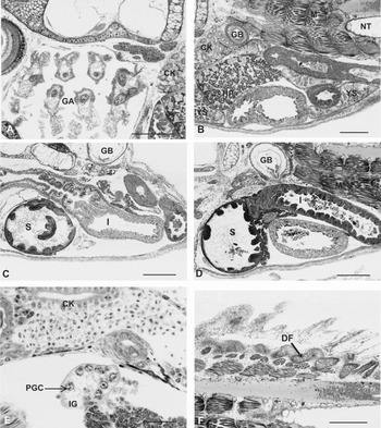
Stages of larval ontogenesis of R. aspera. (A) Fourth day after hatching, cranial portion; (B) fourth day after hatching, medial portion with wide gas bladder; (C) fourth day after hatching, digestive system; (D) medial portion on the fifth day after hatching; (E) indifferent gonad with primordial germ cells on the fifth day after hatching; (F) dorsal fin chondrogenesis on the fifth day after hatching. CK, cranial kidney; DF, dorsal fin chondrogenesis; GA, gill arches; I, intestine; GB, gas bladder; HP, hepato-pancreas; IG, indifferent gonad; NT, notochord; M, miomeres; PGC, primordial germ cell; S, stomach; YS, yolk sac. Scale bars = 200 μm in A, B, C, D and F; 40 μm in E.

Stages of larval ontogenesis of R. aspera. (A) Sixth day after hatching, cranial portion and gill filaments; (B) sixth day after hatching, medial portion; (C) sixth day after hatching, anal fin chondrogenesis; (D) cranial chondrogenesis and encephalon on the seventh day after hatching; (E) gill filaments on the seventh day after hatching; (F) digestive system totally formed on the seventh day after hatching. AF, anal fin chondrogenesis; CK, cranial kidney; E, encephalon; GB, gas bladder; HP, hepato-pancreas; I, intestine; M, miomeres; NC, neurocranium chondrogenesis; NT, notochord; OG, ocular globe; S, stomach; *, retina with cones and rods. Scale bars = 200 μm.

Discussion
Most species in the São Francisco River Basin, including R. aspera, do not reproduce spontaneously in fisheries. This highlights the need for studies that provide additional information regarding the reproduction, breeding and management of these species. The present study provides new information regarding embryogenesis and larval development of R. aspera, as well as information about spawning and oocyte ultrastructure. This particular species has high potential for pisciculture due to its zootechnical characteristics and the quality of its meat.
The results of induced reproduction techniques obtained during this study are similar to the results obtained by Sato et al. (Reference Sato, Fererich-Verani, Verani, Godinho and Sampaio1998). The oocytes of R. aspera are large when compared with oocytes from some Siluriformes species, such as 0.90 ± 0.02 mm in the Pimelodidae Pimelodus maculatus (Luz et al., Reference Luz, Reynalte-Tataje, Ferreira and Zaniboni-Filho2001) and 0.95 mm in Pseudoplatystoma corruscans (Marques et al. Reference Marques, Nakaghi, Faustino, Ganeco and Senhorini2008), but are small when compared with the oocytes of other loricariid species, such as Loricariichthys platymetopus (2.32 ± 0.189 mm), Megalancistrus aculeatus (4.29 ± 0.118 mm) and Hypostomus ternetzy (4.36 ± 0.135 mm) (Suzuki et al., Reference Suzuki, Agostinho and Winemiller2000) and other Siluriformes species, such as the heptapterid Rhamdia quelen (1.47 ± 0.05 mm) (Amorim et al., Reference Amorim, Gomes, Martins, Sato, Rizzo and Bazzoli2009). The oocytes of R. aspera are yellowish, adhesive and with an evident jelly coat. The yellowish colour is characteristic of the oocytes of Siluriformes (Sato et al., Reference Sato, Fenerich-Verani, Nuñer, Godinho, Verani, Godinho and Godinho2003; Marques et al., Reference Marques, Nakaghi, Faustino, Ganeco and Senhorini2008) and is associated with the presence of carotenoid pigments obtained from food. These pigments constitute an endogenous oxygen supply when the respiratory system is inefficient at obtaining exogenous oxygen (Balon, Reference Balon1977; Amorim et al., Reference Gillooly, Charnov, West, Savage and Brown2009).
The jelly coat, observed around the oocyte of R. aspera, is characteristic of Siluriformes species, rare in species of Characiformes (Rizzo et al., Reference Rizzo, Sato, Barreto and Godinho2002) and can also be found in species from the orders Perciformes, Cypriniformes and Cyprinodontiformes (Riehl & Patzner, Reference Riehl and Patzner1998). In Siluriformes, the jelly coat is present in adhesive and non-adhesive eggs, indicating that it bears no relationship with adhesiveness, however it may be that different chemical compositions of the jelly coat may influence egg adhesiveness (Rizzo et al., Reference Rizzo, Sato, Barreto and Godinho2002). The follicular cells acquire synthetic organelles and glycidic molecules rich in carboxylated radicals during oocyte development. Mucosubstances were detected in the vitellogenic oocytes of R. aspera both in the follicular cells and in the jelly coat. These mucosubstances are probably produced by the follicular cells to form the jelly coat and may be related to the adhesiveness of the eggs, as it has been found in other neotropical freshwater teleosts (Andrade et al., Reference Andrade, Bazzoli, Rizzo and Sato2001, Santos et al., Reference Santos, Padilha, Bomcompagni-Júnior, Santos, Rizzo and Bazzoli2006, Normando et al., Reference Normando, Arantes, Luz, Thomé, Rizzo and Bazzoli2009). Mucosubstances were also found at the external layer of the zona radiata of adhesive eggs of the characiforme Serrasalmus brandti (Rizzo & Bazzoli, Reference Rizzo and Bazzoli1991), which may reinforce the relationship between these substances and adhesiveness. Although studies have been indicating this relationship to be true (Riehl & Patzner, Reference Riehl and Patzner1998; Rizzo et al., Reference Rizzo, Sato, Barreto and Godinho2002; present work), more studies concerning the constitution and function of the jelly coat and the relationship between egg surface and adhesiveness are necessary.
The thickness of the zona radiata is related to the environment in which the eggs are deposited. In R. aspera, the zona radiata is thin (4.8 ± 1.0 μm) when compared with other species of Loricariidae, such as in the genera Loricaria and Loricariichthys, whose eggs present a thick zona radiate (9.95 ± 0.25 μm, 9.10 ± 0.57 μm) related to exposure to abrasion when carried on the ventral surface of the male (Suzuki et al., Reference Suzuki, Agostinho and Winemiller2000). This difference may be due to the lower levels of abrasion that the eggs of R. aspera experience, as the species releases its non-buoyant eggs into the water column.
The pattern of segmentation of vertebrate eggs depends on the amount and distribution of the yolk and on its proportion in relation to the cytoplasm that constitutes the blastodisc (Gilbert, Reference Gilbert1991; Ninhaus-Silveira et al., Reference Ninhaus-Silveira, Foresti and Azevedo2006). Fish eggs are classified as telolecitic because they contain a large amount of yolk that is evenly distributed (Ribeiro et al., Reference Ribeiro, Leme dos Santos and Bolzan1995; Leme dos Santos & Azoubel, Reference Leme dos Santos and Azoubel1996; Marques et al., Reference Marques, Nakaghi, Faustino, Ganeco and Senhorini2008). The cleavage of the eggs of R. aspera followed the meroblastic (partial) pattern, restricted to the animal pole as is commonly observed in teleosts (Leme dos Santos & Azoubel, Reference Leme dos Santos and Azoubel1996; Meijide & Guerreiro, Reference Meijide and Guerrero2000; Ninhaus-Silveira et al., Reference Ninhaus-Silveira, Foresti and Azevedo2006; Gomes et al., Reference Gomes, Scarpelli, Arantes, Sato, Bazzoli and Rizzo2007; Amorim et al., Reference Amorim, Gomes, Martins, Sato, Rizzo and Bazzoli2009). Blastopore closure occurred at 9 h and 40 min after fertilization. The identification of this event is important as it indicates that fertilization was successful. It also indicates an effective moment to estimate the fertilization rate (Godinho, Reference Godinho2007) as non-fertilized eggs develop parthenogenetically until prior to blastopore closure.
The embryonic development of R. aspera lasted 45 h and 50 min at 24°C, which is long when compared with the embryonic development in other species of Siluriformes: 18 h at 27°C in P. corruscans (Marques et al., Reference Marques, Nakaghi, Faustino, Ganeco and Senhorini2008); 21 h and 20 min at 23°C in Pimelodus maculatus (Luz et al., Reference Luz, Reynalte-Tataje, Ferreira and Zaniboni-Filho2001) and 25 h and 30 min at 24°C in R. quelen (Amorim et al., Reference Amorim, Gomes, Martins, Sato, Rizzo and Bazzoli2009). Long incubation periods are usually associated with non-migratory species with large eggs and that display parental care behaviour (Sargent et al., Reference Sargent, Taylor and Gross1987; Gillooly et al., Reference Gillooly, Charnov, West, Savage and Brown2002; Goodwin et al., Reference Goodwin, Dulvy and Reynolds2002; Kolm & Ahnesjö, Reference Kolm and Ahnesjö2005). The occurrence of parental care in R. aspera is not well established, being described as a migratory species with no parental care by Suzuki et al. (Reference Suzuki, Agostinho and Winemiller2000). In this study it was observed that R. aspera eggs are adhesive and present a micropyle disc and a thick gelatinous surface layer. These attributes are characteristic of sedentary Siluriformes species that display parental care, as was also indicated by Sato et al. (Reference Sato, Fenerich-Verani, Nuñer, Godinho, Verani, Godinho and Godinho2003).
After hatching, R. aspera larvae presented sharp-pointed projections on their heads that suggest a protective function. These spikes were also identified in R. strigosa larvae (Nakatani et al. Reference Nakatani, Agostinho, Baugartnet, Bialetzki, Sanches, Makrakis and Pavanelli2001). R. aspera larvae are transparent during the first day and pigmentation of the retina began during the second day and was completed during the fourth day, in a similar manner as was reported by Borçato et al. (Reference Borçato, Bazzoli and Sato2004) for the larvae of Leporinus piau. According to Hall et al. (Reference Hall, Smith and Johnston2004), the time of pigmentation of the epithelium of the retina is possibly adaptive and may be related to predation or with the need to develop a functional visual system before the first feeding. The analysis of chromatophores and the pigmentation of the eyes and body are important for taxonomic determination and identification of species (Meijide & Guerreiro, Reference Meijide and Guerrero2000). Fish larvae can present two chromatophore patterns: punctiform and dendritic (Nakatani et al., Reference Nakatani, Agostinho, Baugartnet, Bialetzki, Sanches, Makrakis and Pavanelli2001). In this study, the recently hatched larvae only presented dendritic chromatophores in the yolk sac. Some species, such as Hoplias lacerdae and H. unitaeniatus, have only punctiform chromatophores in their bodies, whereas H. malabaricus has both types (Gomes et al., Reference Gomes, Scarpelli, Arantes, Sato, Bazzoli and Rizzo2007).
The periblast or yolk syncytial layer (YSL) was observed in embryos of R. aspera and was clearly evident in larvae submitted to histology procedures. This layer is the result of incomplete division of the blastomeres and is located between the blastoderm disc and the yolk. Initially, this structure is a ring-shaped layer that surrounds the yolk, which then eventually expands over the entire blastoderm (Kimmel et al., Reference Kimmel, Ballard, Kimmel and Ullmann1995). This layer is important for embryonic development of teleost eggs as all the nutrients in the yolk pass through it to reach the blastoderm (Ninhaus-Silveira et al., Reference Ninhaus-Silveira, Foresti, De Azevedo, Agostinho and Veríssimo-Silveira2007). The YSL also acts as a force for the epiboly movements of the blastoderm (Trinkaus, Reference Trinkaus1984a, Reference Trinkausb, Reference Trinkaus1993; Ninhaus-Silveira et al., Reference Ninhaus-Silveira, Foresti, De Azevedo, Agostinho and Veríssimo-Silveira2007). Knowledge of the characteristics of the YSL has great importance for the development of cryopreservation techniques as it has been considered one of the main obstacles for the cryopreservation of fish embryos (Ninhaus-Silveira et al., Reference Ninhaus-Silveira, Foresti, De Azevedo, Agostinho and Veríssimo-Silveira2007). In addition, the YSL is one of the main barriers for water and cryoprotectors permeability, which restricts the entrance of cryoprotectors into the vitellogenic compartment (Hagedorn et al., Reference Hagedorn, Hsu, Pilatus, Wildt, Rall and Blackband1996, Reference Hagedorn, Kleinhans, Wildt and Rall1997).
On the fifth day after hatching, the undifferentiated gonad, constituted of a few primordial germ cells (PGCs), was observed between the cranial kidney and the digestive tract. In R. aspera, the PGCs are large when compared with somatic cells, have an eccentrically located vesicular nucleus and clear cytoplasm. In Pimephales promelas, on the seventh day after spawning, the gonad organization is also simple, exhibiting few germ cells and a single layer of somatic cells surrounding the gonads that start their differentiation only on the 13th day after fertilization (Uguz, Reference Uguz2008).
The opening of the mouth occurred quickly, during the second day after hatching. However, the digestive tract remained closed until the third day. Only traces of the yolk sac remained on the fourth day and it was totally absorbed on the fifth day, indicating the need for exogenous feeding. The opening of the mouth and digestive tract is essential for the larvae to be capable of ingesting, digesting and assimilating the first food (Borçato et al., Reference Borçato, Bazzoli and Sato2004; Guimarães-Cruz et al., Reference Guimarães-Cruz, Santos, Sato and Veloso-Júnior2009), an important event for pisciculture.
Recently ecloded R. aspera larvae had 3.44 ± 0.08 mm mean total length, a large size when compared with some Siluriformes from the São Francisco River basin, such as 2.56 ± 0.13 mm in P. maculatus (Luz et al., Reference Luz, Reynalte-Tataje, Ferreira and Zaniboni-Filho2001); 2.76 mm in R. quelen (Amorim et al., Reference Amorim, Gomes, Martins, Sato, Rizzo and Bazzoli2009) and 3.30 mm in P. corruscans (Santos & Godinho, Reference Santos and Godinho1994), yet smaller than other larvae such as 4.00 mm in Rhamdia hilarii (Godinho et al., Reference Godinho, Fenerich and Narahara1978) and 7.12 ± 0.62 mm in Lophiosilurus alexandri (Guimarães-Cruz et al., Reference Guimarães-Cruz, Santos, Sato and Veloso-Júnior2009). The growth of R. aspera larvae from the first to the seventh day indicated that the incubation conditions at 24°C were appropriate for their development.
The results obtained regarding oocyte characteristics, embryonic development and larval ontogenesis are essential for a better understanding of the biology of R. aspera. Furthermore, the reproductive parameters obtained constitute a baseline of information for the breeding and management of R. aspera, a species of commercial interest with high potential for pisciculture.
Acknowledgements
The authors thank the partnership of CEMIG-GT/CODEVASF and the Brazilian research foundations of CNPq, CAPES and FAPEMIG for financial support and also Dr Erik Wild for suggestions on the English version.