Introduction
Alzheimer's disease (AD) is the major cause of dementia, affecting some 44 million people globally; this number is expected to almost double in the next 15 years and more than triple by 2050 (Ref. 1). Current therapies to treat AD patients are focused on ameliorating symptoms instead of targeting the underlying causes of the disease. With an aging population and an increase in disease incidence, it is imperative to develop disease-modifying therapies for AD (Ref. 2).
AD is a neurodegenerative disorder associated with progressive damage in brain functions including memory, language, spatial orientation, behaviour and personality. The clinical manifestation of AD is generally preceded by a silent preclinical phase (Ref. Reference Sperling3), the prodromal phase, after which the first clinical symptoms appear as mild cognitive impairment (MCI) (Ref. Reference Albert4); the disease finally results in dementia, which implies the existence of multiple cognitive symptoms severe enough to interfere with daily functioning (Ref. Reference McKhann5). These phases are accompanied by biochemical changes in the brain, which are reflected in the cerebrospinal fluid (CSF) (Ref. Reference Yang6). Extracellular plaques, containing various forms of amyloid-β (Aβ) peptide, and intracellular neurofibrillary tangles, composed of hyper-phosphorylated tau protein, are the two major pathological hallmarks of the AD brain.
Accordingly to the amyloid hypothesis, AD is caused by the progressive accumulation of the Aβ peptide into oligomers and plaques, leading to synaptic loss, neuronal dysfunction and death (Ref. Reference Terry7). This hypothesis has been supported by the findings of several mutations in the Aβ region of the amyloid precursor protein (APP) as well as in other genes related with Aβ processing (PSEN1, PSEN2) in families with autosomal dominant early onset AD (EOAD) (Ref. Reference Benilova, Karran and De Strooper8). These mutations have been shown to increase the production of Aβ in vitro as well as in vivo (Ref. Reference Hardy and Selkoe9). However, in the most prevalent form of the disease, late-onset sporadic AD (LOAD), decreased Aβ clearance rather than increased production has been attributed as the cause triggering the disease process (Ref. Reference Mawuenyega10). The ε4 allele of the APOE gene, which encodes the lipid/cholesterol carrier apolipoprotein E (apoE), has been established as the major risk factor for developing LOAD, whereas the presence of the ε2 allele appears protective and the most common ε3 allele seems to be neutral (Refs Reference Bu11, Reference Liu12). While transporting cholesterol is its primary function, apoE also regulates Aβ metabolism, aggregation and deposition (Ref. Reference Kanekiyo, Xu and Bu13). The presence of apoE4 contributes to the pathogenesis of AD, probably by both loss-of-function in neuroprotection and gain-of-function in neurotoxicity compared with apoE3 (Refs Reference Bu11, Reference Huang and Mucke14). Although several pathways underlying the risk associated with apoE4-linked AD have been defined through in vitro and in vivo studies, the exact mechanisms are still not completely understood (Ref. Reference Kanekiyo, Xu and Bu13).
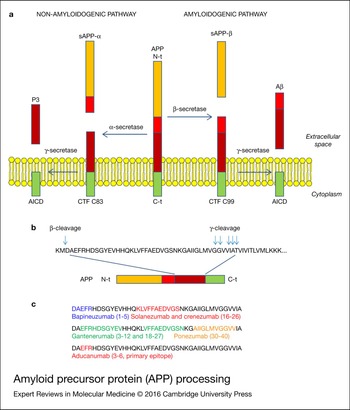
Aβ is a soluble metabolite of ~4 kDa that is produced by the processing of APP, a large transmembrane glycoprotein (Ref. Reference Solomon15). APP can be processed by either of two distinct membrane-anchored proteases, α-secretase (thought to consist of one or more members of the ADAM family of metalloproteases) or β-secretase (an aspartyl protease of the pepsin family, also called β-site APP-cleaving enzyme (BACE-1)) (Ref. Reference Karran, Mercken and De Strooper16). Depending on which enzyme initially processes the APP, two cleavage pathways are possible: the non-amyloidogenic and amyloidogenic, respectively (Fig. 1a). In both cases, the γ-secretase complex, containing a special type of aspartyl protease called presenilin, performs a sequential cleavage to the generated product. The γ-secretase complex is composed of presenilin-1 (PS1) or PS2, nicastrin, anterior pharynx defective phenotype (APH1) and presenilin enhancer 2 (Ref. Reference Wolfe17).
APP processing. (a) α- and γ-secretase act in the non-amyloidogenic pathway, whereas the activity of β-secretase instead of α-secretase defines the amyloidogenic pathway that generates the Aβ peptide. (b) Target proteolytic sites in the amyloidogenic pathway. (c) Aβ epitopes recognised by different mAbs.

In the prevalent non-amyloidogenic pathway, APP is cleaved by α-secretase at a position 83 amino acids from the carboxy (C) terminus, producing a large amino (N)-terminal ectodomain (sAPPα) that is secreted into the extracellular medium (Ref. Reference Kojro and Fahrenholz18). The resulting 83-amino acid C-terminal fragment (C83) is retained in the membrane and subsequently cleaved by the γ-secretase complex, producing a short fragment termed p3 (Ref. Reference Haass19). In this pathway, cleavage by α-secretase occurs within the Aβ region, thereby avoiding Aβ formation.
In the amyloidogenic pathway, the cleavage of APP by β-secretase at a position located 99 residues from the C-terminus generates the release of sAPPβ into the extracellular space, and leaves the resulting 99-amino acid C-terminal fragment (C99) within the membrane, with the newly generated N-terminus corresponding to the first amino acids of Aβ. Subsequent cleavage of this fragment (between residues 38 and 43) by the γ-secretase complex liberates the Aβ peptide (Ref. Reference LaFerla, Green and Oddo20).
γ-secretase cleaves APP at different positions (Fig. 1b), generating a variety of peptides, of which Aβ 43, Aβ 42, Aβ 40, Aβ 39, Aβ 38 and Aβ 37 variants are detected in cell culture and body fluids (Aβ numbering indicates the last C-terminal amino acid residue and the number of amino acids comprising the peptide, with residue 1 being the amino acid residue aspartyl-1 of the Aβ sequence in APP) (Ref. Reference Benilova, Karran and De Strooper8). The cleavage site has a profound influence on the aggregation potential and, consequently, on the pathogenicity of Aβ. Most of the produced Aβ has a length of 40 residues (Aβ 40), but a small fraction (commonly 10%) is the Aβ 42 variant (Ref. Reference Jarrett, Berger and Lansbury21). The Aβ 42 variant is the most hydrophobic and prone to form fibrils, so it is the predominant one in the amyloid plaques seen in AD (Ref. Reference Younkin22), whereas the Aβ 40 variant is preferentially deposited in the wall of blood vessels where it promotes Aβ-type cerebral amyloid angiopathy (CAA) (Ref. Reference Yamada23). In addition, evidence shows that an increase in Aβ 42, leading to changes in the Aβ 40:Aβ 42, ratio is critical to AD neurotoxicity, by specifically increasing the oligomeric forms of Aβ (Ref. Reference Bentahir24).
Although in earlier AD studies it was thought that only the amyloid plaques were toxic, nowadays it is widely accepted that the oligomeric and protofibril forms of Aβ are the crucial toxic species; it has been demonstrated that the levels of these soluble species correlate better with cognitive deficits (Ref. Reference Wang25). In fact, AD pathogenesis is a continuum in which cerebral Aβ accumulation precedes neurodegeneration and neuronal cell death (Ref. Reference Dubois26). It is quite possible that Aβ soluble aggregates (i.e. oligomers) may bind synapses and cause synaptic dysfunction. This may be ahead of plaque deposition. In addition, while diffuse Aβ deposits are often devoid of degenerating neurites, most senile (compacted, fibrillar) plaques contain dystrophic neurites clogged with p-tau and ubiquitin, among others.
Findings on the specific pathogenicity of the oligomeric forms of the Aβ peptide have raised the question of directly attempting to target them. Developing therapies based on the reduction of these species has become a challenge in treating the underlying pathogenic causes of AD. Immunotherapy is one approach that has been advanced recently for its ability to reduce the accumulation of Aβ. In addition, the immune system participates in AD pathogenesis and, among other findings beyond the scope of this review, it has been demonstrated that naturally-produced antibodies against Aβ occur in humans, and AD patients have lower levels of these antibodies compared with healthy individuals (Ref. Reference Weksler27).
Immunotherapy
The primary aim of immunotherapy to treat AD is to reduce the Aβ burden by using antibodies against the Aβ peptide. This can be achieved either by inducing production by the patient (active immunisation) or by administering monoclonal antibodies (mAbs) to the patient (passive immunotherapy).
Active immunotherapy
The basic concept of active immunisation is to prime the immune system to recognise an antigen as a foreign protein in order to generate a response against it (Ref. Reference Spencer and Masliah28). Active Aβ-immunotherapy uses synthetic full-length Aβ peptide, or a fragment. It aims to stimulate B cells to generate specific antibodies in order to sequester amyloid from the brain in the peripheral system, destroy plaques and inhibit further deposit of amyloid into plaques (Ref. Reference Li29).
On one hand, active immunotherapy is attractive because it can induce long-term antibody production while being cost-effective and requiring only a few visits to the doctor (Ref. Reference Lemere30). On the other hand, some disadvantages could be that the polyclonal response is established to varying degrees with variable specificity of the antibodies produced and, in some cases, meaningful titres are not produced. Especially in the older population such as the late-onset AD group, age-related attenuation of the immune system will affect the efficacy of active immunotherapy. In addition, the specificity of the generated antibodies can be tough to predict, and adverse reactions may be persistent and difficult to reverse (Ref. Reference Lannfelt31).
The first Phase IIa clinical trial for an active AD vaccine, AN1792, was performed in 2002 (ELAN & Wyeth). It enrolled patients with moderate-to-severe AD and tested an aggregated full-length Aβ 42 peptide with an adjuvant (QS-21) that preferably promotes T- cell-mediated responses (Ref. Reference Gilman32). Out of the 300 AN1792(QS-21)-treated patients, 59 (20%) developed the predetermined antibody response and, although the study was halted after the first to third doses, assessments were maintained for at least nine additional months. No significant differences were found between antibody responders and the placebo groups based on several cognitive assessments for dementia, but analyses of the z-score composite across the Neurophysiological Test Battery (NTB) revealed differences favouring antibody responders. CSF examinations also showed that tau was decreased in antibody responders, while Aβ levels remained the same. It is noteworthy that the overall incidence of cerebral haemorrhage reported during the study was low and similar between the AN1792(QS-21) and placebo groups. However, one death (cerebral infarct), 205 days after the second dose of AN1792(QS-21) to a non-responder patient without encephalitis, was considered to be related to the treatment. In addition, administrations were halted because of the development of meningoencephalitis in ~6% (18/300) of the treated group, likely because of activation of pro-inflammatory T cells. T cell meningoencephalitis was noted in two postmortem reports on AN1792(QS-21)-treated patients, where depletion of neocortical Aβ was also evident. Although there was no indication of meningoencephalitis during the Phase I studies, a change in the AN1792(QS-21) formulation (addition of polysorbate-80 to prevent precipitation) may have resulted in this complication. As a conclusion, this trial showed that Aβ-immunotherapy is useful in the treatment of AD and revealed the necessity of reformulating the drug in order to avoid severe adverse effects. Considering that activation of Aβ-specific T cells was potentially harmful, second generation vaccines seek to induce a humoral immune response to generate anti-Aβ antibodies while avoiding Aβ T cell epitopes (Ref. Reference Sarazin33). To do so, they have been restricted to the B cell epitopes of the Aβ peptide located at the N-terminus, while avoiding the T cell epitopes located at the C-terminus (which were exposed by polysorbate-80 in the AN1792 trial) (Ref. Reference Spencer and Masliah28). Two of such vaccines, AffitopeAD02 and CAD106, consist of the 1–6 N-terminal fragment of the Aβ peptide, whereas vanutidecridificar (ACC-001) uses the 1–7 N-terminal fragment.
A Phase I clinical trial for AffitopeAD02 (Affiris) using aluminium hydroxide as the adjuvant showed a favourable safety and tolerability profile at 1 year (Ref. Reference Schneeberger34). Phase II trials of AD02 in patients with early AD did not reach either primary or secondary outcome results, but found a new formulation with a greater benefit on the primary outcome. Affiris renamed this formulation to AD04 in 2014, but recently showed that although it was designated as a placebo, it appears to have disease-modifying activity (Ref. Reference Schneeberger35). Similarly, Affitope was redesigned to target modified Aβ peptides, and renamed as AD03 (MimoVax), but a Phase I clinical trial completed in 2011 has not been published and subsequent studies have been terminated for organisational reasons.
In 2012, the results of a Phase I study of CAD106 (Novartis), consisting of the bacteriophage Qβ as a scaffold for presentation of multiple copies of the 1-6 N-terminal fragment of the Aβ peptide, were published (Ref. Reference Winblad36). Although an Aβ antibody response was shown, most patients in both the placebo and treated groups had adverse effects, none of which was thought to be related to CAD106 (Ref. Reference Kingwell37). In the first Phase II trial, a favourable safety profile was seen in subjects with mild AD (Ref. Reference Lannfelt, Relkin and Siemers38).
Vanutidecridificar (ACC-001, Janssen and Pfizer) consists of amino acids 1–7 linked to inactivated diphtheria toxin and formulated with the QS-21 adjuvant (previously used in the AN1792 clinical trial). It was tested in multiple Phase II trials, and a recent paper reports a variety of treatment-emergent adverse events from most subjects during the studies (Ref. Reference Arai, Suzuki and Yoshiyama39). All doses of ACC-001 + QS-21 elicited high, sustained anti-Aβ antibody titres, but QS-21 was necessary for this effect.
Passive immunotherapy
One way to overcome the main drawback of active immunotherapy, i.e. having varying amounts and specificities of the polyclonal antibodies produced by each patient, is to treat the patients with ex vivo produced mAbs (Ref. Reference Spencer and Masliah28). Passive immunotherapy allows for stopping the treatment whenever an adverse effect occurs, apart from targeting a specific epitope. Its main drawbacks are the high cost of mAb production and the necessary long-term administration (Refs Reference Lemere30, Reference Wang, Zhou and Zhou40). For a long time, mAbs have been studied in terms of their use as therapies for different diseases as they have been found to be an excellent paradigm for the design of high-affinity, protein-based binding reagents (Ref. Reference Holliger and Hudson41). They have been successful in the clinic for a number of diseases, including autoimmune disorders (anti-tumour necrosis factor (TNF) Humira and anti-IL-6 Actemra), cancer (anti-HER2 receptor Herceptin and anti-CD20 Rituxan), transplant rejection (anti-CD25 Zenapax and Simulect) (Ref. Reference Waldmann42) and cardiovascular diseases (anti-CD41 ReoPro) (Ref. Reference Holliger and Hudson41).
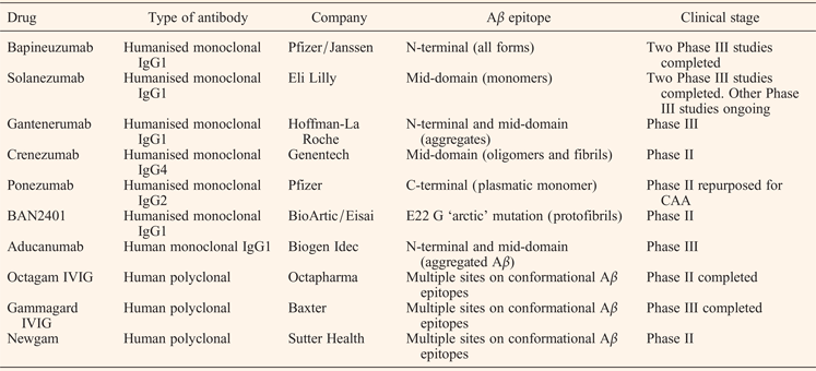
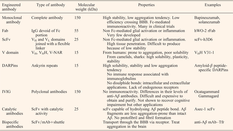
In recent years, several engineered antibodies and derived fragments have been designed to bind the Aβ peptide with the goal of developing a strong disease-modifying treatment (Tables 1 and 2).
Passive immunotherapies for Alzheimer's disease beyond clinical Phase I

Passive immunotherapeutic approaches that target Aβ peptide
Complete mAbs
Full-length mAbs were the first molecules tested for passive immunotherapy to treat AD. Some of them showed promising results in vitro and in AD mouse models, and advanced to clinical trials (Table 1). At the moment, none of them have passed Phase III, but the experience of these trials has led us to a better knowledge of the disease and has given important clues to develop more appropriate therapies.
Bapineuzumab (AAB-001 & AAB-003, Pfizer/Janssen)
In 2000, Bard et al. demonstrated that peripheral administration of an IgG1 mAb specific for the five N-terminal residues of Aβ (Fig. 1c), 3D6 mAb, into AD Tg-mice resulted in transfer of the antibody into brain, binding of the antibody to plaques and induction of the Fc receptor-mediated microglial-phagocytosis of Aβ deposits (Ref. Reference Bard43). 3D6 mAb binds all forms of the Aβ peptide (monomeric, oligomeric and fibrillar).
This antibody is the precursor of the humanised bapineuzumab (AAB-001), which has been tested up to Phase III (Ref. Reference Salloway44). In a Phase I study, bapineuzumab appeared safe and well-tolerated overall in patients with mild-to-moderate AD, but MRI monitoring showed microhaemorrhages in three out of ten patients on the highest dose. This finding led to the new term amyloid-related imaging abnormalities (ARIA) and, more specifically, ARIA-oedema/effusions (ARIA-E) to refer to vasogenic oedema and ARIA-H to refer to microhaemorrhage (Ref. Reference Sperling45). ARIAs are thought to result from transient leakiness of cerebral vessels following vascular amyloid clearance.
In the subsequent Phase II trial, the highest dose was lowered (from 5 to 2 mg/kg). Although no significant differences were seen by two different cognitive assessments (ADAS-Cog and DAD), a considerable reduction in amyloid was shown by PIB-PET (positron-emission tomographic imaging using Pittsburgh compound B) as well as reduced CSF phospho-tau. However, the occurrence of ARIA-E in APOE ε4 allele carriers led to recruiting APOE ε4 and non-APOE ε4 carriers separately for Phase III. CSF analyses showed a reduction in phospho-tau with treatment only in the APOE ε4 group, while the Aβ concentration was maintained in all the cohorts. Volumetric magnetic resonance imaging (MRI) did not show any differences between treated and untreated groups, but amyloid PET scans demonstrated the clearance of some fibrillar cerebral Aβ. Diffusion tensor imaging (DTI, a variant of MRI) showed that ARIA-E occurred in at least 5% of the bapineuzumab-treated patients, especially in the APOE ε4 carrier cohort, and this led to the discontinuation of the study at the highest dose. Diminished levels of CSF phospho-tau were also seen exclusively in this cohort. These facts were attributed to the increased Aβ burden, including vascular amyloid, in APOE ε4 carriers. Because bapineuzumab did not improve clinical outcomes in patients with AD, despite treatment differences in biomarkers observed in APOE ε4 carriers, the trial was stopped in August 2012. The commonly cited explanation for the failure of this trial points to the recruitment of patients with mild-to-moderate AD, a stage at which brain amyloid deposition has been occurring for many years. Thus, the main thing learned from this study is that clinical trials for AD must be performed on patients in the prodromal phase.
Pfizer and Janssen have been working on an improved version of bapineuzumab (AAB-003), which has been tested in two recently completed Phase I trials (https://clinicaltrials.gov/;accessed2015-10-01). This version has a redesigned Fc region, aiming to prevent ARIAs (Ref. Reference Black46).
Solanezumab (LY2062430, Eli Lilly)
In 2001, DeMattos and colleagues demonstrated that a humanised IgG1 mAb targeted to an internal epitope of Aβ (residues 16–26) (Fig. 1c), m266, reversed memory deficits in APP-transgenic mouse models while keeping amyloid plaques in place, opening the possibility of targeting the soluble pool of Aβ (Ref. Reference DeMattos47). m266 binds soluble monomeric forms of the Aβ peptide.
This antibody is the precursor of solanezumab, which is being tested in Phase III clinical trials. In Phase I, solanezumab was well tolerated in healthy volunteers and patients with mild-to-moderate AD. MRI showed no evidence of inflammation, vasogenic oedema, or microhaemorrhage. Biomarker studies found changes in plasma and CSF Aβ 40, Aβ 42 and N-terminally truncated Aβ, and plasma pyro-Glu Aβ, but not total tau and phospho-tau in CSF. In the subsequent Phase II trial, doses were increased and the antibody's safety and tolerability were confirmed, while dose-dependent increases in various Aβ species in plasma and CSF were found. However, the desired cognitive improvement was not achieved until Phase III, carried out with mild AD patients (33% reduction in the rate of decline) (Ref. Reference Doody48). The benefit appeared after some months of treatment, grew over time, and was consistent with a disease-modifying effect. However, this benefit was not better than that of the palliative acetylcholinesterase inhibitor drugs. Other Phase III trials are ongoing and are expected to be completed at the end of 2016.
Apart from showing good safety and some clinical benefit, solanezumab has shown similar efficacy in patients with or without the APOE ε4 allele (Ref. Reference Samadi and Sultzer49). For these reasons, the Dominantly Inherited Alzheimer's Network (DIAN) is planning to supervise a Phase II/III trial to test solanezumab (and gantenerumab, described below) in asymptomatic and very mildly symptomatic carriers of autosomal-dominant Alzheimer's disease (ADAD) mutations (APP, PSEN1 and PSEN2), ranging from 30 to 50 years of age. At the moment, cross-sectional measurements by PET and MRI and CSF and plasma biomarkers levels assessed in 54 non-treated PSEN1 E280A kindred members (age range 20–59 years) have just been published (Ref. Reference Fleisher50). Compared with non-carriers, PSEN1 E280A carriers had significantly lower precuneus cerebral metabolic rates for glucose, a smaller hippocampal volume, lower CSF Aβ 42, higher CSF total tau and phospho-tau181, and higher plasma Aβ 42 levels. Another prevention study in which solanezumab will be tested is the Alzheimer's Disease Cooperative study A4, in which asymptomatic or very mildly symptomatic people aged 65–85 years and who have evidence of brain amyloid deposition by PET are being recruited.
Gantenerumab (Hoffman-La Roche)
In 2012, Bohrmann et al. developed the first fully human IgG1 mAb for Aβ-immunotherapy, gantenerumab (Ref. Reference Bohrmann51). It was optimised in vitro for binding with sub-nanomolar affinity to a conformational epitope shown by Aβ fibrils using HuCAL(®) phage display technology. This conformational epitope is made by both the N-terminal and the mid-region of Aβ. Gantenerumab binds first to the N-terminal epitope and then to the mid-region epitope (Fig. 1c), but only when the peptide is folded in a particular way such that the mid-region is close to the N-terminus. Epitope mapping by pepspot analysis using overlapping decameric Aβ peptides revealed peptides spanning residues 3–12 and 18–27 as those showing the strongest signals (Fig. 1c). It is noteworthy that the crystal structure of the Fab (fragment antigen-binding)-Aβ (1–11) complex exhibited a novel orientation of the N-terminal Aβ bound to the complementarity determining regions (CDRs). This makes sense because, in contrast to what happens with bapineuzumab, the first two residues of the Aβ peptide are not included within the epitope.
Gantenerumab preferentially interacts with aggregated brain Aβ, both parenchymal and vascular. In a mouse model of AD, gantenerumab showed sustained binding to cerebral amyloid and reduced small amyloid plaques by recruiting microglia and preventing new plaque formation, while plasma Aβ remained the same (Ref. Reference Bohrmann51).
Phase I clinical trials in mild-to-moderate AD patients treated with gantenerumab reduced the brain amyloid load, but two patients in the high dose group experienced ARIA-E at sites with the highest level of amyloid reduction, as seen by MRI (Ref. Reference Ostrowitzki52). The Phase II study enrolled people aged 50 and older whose memory function tests were below normal and who were positive for amyloid on PET assessments. Although this phase was extended to Phase III, an interim futility analysis made Roche discontinue this trial in December 2014. Based on the inclusion criteria, three different Phase III trials are currently recruiting patients: in the prodromal phase, with mild AD and carriers of the FAD mutation mentioned above for the DIAN-TU initial study.
Crenezumab (MABT5102A, Genentech)
Crenezumab (MABT) was designed on an IgG4 scaffold instead of IgG1 to reduce the risk of microglial-mediated pro-inflammatory effects in brain, including vasogenic oedema. When compared in a mouse model with a human IgG1 wild-type subclass, containing the same antigen-binding variable domains and with equal binding to Aβ, MABT5102A showed reduced activation of stress-activated p38MAPK in microglia and induced less release of the pro-inflammatory cytokine TNFα. This antibody binds to protofibrillar species, covering oligomers, and at the same time it avoids aggregation and promotes disaggregation of amyloid plaques (Ref. Reference Adolfsson53). Remarkably, the central Aβ residues recognised by crenezumab almost perfectly overlap with those recognised by solanezumab, explaining the observed shared cross-reactivity of these mAbs (Ref. Reference Crespi54) (Fig. 1c). It is worthy to note that, as mentioned above, solanezumab only recognises monomers; this apparent contradiction could be explained by the fact that, although the CDRs of solanezumab and crenezumab are highly homologous, positions 33 (CDR1; Ser in solanezumab, Tyr in crenezumab) and 36 (just outside L1; Phe in solanezumab, Gly in crenezumab) are rather different.
Phase I trials showed good safety data for mild-to-moderate AD patients, and Phase II testing with higher doses was completed in spring 2014 and continued into an open-label extension trial that will run until February 2017. It is also being evaluated as part of the Alzheimer Prevention Initiative, in which pre-symptomatic carriers of autosomal-dominant presenilin mutations, such as PSEN1 E280A, were recruited for a Phase II trial set to run until 2020.
Ponezumab (PF-04360365RN1219, Pfizer)
Ponezumab is a humanised IgG2 mAb targeted to the Aβ C-terminus (residues 30–40) (Ref. Reference La Porte55) (Fig. 1c), which makes it efficient at binding plasmatic Aβ. It also contains two mutations (A33S, P331S) that eliminate effector function, and thus it is immunologically inert. Early clinical data pointed to good safety profile with increased plasma Aβ 40; however, no improvement in cognitive impairment was observed in two completed Phase II trials. Because it is more appropriate to recognise Aβ 40 than Aβ 42, it was repurposed for treating CAA and a Phase II clinical trial is ongoing.
BAN2401 (BioArctic/Eisai)
BioArctic Neuroscience has produced BAN2401, a humanised version of the IgG1 mAb158, which is a conformation-dependent antibody with the ability to recognise a unique structure in the Aβ protofibril instead of a linear epitope. This is so because the mAb was derived from an epitope containing the E22G ‘arctic’ mutation in APP, which leads to a form of AD that is featured by particularly high levels of Aβ protofibrils. Recently, it has been demonstrated that it selectively reduces Aβ protofibrils in the brain and CSF of Tg-ArcSwe mice (Ref. Reference Tucker56).
A Phase I/IIa clinical safety study has just been completed and no serious adverse events were observed. Phase IIb was started in January 2013 in patients with MCI and early AD (Ref. Reference Lannfelt57).
Aducanumab (BIIB037, Biogen Idec)
After many failures in passive Aβ immunotherapy-based clinical trials, the AD community begun to question the amyloid cascade hypothesis. As an example, and among other amyloid-independent pathways likely involved in AD development, several researchers claim that early-onset AD-associated mutations in presenilins cause, apart from obvious catalytic impairment, a Ca2+ overload of endoplasmic reticulum (ER) stores; this leads to instability in mature synaptic spines and subsequent memory loss (Ref. Reference Popugaeva and Bezprozvanny58).
In March 2015, Biogen first reported the effectiveness of aducanumab (BIIB037), a fully human monoclonal IgG selective for Aβ aggregates that recognises a conformational epitope. Similarly to bapineuzumab, aducanumab recognises Aβ's N-terminal residues 3–6 (Fig. 1c); however, its slightly wider binding pocket recognises a structural epitope that is present on aggregated Aβ but absent from monomers.
A dose-dependent reduction of Aβ and stabilisation of cognition were reported in a Phase I clinical trial with Aducanumab. However, ARIA-E occurrence was also dose-dependent and observed in APOE ε4 allele carriers (Ref. Reference Sevigny59). So, the company tried an intermediate dose and reported decreased levels of amyloid at the most recent Alzheimer's Association International Conference in Washington D.C. (AAIC, July 2015). Although the study failed to significantly slow mental decline, it showed glimmers of promise (Refs Reference Underwood60, Reference Reardon61) and a Phase III trial at two different doses is currently recruiting 2700 participants.
With the aim of improving properties as efficacy and safety of the mAbs studied to date, some other immunotherapeutic approaches targeting Aβ peptide have been developed. These are based on natural polyclonal IgGs and on antibody fragments derived from those mAbs mentioned above (Table 2).
Intravenous immunoglobulin (IVIG)
Intravenous immunoglobulin (IVIG) products are prepared from purified plasma immunoglobulins from large numbers (>10 000) of healthy donors (Ref. Reference Loeffler62), and have been established as safe for human use in immunodeficiency and certain autoimmune conditions. The idea of passive immunisation with IVIG for AD arose after the discovery that AD patients had lower levels of Aβ antibodies than controls of the same age (Refs Reference Weksler27, Reference Dodel63). Natural polyclonal anti-Aβ antibodies have been detected in various IVIG preparations (Refs Reference Dodel63, Reference Weksler, Pawelec and Franceschi64). In contrast to mAbs that recognise only one specific linear or conformational epitope in Aβ, polyclonal anti-Aβ antibodies recognise a variety of different linear and conformational epitopes (Ref. Reference Dodel65). The main drawback of this approach is that these drugs differ in their levels of anti-Aβ antibodies, and obtaining purified and enriched products could be expensive.
One of the potential mechanisms of action proposed for IVIG in AD is the direct binding of Aβ by natural anti-Aβ antibodies. IVIG has been shown in vitro to disaggregate preformed Aβ fibrils, promote Aβ phagocytic removal (Ref. Reference Istrin, Bosis and Solomon66), protect against Aβ neurotoxicity (Refs Reference Dodel65, Reference Szabo, Relkin and Weksler67) and prevent the formation of Aβ soluble oligomers (Ref. Reference Klaver68). However, studies in mouse models of AD have produced controversial results as to whether IVIG can reduce Aβ in the brain (Ref. Reference Loeffler62).
Pilot studies with the IVIG preparations such as octagam (Octapharma) and gammagard (Baxter) in individuals with mild-to-moderate AD suggested the stabilisation of cognitive functioning, and a Phase II trial with gammagard reported similar findings. Although subsequent reports from octagam's Phase II trial and gammagard's Phase III trial found no evidence for slowing AD progression (Ref. Reference Loeffler62), the effect of a similar drug, newgam, in MCI patients is being evaluated in a Phase II study (https://clinicaltrials.gov/. Accessed 2015-10-01).
In any case, IVIG looks promising because, apart from the direct effect of IVIG, other molecules could be involved in the beneficial effect of the preparations. This is the case for albumin, which is known to bind most of the circulating Aβ. Hence, the potential mobilisation of plasma Aβ through plasma exchange could in turn translate into the mobilisation of brain Aβ (Ref. Reference Boada69). Based on this hypothesis, Grifols is conducting the AMBAR study to evaluate albumin and immunoglobulin in AD (AMBAR) (https://clinicaltrials.gov/. Accessed 2015-10-01).
Other interesting properties of IVIG are related to its immunomodulatory and anti-inflammatory effects (Ref. Reference Dodel70). IVIG might influence inflammatory processes in the brain, including the modulation of microglial activation and an increase in microglial phagocytosis of fibrillar Aβ (Refs Reference Istrin, Bosis and Solomon66, Reference Magga71). In addition, it has been shown that IVIG can inhibit complement activation (Ref. Reference MacHimoto72), neutralise inflammatory cytokines (Ref. Reference Abe73), and modulate chemokine expression (Ref. Reference Pigard74) and regulatory T cell subsets (Ref. Reference Kessel75). Moreover, IVIG has plenty of components that could be helpful in AD treatment. For example, anti-RAGE (receptor for advanced glycation end-products) antibodies have been reported in IVIG preparations. This receptor for AGEs is present in the blood–brain barrier (BBB) (Ref. Reference Schmidt76), where it transports Aβ into the brain (Ref. Reference Deane77), so these antibodies could reduce Aβ influx into the brain. Likewise, soluble lipoprotein-receptor-related protein (sLRP) has also been reported in IVIG. Low-density LRP1 is the main receptor for transporting Aβ out of the brain (Ref. Reference Shibata78) and its expression on the BBB is reduced in AD (Ref. Reference Deane, Sagare and Zlokovic79). sLRP binds 70–90% of plasma Aβ, preventing it from entering back the brain (Ref. Reference Sagare80), such that increasing peripheral blood sLRP levels through IVIG might help to lower brain Aβ levels (Ref. Reference Loeffler62).
Further studies are needed to better know which molecules present in IVIG preparations could help in AD treatment and to design less cost strategies to produce IVIG enriched in these potential modulators.
Fabs and single-chain Fvs
Fabs were initially obtained after release of the Fc fraction by papain digestion above the hinge region of mAbs, so two Fab fragments are generated. Similarly, F(ab′)2 are generated by pepsin digestion below the hinge region, so the two Fabs are kept together by a disulphide bridge. Single chain Fvs (scFvs) are a recombinant format in which the VH and VL domains of immunoglobulins are joined with a flexible polypeptide linker that prevents dissociation. Antibody Fab and scFv fragments, comprising both VH and VL domains, usually retain the specific, monovalent, antigen-binding affinity of the parent IgG, while showing improved pharmacokinetics regarding tissue penetration (Ref. Reference Holliger and Hudson41). In addition, and as is desirable for a therapeutic agent against a brain disease, these fragments do not induce Fc-mediated activation of microglia.
Few recombinant Fab fragments (rFab) with immunotherapeutic potential for treating AD have been developed. In 2005, Tamura et al. showed that a F(ab′)2 fragment of an IgG1 mAb targeting the N-terminal 1–13 amino acids of the Aβ peptide reduced amyloid deposition in the Tg2576 mice, whether the fragment was injected intracranially or intraperitoneally (Ref. Reference Tamura81). Later, Robert et al. generated a WO-2 hybridoma-derived rFab that inhibited the formation of amyloid fibrils in vitro and protected neuroblastoma cells from oligomer-mediated neurotoxicity (Ref. Reference Robert and Wark82).
On the other hand, several Aβ-specific scFvs have been developed for several purposes, such as interfering with Aβ aggregation (Refs Reference Liu83, Reference Frenkel, Solomon and Benhar84, Reference Lafaye85, Reference Robert86), attenuating Aβ neurotoxicity (Refs Reference Frenkel, Solomon and Benhar84, Reference Lafaye85, Reference Robert86, Reference Habicht87), increased specificity for the oligomer conformation (Ref. Reference Meli88), inhibiting β-secretase cleavage (Ref. Reference Paganetti89), and having α-secretase-like activity (Ref. Reference Rangan90) or Aβ-hydrolysing activity (Ref. Reference Taguchi91).
With this latter aim, in 2008 Taguchi et al. described a phage display-derived scFv capable of hydrolyzing peptide bonds within Aβ molecules via serine protease-like activity (Refs Reference Robert and Wark82, Reference Taguchi92). Aβ fragments were less aggregation-prone than intact Aβ. Such a catalytic function may reduce the negative effects of conventional Aβ-binding IgGs, like Aβ deposition in blood vessels and the subsequent occurrence of cerebral microhaemorrhages (Ref. Reference Taguchi92). One of the main properties of this type of molecule, commonly named catalytic antibodies, must be a high specificity for binding to Aβ, thus avoiding the proteolysis of other proteins. For this purpose, in 2010 Kasturirangan et al. improved the specificity for Aβ of the previously designed scFv using affinity maturation (Ref. Reference Kasturirangan and Sierks93).
In terms of interfering with Aβ aggregation and attenuating Aβ neurotoxicity, the single-chain variable fragment scFv-h3D6, derived from the mAb bapineuzumab, has been shown to be a promising approach to treat AD. It has been found to be effective in the triple transgenic 3xTg-AD mouse model of AD at both the behavioural and molecular levels (Refs Reference Giménez-Llort94, Reference Esquerda-Canals95). Prior to the treatment, 5-month-old female animals, corresponding to the early stages of the disease, showed the first behavioural and psychological symptoms of dementia-like behaviours. Cognitive deficits included short- and long-term learning and memory deficits, and high navigation speed. After a single intraperitoneal dose of scFv-h3D6, the swimming speed was the same as the control non-transgenic mice and learning and memory deficits were ameliorated. Western analysis of brain areas extracts from treated 3xTg-AD animals revealed a global decrease in Aβ oligomers in the cortex and the olfactory bulb, not seen in the hippocampus, plausibly because the study was performed at an early stage of disease progression. Indeed, scFv-h3D6 showed great potential in treating other molecular features of AD. In the untreated 3xTg-AD animals, increases in the concentrations of both apoJ and apoE in the cortex, as well as apoE in the hippocampus were observed. Treatment significantly recovered the non-pathological levels of these apolipoproteins (Ref. Reference Giménez-Llort94).
V domains
The development of human VH and VL single-domain antibody therapeutics as stand-alone affinity reagents has a great interest because of their small size and apparent lack of immunogenicity, but they are highly prone to aggregation and exhibit poor solubility (Ref. Reference Kim96). The discovery of a novel type of antibody, heavy-chain antibodies (hcAbs), in the immune system of camelid species (camels and llamas) and cartilaginous fish (wobbegong and nurse sharks) (Ref. Reference Holliger and Hudson41), lead to the engineering of stable single-domain antibodies (sdAb) for applications such as immunotherapy. SdAbs, also called nanobodies, represent the smallest antigen-binding units with a molecular size of 15 kDa, compared with single-chain antibody fragments (scFvs, 25 kDa) to Fabs (55 kDa) and to whole IgG antibodies (150 kDa).
HcAbs are homodimers devoid of light chains and have only the three N-terminal domains of the heavy chain (instead of the common four) in camelid species (75 kDa) and three additional domains in sharks (175 kDa). Interestingly, their VH domains can exist as autonomous binding fragments. Several studies have revealed the aggregation resistance and the monomeric character of these domains, called VHH in camelids and V-NAR (new antigen receptor) in sharks, in contrast to conventional VH domains (Ref. Reference Hamers-Casterman97). Furthermore, the CDRs of VHHs and V-NARs, especially CDR3 (Ref. Reference Muyldermans, Cambillau and Wyns98), are statistically longer than for conventional murine and human antibodies, improving penetration into cavities within target antigens (Ref. Reference Holliger and Hudson41). Small size and increased plasticity, solubility and stability appear to endow VHHs and V-NARs with unique potential (Refs Reference Lafaye85, Reference Paraschiv99).
In 2009, Lafaye et al. described the generation of novel camelid single-chain binding domains that specifically recognised low molecular weight oligomers. They obtained three VHH specific for Aβ by phage display from an immunised alpaca. Two of them were able to selectively recognise intraneuronal Aβ oligomers and one, VHH V31-1, also prevented Aβ-induced neurotoxicity and inhibited fibril formation (Ref. Reference Lafaye85).
DARPins
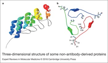
Recently, Hanenberg et al. have shown that the novel class of designed ankyrin repeat proteins (DARPins) presents an attractive alternative to the immunoglobulin scaffold for AD treatment (Ref. Reference Hanenberg100). DARPins are built from several ankyrin repeat modules that are tightly packed and capped by terminating repeats that shield the hydrophobic core (Fig. 2a). This structure has been demonstrated to show important advantages as compared with immunoglobulin-based (IgG) scaffolds. On one hand, their small size and their compact modular architecture result in high stability and solubility with low aggregation tendency (Refs Reference Binz101, Reference Binz102). Indeed, DARPin binding activity measured both at room temperature and in human serum at 37 °C (ex vivo) revealed half-lives of more than 60 days (Ref. Reference Stumpp, Binz and Amstutz103). Moreover, the absence of redox-sensitive disulphide bonds has enabled DARPins to be used for both intracellular and extracellular applications (Refs Reference Amstutz104, Reference Kohl105). Finally, the lack of specific endogenous receptors allows for improved fine-tuning of tissue distribution and clearance (Ref. Reference Zahnd106).
Three-dimensional structure of some non-antibody-derived proteins. (a) DARPin D1 (pdb 4DUI) is a scaffold protein built from several ankyrin repeat modules. The ribbon representation is coloured as the secondary structure succession. (b) Aβ 40 trimers (pdb 2M4J) show the five N-terminal residues (D1-L5) exposed to the solvent, whereas those in the C-terminus (V39–V40 are labeled) are buried in the hydrophobic core. The β-sheet spans residues L17-A21. Each monomer is arbitrarily coloured.

In a recent publication, it was demonstrated that Aβ peptide-specific DARPins, selected for their capacity for binding Aβ (residues 1–28) from libraries through ribosome display, were able to delay Aβ aggregation and prevent Aβ-mediated neurotoxicity in vitro. More importantly, DARPins improved cognitive performance and reduced soluble Aβ levels in 11-month-old Tg2576 mice (Ref. Reference Hanenberg100).
Which is the best target?
During the 1990s, several research groups showed that neuronal injury in AD was caused by soluble aggregated Aβ species (Refs Reference Pike107, Reference Busciglio, Lorenzo and Yankner108) and not only by amyloid plaques as had been thought previously, so soluble Aβ became an interesting target for AD disease-modifying treatment. Since evidence suggests that therapeutic interventions that can reduce Aβ fibrils at the cost of augmenting non-fibrillar Aβ assemblies, including Aβ oligomers, could be harmful (Ref. Reference Cheng109), targeting Aβ oligomers rather than amyloid plaques has been recommended (Ref. Reference Walsh and Selkoe110). This does not mean that the reduction of Aβ fibrils without augmenting oligomers is not worthy, as the recent findings with aducanumab demonstrate (see above).
Previously, it had been reported that antibodies generated against the N-terminus of Aβ, i.e. the B cell epitope, were able to inhibit Aβ aggregation, neutralise Aβ neurotoxicity and disaggregate pre-existing Aβ fibrils, and were most effective in reducing the brain amyloid burden (Refs Reference Bard43, Reference Horikoshi111, Reference Bussière112). As described recently by Lu et al. using seeded fibril growth from human brain extracts and data from solid-state nuclear magnetic resonance and electron microscopy, Aβ 40 peptide monomers were found to aggregate in oligomers with multiples of three units (trimers, hexamers, nonamers and dodecamers), where the N-termini (DAEFR, residues 1–5) are exposed to the solvent, while the highly hydrophobic C-termini are buried in the trimer core (Fig. 2b). Likewise, the 0.5 nm twisted hydrophobic package of the trimers leads to the formation of amyloid fibrils, in such a way that all of the N-termini within the fibril are solvent-exposed (Ref. Reference Lu113). Although this structure has not still been described for the Aβ 42 peptide, it offers a putative mechanism explaining why molecules designed to bind Aβ should be targeted to the N-terminus and suggests the reason why these have been described as the most effective interventions in terms of clearing Aβ oligomers.
How do all these molecules reduce the Aβ burden in brain?
Once antibodies have bound Aβ, they can inhibit its aggregation by blocking the oligomers, resulting in protection from their neurotoxic effects, reducing the Aβ burden and ameliorating AD symptoms. However, the mechanisms behind Aβ clearance, and how and where it takes place, are not known.
Earlier studies using AD mouse models suggested Fc receptor-mediated phagocytosis by microglia as the mechanism of Aβ clearance (Refs Reference Bard43, Reference Schenk114), assuming that antibodies cross the BBB and bind Aβ species in the brain (Refs Reference Bard43, Reference Wilcock115). In addition, expression of the neonatal Fc receptor (FcRn) at the BBB may mediate ‘reverse transcytosis’ of IgG in the brain-to-blood direction (Ref. Reference Schlachetzki, Zhu and Pardridge116). However, topical administration of a F(ab’)2 against Aβ also cleared amyloid deposits in an AD mouse model (Ref. Reference Bacskai117). In addition, the extent of amyloid clearance by passive Aβ immunisation in an Fc receptor-deficient AD mouse model was similar to that in an Fc receptor-sufficient AD mouse model (Ref. Reference Das118). These results indicated that amyloid clearance by Aβ immunisation does not require Fc receptor-mediated phagocytosis (Ref. Reference Kou119). Consequently, single-chain antibodies (scFv) and other structures devoid of the Fc fragment are becoming attractive therapeutic strategies, as they provide an alternative, non-inflammatory approach to facilitate Aβ clearance. Owing to the lack of the Fc fragment, they are incapable of directly activating microglia and could prevent meningoencephalitis, vasogenic oedema and cerebral microhaemorrhages that may occur with the use of complete antibodies (Ref. Reference Wang120).
There is also an alternative hypothesis to explain how the Aβ peptide is cleared by immunotherapy, the ‘sink hypothesis’, which states that anti-Aβ antibodies in the blood can bind and sequester plasma Aβ in the periphery, preventing it from influxing back into the brain, thus inducing a shift in the concentration gradient of Aβ over the BBB; this then favours the net transport of the peptide out of the brain (Ref. Reference DeMattos47). As mentioned above, the RAGE receptor is thought to be a primary transporter of β-amyloid across the BBB into the brain from the systemic circulation, while the low-density LRP1 mediates transport of β-amyloid out of the brain.
On the other hand, some researchers have pointed out that the poor clinical success of several anti-Aβ therapies might be because of the low rate of transport of the drug across the BBB. Previous work showed that an intact BBB could prevent 98% of small molecules and almost 100% of large molecules (>400 Da) from entering the brain (Ref. Reference Li29), so only a small percentage of antibodies (~0.1%) crosses the BBB (Ref. Reference Lemere30). Assuming that antibodies need to get into the brain to be effective, there have been several studies designed to find ways to improve antibody penetration into the brain.
Molecules designed to cross the BBB
One way to improve penetration into the brain is to target receptors on the BBB to induce active transport or transcytosis of proteins from the blood into the central nervous system (CNS) (Ref. Reference Boado121).
The transferrin receptor (TfR), a type II transmembrane protein highly expressed on brain endothelial cells (Refs Reference Kissel122, Reference Jefferies123), undergoes transcytosis at the BBB allowing the entry of iron (Refs Reference Fishman124, Reference Roberts, Fine and Sandra125). Although delivery of drug–Tf conjugates and TfR antibody conjugates have had some success (Refs Reference Dufes, Al Robaian and Somani126, Reference Yu and Watts127), many limitations have also surfaced, including evidence that high-affinity TfR antibodies remain trapped within the brain vasculature (Ref. Reference Manich128).
Recently, Niewoehner et al. designed a shuttle version of an anti-Aβ antibody using a monovalent binding mode to the TfR. This strategy increased β-amyloid target engagement in the PS2APP mouse model by 55-fold compared with the parent antibody. These authors also provided evidence that the monovalent binding mode facilitated transcellular transport, whereas bivalent binding mode led to lysosome sorting. This indicates that monovalent binding to the receptor might engage the sorting pathway normally used to transport monomeric transferrin and might lead to more efficient transport across brain endothelial cells (Ref. Reference Niewoehner129).
In the past year, Bien-Ly et al. have shown that bispecific antibodies against TfR and β-secretase (BACE1) traverse the BBB and effectively reduce brain Aβ levels. They have indeed proposed that the lower affinity for TfR enhances uptake into the brain by facilitating dissociation from TfR (Ref. Reference Yu130).
Another approach to ensure that antibodies targeted to Aβ are trafficked to the brain is to induce expression by the patient, not by driving an immune response as in active immunotherapy, but rather by delivering the gene encoding the antibody. In this strategy, an adeno-associated virus (AAV) is injected into the patient and integrates its DNA, leading to the continuous production of an anti-Aβ mAb or scFv over an extended period of time. The AAV is the most promising vector for gene therapy because of its non-toxicity and long-term gene expression (Ref. Reference Robert86), and, in contrast to active immunotherapy, the use of inducible promoters allows intervention in case an adverse reaction occurs. In 2013, Shimada et al. examined the effect of delivering an anti-Aβ mAb-expressing AAV vector to Tg2576 mice. Prophylactic treatment of young (5 months) and therapeutic treatment of older (10 months) animals resulted in a significant and prolonged decrease in the amount of Aβ peptide that accumulated in the brain. A single dose of the Aβ mAb-expressing AAV vector resulted in the production of an Aβ mAb that persisted throughout the 64-week experimental period, with no evidence of microhaemorrhages or inflammation (Ref. Reference Shimada131).
In another study, which combined both approaches explained above, the focal stereotactic infusion of an rAAV vector expressing an ER-targeted anti-Aβ scFv into the hippocampus of young adult triple-transgenic AD (3xTg-AD) mice resulted in significant suppression of amyloid and tau pathologies, indicating specific subcellular targeting of these therapeutics (Ref. Reference Sudol132).
Thus, in order to diminish the therapeutic doses and guarantee safe and effective delivery of the drug, therapy development must not only to target Aβ-oligomers and address the mechanism behind their clearance, but also maximise the amount of therapy that reaches the brain.
Conclusions
The great number and variety of therapies designed to treat AD mirror the need to find a solution to cure this devastating disease. Aβ-immunotherapy has emerged as a promising strategy to this purpose, albeit some serious drawbacks have appeared during clinical trials. Active immunotherapy is still in Phase II, whereas passive immunotherapy for several full-length mAbs has reached Phase III. Bapineuzumab did not improve clinical outcomes in patients with AD, despite differences in biomarkers observed in treated APOE ε4 carriers, a cohort in who ARIA-E occurred. To prevent ARIAs, an improved version of bapineuzumab was designed and tested in two recently completed Phase I trials. In contrast, solanezumab showed a slight benefit regarding cognitive decline after some months of treatment, without differences based on APOE ε4 allele expression. Because a good safety profile was obtained, DIAN is supervising a Phase II/III trial in asymptomatic and very mildly symptomatic carriers of ADAD mutations. Another prevention study using solanezumab is the Alzheimer's Disease Cooperative study A4, in which asymptomatic or very mildly symptomatic people who have evidence of brain amyloid deposition by PET are being recruited. Gantenerumab and crenezumab, currently in Phases II and III, respectively, will be also evaluated as a part of the Alzheimer Prevention Initiative. Aducanumab was found to be effective in reducing cognitive decline at the intermediate dose, and in decreasing brain levels of amyloid at the non-safe higher dose. Therefore, the intermediate dose is being assayed in an ongoing Phase III trial. Finally, ponezumab has been repurposed for treating CAA, BAN2401 is in Phase II, and IVIG preparations trials found no evidence of slowing AD progression but are still under study. As a summary, at the moment, two mAbs, i.e. solanezumab and aducanumab, have already shown some clinical/cognitive benefit in mild AD patients.
After all the clinical trials carried out in patients with AD, the main conclusion is that they must be performed with patients in the prodromal phase, as patients with MCI show irreversible effects of the disease. Consequently, therapies in development must be designed to treat patients from the very early stages of the disease, which indicates the necessity to continue working on the worldwide establishment of validated CSF biomarker signatures and neuroimaging protocols.
Antibody-derived molecules in pre-clinical stages such as Fabs, scFvs, V domains and DARPins are promising approaches because they lack the Fc region. Although earlier studies suggested Fc receptor-mediated phagocytosis by microglia as the mechanism of clearance of opsonised-Aβ, later results indicated that this is not the case. Avoiding Fc-mediated activation of microglia could prevent meningoencephalitis, vasogenic oedema and cerebral microhaemorrhages that may occur with the use of complete antibodies. In any case, much more research is needed before clinical trials are initiated.
Finally, it must be taken into account that one of the main obstacles to Aβ immunotherapy is the ability of the drug to cross the BBB. To overcome this drawback, transcytosis by receptors highly expressed on brain endothelial cells is being explored, as well as AAV-based gene therapy.
Although much more needs to be done to completely understand the multiple aspects of AD, including its relationship with cholesterol metabolism and the role of the immune system, our knowledge of the pathogenesis of the disease has been widely improved and we are getting closer to finding a disease-modifying drug that could prevent the development of this disease, which is an urgent necessity in today's society.
Acknowledgements and funding
Instituto de Salud Carlos III [FIS-PI113-01330], Generalitat de Catalunya [SGR-GRC-2014-00885], Generalitat de Catalunya/FEDER [2014-PROD00032] and a PIF-UAB student grant (LMG).
FURTHER RESOURCES AND CONTACTS
ALZFORUM http://www.alzforum.org/
A networking organisation with the goal of finding a cure for Alzheimer Disease. News, webinars, databases, papers, professional resources, and general information about AD are available.
ALZHEIMER'S DISEASE INTERNATIONAL http://www.alz.co.uk/
A global Foundation with the aim of providing information, solutions and knowledge about AD. Its annual reports are of great interest to AD researchers.
DOMINANTLY INHERITED ALZHEIMER NETWORK (DIAN) http://www.dian-info.org/
DIAN is an international research partnership of leading scientists determined to understand a rare form of AD that is caused by a gene mutation. Understanding this form of AD may provide clues to decoding other dementias and developing dementia treatments.
ALZHEIMER'S PREVENTION INITIATIVE (API) http://banneralz.org/research-plus-discovery/alzheimers-prevention-initiative.aspx
An international collaborative initiative formed to launch a new era of AD prevention research by evaluating the most promising therapies in cognitively normal people who, based on their age and genetic background, are at the highest imminent risk of developing AD symptoms.