Case report
We report the case of a 4-month-old girl, born at term, whose history was notable for failed critical congenital heart disease (CHD) screening test in the neonatal nursery. The right upper extremity oxygen saturation was 92% and the right lower extremity saturation was 93%, on initial and two repeat measurements. An echocardiogram at that time was interpreted as normal. The patient subsequently had an 8-day hospital admission for viral bronchiolitis and failure to wean off oxygen despite a repeat echocardiogram showing only a small patent foramen ovale with trivial left-to-right shunting.
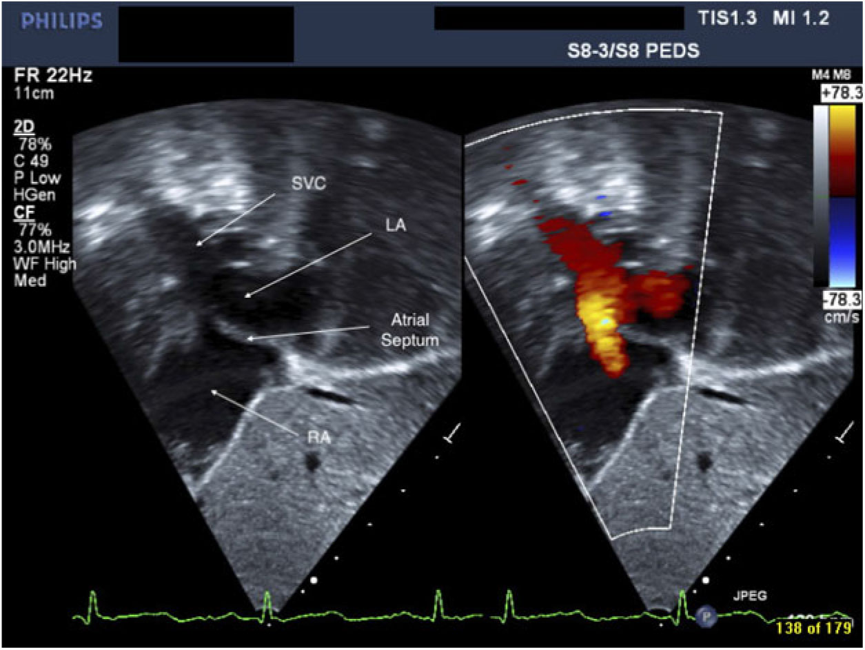
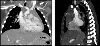
The patient presented 1 month later to cardiology clinic for persistent hypoxemia, with a repeat echocardiogram. The oxygen saturations on room air were 89–91%, corrected to 91–93% with 3 liters per minute nasal cannula, without any pre- or post-ductal saturation difference and without any signs of respiratory illness. The repeat echocardiogram (Fig 1, Video S1) was notable for a right superior vena cava straddling the atrial septum, with aliasing flow into the right atrium, dominant flow into the left atrium, small superior sinus venosus atrial septal defect, and partial anomalous pulmonary venous return of the right upper pulmonary vein to the superior vena cava. The majority of blood flow from the superior vena cava was directed towards the left atrium. The finding was confirmed by computed tomography angiography (Fig 2). She was discharged on aspirin and later underwent a Warden procedure resulting in complete resolution of hypoxemia.
Echocardiogram showing the superior vena cava (SVC), with the majority of flow into the left atrium (LA), and aliasing flow into the right atrium (RA).

Coronal (left) and sagittal (right) contrast-enhanced CT angiography showing the right upper pulmonary veins (RUPV) draining into the right superior vena cava (RSVC), which is draining with preferential flow into the left atrium (LA), and minimal flow into the right atrium (RA).

Discussion
Sinus venosus atrial septal defect is a well-known entity, but rarely presents with cyanosis. It consists of a communication between the superior vena cava, right atrium and left atrium at the atrial level, and often involves partial anomalous pulmonary venous return of the right pulmonary veins into the superior vena cava, which is seen in 80–90% of cases.Reference Hoey, Lewis and Yusuf 1 Of those cases where partial anomalous pulmonary venous return is not reported, it is likely that it may have been missed on echocardiogram, as all documented cases that have undergone surgery or autopsy have shown partial anomalous pulmonary venous return.Reference Van Praagh, Geva and Lock 2 In most cases left to right shunts are present.Reference Hoey, Lewis and Yusuf 1 Our case presentation, with cyanosis due to a right to left shunt, is quite rare, and was due to the right superior vena cava which is straddling the atrial septum.
The embryological basis behind a sinus venosus atrial septal defect was initially thought to be due to malalignment of the atrial septum or leftward cephalic deviation of the right horn of the sinus venosus,Reference Van Praagh, Geva and Lock 2 but this failed to account for the constant presence of partial anomalous pulmonary venous return. More recent studies have discovered that sinus venosus atrial septal defect, biatrial right superior vena cava and right superior vena cava drainage isolated to the left atrium are all cases of unroofing of the anterior wall of the right upper and lower pulmonary veins through the posterior wall of the right superior vena cava. Where this occurs near the right atrium–superior vena cava junction, it creates the appearance of an interatrial communication but no true atrial septal defect exists.Reference Vassallo, Pascotto and Pisacane 3 In this low-type sinus venosus atrial septal defect, unroofing of the right pulmonary veins into the sinus venosus portion of the right atrium is more accurately a sinopulmonary venous defect.Reference Van Praagh, Geva and Lock 2 In a high-type sinus venosus atrial septal defect, the pulmonary veins unroof into the right superior vena cava proper, away from the sinus venosus, and is more accurately a cavopulmonary venous defect.Reference Van Praagh, Geva and Lock 2 Thus sinus venosus atrial septal defect is a misnomer and the interatrial communication is really the orifice of the left atrium as it communicates with the uninterrupted right pulmonary venous return. The right superior vena cava drains into the unroofed partial anomalous pulmonary venous return defect, through the anomalous pulmonary veins and into the left atrium. Where left atrium flow dominates, the right superior vena cava–right atrium junction becomes stenotic or atretic with resultant right superior vena cava return mostly or entirely into the left atrium.Reference Van Praagh, Geva and Lock 2 Cases with biatrial right superior vena cava drainage are rare, documented only as case reports in the literature.Reference Van Praagh, Geva and Lock 2 , Reference Shapiro, Al-Sadir and Campbell 4
In this patient’s case, aliasing flow on echocardiogram of the superior vena cava into the right atrium suggested narrowing of the right atrium–superior vena cava junction, with concomitant lower velocity flow into the left atrium, suggesting straddling across the atrial septum, with the vast majority of blood flow shunted to the left atrium (Video S1). One can further corroborate an aberrant superior vena cava connection with agitated saline contrast injection on echocardiogram. Saline must be injected into the upper extremities to elucidate upper venous anatomy. Upper extremity injection will show abnormal simultaneous flow into the left and right heart, suggesting a right to left shunt due to aberrant superior venous return.Reference Truman, Rao and Kulangara 5 When an isolated anomalous left superior vena cava to left atrium or left superior vena cava to unroofed coronary sinus is suspected, with normal right superior vena cava, injection must be done in the left upper extremity. Cross axial imaging with computed tomography angiography and magnetic resonance angiography are better able to visualize the full anatomy of the superior vena cava and partial anomalous pulmonary venous return.Reference Sonavane, Milner and Singh 6 Cardiac catheterization may be performed where diagnosis is uncertain, and is traditionally considered the gold standard.
Complications of aberrant superior vena cava drainage into the left atrium include the risks of cyanosis, paradoxical emboli (even with peripheral intravenous lines), declining intellectual capacity, effort intolerance, and brain abscesses, and thus surgical correction is indicated. A cohort of 83 children with brain abscesses revealed 50% had CHD.Reference Wong, Lee and Wang 7 Multiple case reports have also demonstrated that otherwise asymptomatic adults with mild cyanosis and history of recurrent cryptogenic brain abscesses may have an underlying right to left shunt from an anomalous left superior vena cava.Reference Van Praagh, Geva and Lock 2 , Reference Erol, Metin and Alehan 8 – Reference Menachem, Sundaram and Rhodes 10 This highlights the importance of early diagnosis and intervention.
The Warden procedure, as was done in our case, is well described, with some modifications since its inception by Dr. Warden in 1984.Reference Ling, Gan and Sandeep 11 After median sternotomy, the superior vena cava is directly cannulated above the anomalous pulmonary venous connection. The superior vena cava is divided. The anomalous pulmonary venous drainage is baffled through the right atrium, through the atrial septal defect, and into the left atrium. The cephalic portion of the superior vena cava is then sewn to the right atrium appendage.Reference Ling, Gan and Sandeep 11
While this patient’s CHD was not critical, a congenital cardiac abnormality was first suspected due to critical CHD screening. Optimal screening is done between 6 and 24 hours of life, where sensitivity and specificity are high and the risk of deterioration is low, given that up to 9% of neonates with CHD may present with circulatory collapse when screened >24 hours of life.Reference Narayen, Blom and Ewer 12 This screening modality has the added benefit of detecting pathologies other than critical congenital heart disease, including infection, pulmonary disease and non-critical CHD in up to 80% of false-positive cases.Reference Narayen, Blom and Ewer 12 While cut-offs differ, the test is generally considered positive when pulse oximetry is <95% in any limb, or there is a difference >2–3% between extremities despite three measurements spaced 1 hour apart. Pulse oximetry screening has proven to be cost-effective, safe, reproducible, and life-saving.Reference Narayen, Blom and Ewer 12
In conclusion, our case demonstrates that superior sinus venosus defect can rarely present with cyanosis. Also elucidated is the lesion’s embryological basis, and the role of echocardiogram and cross-axial imaging to complete a comprehensive workup leading to definitive operative repair.
Supplementary Material
To view supplementary material for this article, please visit https://doi.org/10.1017/S1047951119000234
Financial Support
This research received no specific grant from any funding agency, commercial or not-for-profit sectors.
Conflict of Interest
None.
Ethical Standards
This article does not contain any studies with human participants or animals performed by any of the authors.