Case report
Infants of diabetic mothers are at risk for transient hypertrophic cardiomyopathy. Although poor maternal glycaemic control is associated with a thick neonatal interventricular septum, studies have shown that infants of diabetic mothers with tight glycaemic control can suffer from hypertrophy as well.Reference Ullmo, Vial and Di Bernardo 1
Classically, there is asymmetric hypertrophy of the interventricular septum, which may lead to left ventricular outflow tract obstruction caused by systolic anterior motion of the mitral valve. Neonates are usually asymptomatic, but 5–10% of infants develop impaired cardiac output due to left ventricular outflow tract obstruction, which worsens with increasing septal hypertrophy.Reference Hay 2
Spontaneous regression of hypertrophy occurs as plasma insulin concentrations normalise within the first few months to a year of life. Medical therapies including β-blockers and fluid resuscitation have been found to help support cardiac output. Limited literature exists about the use of extracorporeal membrane oxygenation as a supportive therapy for patients with poor cardiac output refractory to maximal medical management.
Case
A 4-kg, large-for-gestational age, male was born via emergent C-section because of poor biophysical profile at 37 weeks gestation to a 32-year-old G2P1 mother with insulin-dependent diabetes. Maternal medications included metformin and insulin; glycated hemoglobin during pregnancy was not documented. There was no family history of CHD or familial hypertrophic cardiomyopathy. Apgars were 1, 5 and 8 at 1, 5, and 10 minutes of life, respectively. He required intubation shortly after delivery for persistent hypoxia despite 100% supplemental oxygen.
On day of life one, he had severe hypotension and worsening lactic acidosis. On examination, he had a loud systolic ejection murmur, hepatomegaly, and poor perfusion. He was started on a prostaglandin infusion to improve cardiac output. There was >10% difference in pre- and post-ductal oxygen saturations; inhaled nitric oxide and 100% supplemental oxygen were administered because of concern for persistent pulmonary hypertension.
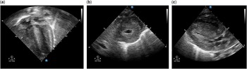
An echocardiogram revealed severe biventricular hypertrophy with dynamic left ventricular outflow tract obstruction and near cavity obliteration of the left ventricle (Fig 1). There was systolic anterior motion of the mitral valve towards the left ventricular outflow tract during systole and mitral regurgitation due to incomplete mitral leaflet apposition during systole. The interventricular septum measured 14–16 mm, (normal for size is 3 mm).Reference Overbeek, Kapusta and Peer 3 There was a large, unrestrictive patent ductus arteriosus, with right-to-left shunting. Despite aggressive fluid resuscitation, supplemental oxygen, inhaled nitric oxide, prostaglandin, vasopressin, norepinephrine, and milrinone infusions, the baby experienced ongoing hypotension and hypoxaemia.
Transthoracic echocardiogram at day one of life. ( a ) Apical four-chamber view. ( b ) Parasternal short-axis view. ( c ) Parasternal long-axis view. All images are obtained at end diastole.

On day of life two, his clinical status continued to decline with the development of severe anasarca, worsening lactic acidosis (peak lactate 13 mmol/L), and end-organ dysfunction as evidenced by transaminitis (alanine aminotransferase 63 U/L, aspartate aminotransferase 677 U/L), coagulopathy (international normalized ratio 3.8, prothrombin time 37.5 s, partial thromboplastin time 60.4 s, fibrinogen 151 mg/dL), and oliguric renal failure (blood urea nitrogen 3 mg/dL, Cr 0.9 mg/dL). To support cardiac output, he was placed on venous-arterial extracorporeal membrane oxygenation at ~48 hours of life using an 8-Fr cannula in his right carotid artery and a 10-Fr cannula in his right internal jugular vein.
On venous-arterial extracorporeal membrane oxygenation, he was weaned off vasopressin and norepinephrine infusions, but remained on milrinone for its lusitropic effects. A peritoneal catheter was placed to drain ascites, and then used for peritoneal dialysis and fluid removal. Within 48 hours of extracorporeal membrane oxygenation support, his urine output and kidney function improved, his lactic acid levels returned to normal, and his transaminitis and coagulopathy resolved. Infection and metabolic disease were ruled out. Genetic testing for Pompe disease was negative.
Serial echocardiograms during the first week of life showed regression of ventricular hypertrophy, left ventricular outflow tract obstruction, and resolution of right-to-left shunting. After 7 days of extracorporeal membrane oxygenation support, on day of life nine, he was successfully decannulated. He was managed on milrinone and esmolol infusions to promote diastolic relaxation and to optimise diastolic filling time, respectively.
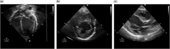
After successful extubation, he required non-invasive positive pressure ventilation that was gradually weaned over the next week. He was weaned off milrinone at 3 weeks of life and transitioned to oral propranolol from esmolol. At that time, his echocardiogram demonstrated near resolution of the severe biventricular hypertrophy with a septal measurement of 5–6 mm and no evidence of residual left ventricular outflow tract obstruction (Fig 2). His only discharge medication was propranolol at 1 month of age.
Transthoracic echocardiogram at 4 weeks of age. ( a ) Apical four-chamber view. ( b ) Parasternal short-axis view. ( c ) Parasternal long-axis view. All images are obtained at end diastole.

Discussion
Infants of diabetic mothers may be born with hypertrophic cardiomyopathy, but the disease is usually transient in nature with most being asymptomatic and haemodynamically stable. The hypertrophy regresses significantly within the first 2 weeks of life and usually completely resolves by 6 months of age.Reference Hay 2
Approximately 5% of these infants, however, will experience low cardiac output secondary to left ventricular outflow tract obstruction related to severe left ventricular hypertrophy. Typical management of these infants includes maintaining intravascular volume for adequate preload and β-adrenergic blockade to slow the heart rate and maximise diastolic filling time. Rarely, in the case of severe haemodynamic instability from left ventricular outflow tract obstruction and low cardiac output, further haemodynamic support may be warranted. There are limited reports on the use of venous-arterial extracorporeal membrane oxygenation for this indication.Reference Robinson, Eshaghpour, Ewing and Baumgart 4 – Reference Nolent, Renolleau and Hallalel 7 Similar to the case described by Goldberg et alReference Goldberg, Mery and Griffiths 6 , our case had evidence of persistent pulmonary hypertension contributing to hypoxaemia, which was improved after <1 week of extracorporeal membrane oxygenation.
The decision to support a patient with venous-arterial extracorporeal membrane oxygenation must take into account the significant risks known to be related to bleeding and/or thromboembolism. A major neurologic morbidity – seizure, ischaemic stroke, or intracranial haemorrhage – is especially high in the neonatal population. In a patient who is experiencing end-organ dysfunction from low cardiac output despite maximal medical management, it is reasonable to temporarily support the patient on venous-arterial extracorporeal membrane oxygenation, as the benefit would hopefully outweigh the risks. In our case, the severe hypertrophy and left ventricular outflow tract obstruction resolved enough to decannulate the patient after 1 week of support with resolution of all end-organ dysfunction and no extracorporeal membrane oxygenation complications.
Acknowledgements
None.
Financial Support
The research received no specific grant from any funding agency or from commercial or not-for-profit sectors.
Conflicts of Interest
None.