Introduction
The chemical composition of follicular fluid plays an important role in oocyte fertilizability and also in the resultant embryo development because developing oocytes are in direct proximity to this microenvironment (Fortune, Reference Fortune1994; Józwik et al., Reference Józwik, Józwik, Teng and Battaglia2006; Revelli et al., Reference Revelli, Piane, Casano, Molinari, Massobrio and Rinaudo2009). Urea can be transported by passive diffusion through the blood–follicle barrier (Józwik et al., Reference Józwik, Józwik, Teng and Battaglia2006), indicating a substantial correlation between urea concentration in follicular fluid and plasma in both humans and cows (Hammon et al., Reference Hammon, Holyoak and Dhiman2005; Józwik et al., Reference Józwik, Józwik, Teng and Battaglia2006). Non-renal causes of increased blood urea nitrogen (BUN), therefore, include increased protein intake associated with higher urea levels in the female reproductive tract (FRT) of healthy dairy cows.
A body of evidence suggests a relationship between high levels of BUN and a reduction in fertility, even in healthy dairy cows (Ferguson et al., Reference Ferguson, Galligan, Blanchard and Reeves1993; Butler et al., Reference Butler, Calaman and Beam1996; Sinclair et al., Reference Sinclair, Kuran, Gebbie, Webb and McEvoy2000; De Wit et al., Reference De Wit, Cesar and Kruip2001). Cumulus–oocyte complexes (COCs) collected from heifers offered a high-NH3-generating diet (high levels of BUN) and had a reduced ability to develop to the blastocyst stage (Santos et al., Reference Santos, Marques, Antunes, Chaveiro, Andrade, Borba and da Silva2009). Urea in in vitro oocyte maturation impaired the segregation of chromatids and the extrusion of the polar body (De Wit et al., Reference De Wit, Cesar and Kruip2001), resulting in decreased oocyte fertilizability, cleavage, and developmental rate.
Transcription factors such as CDX2 and OCT4 are necessary for the first cell fate decision to form a trophectoderm or inner cell mass (ICM) (Arnold & Robertson, Reference Arnold and Robertson2009). Developmentally, these transcription factors should be strictly regulated to govern cell lineage allocation and the pattern of the early embryonic development in mammals (Wang et al., Reference Wang, Miyazaki and Smith2000). For example, to sustain the pluripotent state in vitro, the precise level of OCT4 should be balanced (Wang et al., Reference Wang, Miyazaki and Smith2000). Reduced OCT4 expression results in induced trophectoderm differentiation, however its overexpression leads to differentiation into endoderm and mesoderm (Wang et al., Reference Wang, Miyazaki and Smith2000). Any upregulation or downregulation of OCT4 expression or any changes in the OCT4:NANOG ratio results in a divergent developmental programme (Wang et al., Reference Wang, Miyazaki and Smith2000; Theunissen et al., Reference Theunissen, van Oosten, Castelo-Branco, Hall, Smith and Silva2011; Muñoz-Descalzo et al., Reference Muñoz-Descalzo, Rué, Garcia-Ojalvo and Martinez Arias2012).
It has been shown that the zona pellucida (ZP), an extracellular matrix surrounding the mammalian oocyte, is involved in supporting communication between oocytes and follicle cells during oogenesis and also in protecting oocytes (Wassarman et al., Reference Wassarman, Chen, Cohen, Litscher, Liu, Qi and Williams1999; Gook et al., Reference Gook, Edgar, Borg and Martic2008). Female mice that were null for ZP3 (ZP–/–) showed retarded follicular development and their ovaries contained lower numbers of graafian follicles, suggesting a role for ZP proteins produced around growing oocytes in follicle development (Wassarman et al., Reference Wassarman, Chen, Cohen, Litscher, Liu, Qi and Williams1999; Gook et al., Reference Gook, Edgar, Borg and Martic2008). In addition, ZP proteins ZP2 and ZP3 are shared in mammals (Pimenta et al., Reference Pimenta, Sardinha, Marques, Domingos, Baptista, Barbas, Martins, Mesquita, Pessa, Soares, Viegas, Cabrita, Horta, Fontes, Prates and Pereira2013) and make up 80% of the total ZP (Wassarman, Reference Wassarman2008). The presence of both ZP2 and ZP3 is critical for the general assembly of the ZP (Wassarman, Reference Wassarman2008); sperm cells first bind to ZP3 to accomplish the acrosome reaction, then can bind to ZP2 (as the secondary receptor for acrosome-reacted sperm), penetrate the ZP, and fuse with the oocyte plasma membrane (Callebaut et al., Reference Callebaut, Mornon and Monget2007).
Two domains of the ZP3 and ZP2 proteins, the N-terminal (ZP-N) and C-terminal (ZP-C), have been identified that are essential for dimerization of ZP proteins (Monné et al., Reference Monné, Han, Schwend, Burendahl and Jovine2008; Han et al., Reference Han, Monné, Okumura, Schwend, Cherry, Flot, Matsuda and Jovine2010). The tight association of ZP-C and ZP-N is essential to ensure the formation of a complete ZP (Callebaut et al., Reference Callebaut, Mornon and Monget2007). Importantly, it has been shown that ZP-N is involved in other specific functions such as in zona pellucida filament formation (Jovine et al., Reference Jovine, Janssen, Litscher and Wassarman2006; Wassarman, Reference Wassarman2008; Monné et al., Reference Monné, Han, Schwend, Burendahl and Jovine2008). Koyama et al. (Reference Koyama, Hasegawa, Inoue and Isojima1991) showed that using ZP2 antisera against the N-terminal of ZP-N domain, inhibited the penetration and binding of sperm into the oocyte. Many pathological mutations fall within the ZP-N domain (Monné et al., Reference Monné, Han, Schwend, Burendahl and Jovine2008), it may work as an independent unit to show a specific function in reproductive processes (Jovine et al., Reference Jovine, Janssen, Litscher and Wassarman2006).
In the current study, we used the ZP-N domain of the ZP3 or ZP2 protein for molecular docking with urea and primarily focused on the interaction of urea with the ZP-N domain of ZP3. Urea has a strong interaction with the backbone and side chains of proteins, causing denaturation (Caflisch & Karplus, Reference Caflisch and Karplus1999; Hua et al., Reference Hua, Zhou, Thirumalai and Berne2008). We, therefore, hypothesized a possible interaction between urea and the ZP-N domain of ZP3 or ZP-2. The supplementation of maturation medium with urea may alter oocyte competence and induce an imbalanced expression of transcription factors in the resultant blastocyst. Oocytes and early embryos, in vivo in contrast with in vitro, are exposed to various follicular and oviductal components such as hormones and cytokines that may interact with urea (Matoba et al., Reference Matoba, Bender, Fahey, Mamo, Brennan, Lonergan and Fair2014; Kowsar et al., Reference Kowsar, Marey, Shimizu and Miyamoto2016).
Materials and methods
Computational and molecular docking (in silico study)
Using AutoDock4.2 via the genetic algorithm (GA) procedure (Morris et al., Reference Morris, Huey, Lindstrom, Sanner, Belew, Goodsell and Olson2009; Barzegar et al., Reference Barzegar, Mansouri and Azamat2016), the urea molecule was docked into a PDB structure of: (1) the mammalian ZP-N domain of ZP3, crystal form I (PDB code: 3D4C); (2) the ZP-N domain of mouse ZP2 (PDB code: 5II6); (3) the full-length chicken sperm receptor ZP3 (PDB code: 3NK3) and (4) the predicted full-length mammalian sperm receptor ZP3. The files required to perform docking simulation were prepared utilizing the AutoDock Tools package 1.5.6 (Morris et al., Reference Morris, Huey, Lindstrom, Sanner, Belew, Goodsell and Olson2009). The binding sites of urea/protein complexes were regulated with a grid size that was set at 54 × 52 × 52 points for the urea/ZP-N domain of the ZP3 complex (PDB code: 3D4C), 58 × 52 × 78 points for the urea/ZP-N domain of mouse ZP2 complex (PDB code: 5II6), 82 × 56 ×110 points for the full-length chicken sperm receptor ZP3 (PDB code: 3NK3), and 68 × 82 × 66 points for the predicted full-length mammalian sperm receptor ZP3 along xyz axes and through a grid spacing of 0.375 Å, to cover the full active site of the ZP3 ZP-N domain and 1 Å for other proteins. To investigate the free energy and behaviour of urea binding to ZP-N, docking procedures were performed six consecutive times, such that each time the previous docked urea/ZP-N complex was used as the template (macromolecule) for the next docking run under the same conditions. To find the best free energy that indicated the strongest binding of urea to ZP-N, the total number of docking runs was set at 300.
Because the whole molecule of mammalian ZP3 (containing both ZP-N and ZP-C domains) was not determined, the full-length chicken sperm receptor ZP3 (PDB ID: 3NK3) was used to predict the whole mammalian ZP3 molecule using I-TASSER software (Roy et al., Reference Roy, Kucukural and Zhang2010; Zhang Reference Zhang2007).
Production of bovine embryos (in vitro study)
Oocyte collection, in vitro oocyte maturation and fertilization, and in vitro embryo culture have been described previously (Hernandez-Ledezma et al., Reference Hernandez-Ledezma, Sikes, Murphy, Watson, Schultz and Roberts1992; Kimura et al., Reference Kimura, Spate, Green and Roberts2004; Green et al., Reference Green, Harvey, Spate, Kimura, Thompson and Roberts2016). Briefly, bovine ovaries were obtained from a local slaughterhouse, transported to the laboratory within 2 h at approximately 30°C in a thermos box containing 0.9% NaCl, 0.1% penicillin and streptomycin, washed in 30°C sterile 0.9% NaCl containing 0.1% penicillin and streptomycin, and stored in sterile saline until aspiration. Cumulus–oocyte complexes were only aspirated from follicles ranging from 4 to 8 mm in diameter as these oocytes, occupying the medium-sized (> 4–8 mm) follicles, have been shown to be influenced by a key nutritionally sensitive process (Sinclair et al., Reference Sinclair, Kuran, Gebbie, Webb and McEvoy2000). Approximately 540 bovine COCs graded as class 1 or class 2 only according to a previous study (Leibfried & First, Reference Leibfried and First1979) were selected under a stereomicroscope (Olympus, Tokyo, Japan) and washed in HEPES–TCM-199 (Invitrogen, Carlsbad, CA, USA) supplemented with 50 mg/ml kanamycin (Sigma-Aldrich), 50 mg/ml heparin (Sigma-Aldrich). Cumulus–oocyte complexes were then washed twice in HEPES–TCM-199 and randomly incubated in maturation medium in groups of 10 in bicarbonate-buffered TCM-199 supplemented with 10% (w/v) fetal bovine serum (FBS; BioWhittaker, Walkersville, MD, USA) and 10 IU/ml follicle-stimulating hormone (FSH; Sigma-Aldrich) in 35 mm cell culture dishes (Falcon brand, BD Biosciences, San Jose, CA, USA) for 24 h at 38.5°C under 5.5% CO2 and 20% O2, balanced N2 and maximum humidity. The maturation medium was free from bovine serum albumin (BSA) to avoid any possible interaction with urea in the medium. To see how embryo development was affected by urea-exposed oocytes, urea (Sigma-Aldrich) was added to the maturation medium at 0 mg/dl (control group, n = 143 COCs, five replicates), 20 mg/dl (n = 184 COCs, five replicates), or 40 mg/dl (n = 213 COCs, five replicates). These urea concentrations were equivalent to 0, 9.3 and 18.7 mg/dl BUN, respectively. The urea levels used in our study are usually observed in serum of healthy dairy cows fed low or high protein diets (Ferguson et al., Reference Ferguson, Galligan, Blanchard and Reeves1993). After in vitro maturation, urea-exposed oocytes and control oocytes (n = 10/droplet) were fertilized with sperm from a single sire (Spermex, GmbH) at a final concentration of 1 × 10⁶/ml in in vitro fertilization medium supplemented with amino acids (50 μl) in 35 mm cell culture dishes (Falcon brand, BD Biosciences, San Jose, CA, USA) at 38.5°C under 5% CO2 in the air. Zygotes were recovered after 24 h, cumulus cells were manually stripped and the resultant embryos transferred into cell culture dishes (n = 20 per droplet) containing synthetic oviductal fluid (SOF) medium supplemented with amino acids and 4 mg/ml BSA. Embryos were then cultured in an incubator at 38.5°C under 5% O2 and 6% CO2 for 60 h. Developing embryos were scored using a stereomicroscope (Olympus, Tokyo, Japan) at the zygote, 8–16- or 2–8-cell stage, blastocyst, and hatching stages at days 1, 3, 7 or 9 post-insemination, respectively. Pools of seven embryos in five replicates were collected and stored in RNeasy lysis buffer (Qiagen) supplied with 2-mercaptoethanol (Sigma-Aldrich) at −70°C for qRT-PCR analysis. The criterion for the fertilization was the observation of two pronuclei (2-PN) in zygotes 18 h after insemination using a phase-contrast microscope (Nagano et al., Reference Nagano, Katagiri and Takahashi2006; Son et al., Reference Son, Lee and Lim2005). Oocytes with cytoplasmic degeneration by day 3 after insemination were considered as the degenerated oocytes (Makita et al., Reference Makita, Ueda and Miyano2016).
Scanning electron microscopy
For scanning electron microscopy analysis, COCs were cultured in maturation medium supplemented with different levels of urea (0, 20, 40 mg/dl). After 24 h, COCs were denuded from cumulus cells by pipetting in maturation medium. Then, oocytes were washed in phosphate-buffered saline to eliminate the culture medium. Oocytes were fixed for 2 h in 2.5% glutaraldehyde in 0.2 mol/l cacodylate buffer on 12 mm circular coverslips placed in Petri dishes containing a fixative solution. Coverslips were coated with poly-l-lysine solution (Sigma P-8920) (1:10 poly-l-lysine solution for 15–30 min). Next, using an ascending series of ethanol solutions, the oocytes were dehydrated and dried by immersing in hexamethyldisilazane (HMDS, Sigma-Aldrich) for 10 min and then air dried in a desiccator at room temperature (Jusman et al., Reference Jusman, Ng and Abu Osman2014). The dried samples were mounted on stubs, sputter-coated with a 10 nm layer of gold, and were checked under a Philips XL 30 scanning electron microscope (Eindhoven, The Netherlands).
RNA extraction, reverse transcription, quantitative analysis of transcripts by qRT-PCR
RNA extraction and reverse transcription were done as previously reported (Moulavi et al., Reference Moulavi, Hosseini, Hajian, Forouzanfar, Abedi, Ostadhosseini, Asgari and Nasr-Esfahani2013). Briefly, total RNA was extracted using an RNeasy Micro kit (Qiagen, Mississauga, Ontario, Canada), then treated with DNase I (Ambion, Streetsville, Ontario, Canada) according to the manufacturer's manual. The WPA Biowave spectrophotometer (Cambridge, United Kingdom) was used to examine the quality and quantity of RNA. Reverse transcription was carried out at 25°C for 10 min, 42°C for 1 h and 70°C for 10 min. qRT-PCR was carried out using 1 µl of cDNA (50 ng), 5 µl of the SYBR Green/0.2 µl ROX qPCR Master Mix (2×) (Fermentas, Germany) and 1 µl of forward and reverse primers (5 pM) adjusted to a total volume of 10 ml using nuclease-free water. ACTB was used as the reference gene. The abundance of ACTB was constant throughout embryo developmental stages assessed. Each replicate of qRT-PCR was repeated three times to minimize technical errors. Primer sequences used for qRT-PCR were as follows: 5′-CCTTCTTTGAGTTCGGAG-3′, forward, and 5′-CCTTCAGAGACAGCCAG-3′, reverse, for BCL2 (AC_000181.1, 121 bp, Tm: 60°C); 5′-AGCGAGTGTTC-TGAAGCG-3′, forward, and 5′-CCCAGTTGAAG-TTGCCGT-3′, reverse, for BAX (AC_000175.1, 182 bp, Tm: 61°C); 5′-GGTGTTTGGTGAACTCTC-3′, forward, and 5′-CATTGATTGTTCCAAGGCT-3′, reverse, for NANOG (AC_000162.1, 98 bp, Tm: 53°C); 5′-GGAAAGGTGTTCAGCCA-3′, forward, and 5′-TTCTCGTTGTTGTCAGC-3′, reverse, for OCT4 (NC_000006.12, 131 bp, Tm: 62°C); 5′-GAAGCAGAA-TAAGAATCGG-3′, forward, and 5′-TTTGAAGAGT-CGTCTGGAA-3′, reverse, for DNMT1 (AC_000164.1, 125 bp, Tm: 53°C); 5′-CCATCGGCAATGAGCGGT-3′, forward, and 5′-CGTGTTGGCGTAGAGGTC-3′, reverse, for ACTB (AC_000182.1, 146 bp, Tm: 60°C).
Evaluation of sensitivities of developmental parameters to urea
Linear and second-order polynomial basic functions were used to examine the sensitivity of developmental competence by the following relationships:
 $$\begin{equation}
f(U) &=& {A_0} + {A_1}U
\end{equation}$$\\
 $$\begin{equation}
f(U) &=& {B_0} + {B_1}U + {B_2}{U^2}
\end{equation}$$
 $$\begin{equation}
{a_i} = \frac{{A_1^i}}{{{{\sum\nolimits_{i = 1}^n {\left( {\sqrt {{\rm{A}}_1^{\rm{i}}} } \right)} }^2}}}
\end{equation}$$
where Ai 1 is the coefficient of the linear function for ith developmental competence index and n is the total numbers of developmental indexes (n = 8). The positive and negative values for the selectivity factor ai are detonated by the increase or decrease of developmental parameters (cleavage rates, blastocyst rate, hatching rate, and degenerated oocytes) regarding the increasing urea concentration, respectively.
Statistical analysis
Using the Anderson–Darling test (EasyFit software, version 5.6, MathWave Technologies, Spokane, WA, USA), the normality of data, including gene expression data, was confirmed before analysis. Statistical analysis was performed using StatView 5.0 software (SAS Institute Inc., Cary, NC). The data were analyzed using one-way analysis of variance (ANOVA) using a PROC MIXED procedure; Tukey's multiple comparison post hoc test. Data are presented as mean ± SEM and all results were considered as statistically significant at P < 0.05. These data were collected by a minimum of five replicates with 35–40 embryos per replicate.
Results
Predicted urea/ZP docking
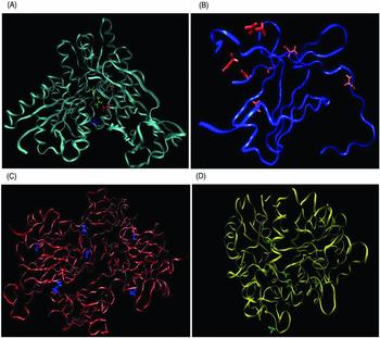
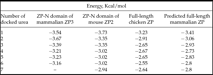
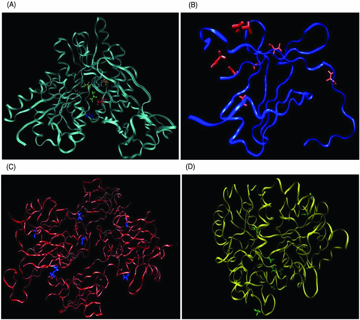
Molecular docking showed that urea molecules surround the active site of: (1) ZP-N domain of ZP3 (Fig. 1A); (2) ZP-N domain of ZP2 (Fig. 1B); (3) full-length chicken sperm receptor ZP3 (Fig. 1C); and (4) predicted full-length mammalian sperm receptor ZP3 (Fig. 1D). Also, docking procedures were run six or seven consecutive times. Each time the previous docked urea/ZP complex was used as the template (macromolecule) for the next docking run. The binding energy values for the interaction of each run achieved from 300 runs are shown in Table 1. The results showed that the binding energy values for urea interaction with all studied proteins, ZP-N domain of both ZP3 and ZP2 proteins, full-length chicken sperm receptor, and predicted full-length mammalian sperm receptor, were negative (Table 1).
Molecular docking as a key approach is widely used in structural molecular biology to predict how a ligand binds to a protein (Morris & Lim-Wilby, 2008). AutoDock4.2 via the genetic algorithm (GA) procedure was conducted to predict urea binding to: (A) the zona pellucida (ZP)-N domain of ZP3 (PDB code: 3D4C); (B) the ZP-N domain of ZP2 (PDB code: 5II6); (C) the full-length chicken sperm receptor ZP3 (PDB code: 3NK3); and (D) the predicted full-length mammalian sperm receptor ZP3 (predicted using I-TASSER software). We ran the docking procedure six consecutive times such that each time we used the previous docked urea/ZP protein complex as the template macromolecule for the next docking run. As shown in (A), across six consecutive runs, the urea molecule is shown in black for the first run, then shown by blue, red, grey, orange, and green (the last run), respectively. Docking showed a favorable binding activity of urea towards the ZP-N domain of ZP3 and that of ZP2, the full-length chicken sperm receptor ZP3, and the predicted full-length mammalian sperm receptor ZP3.
Binding energies (Kcal/mol) obtained from docking of urea molecules with ZP proteins using AutoDock4.2 via the genetic algorithm procedure

Atomic interactions between urea and ZP-N domain of ZP3
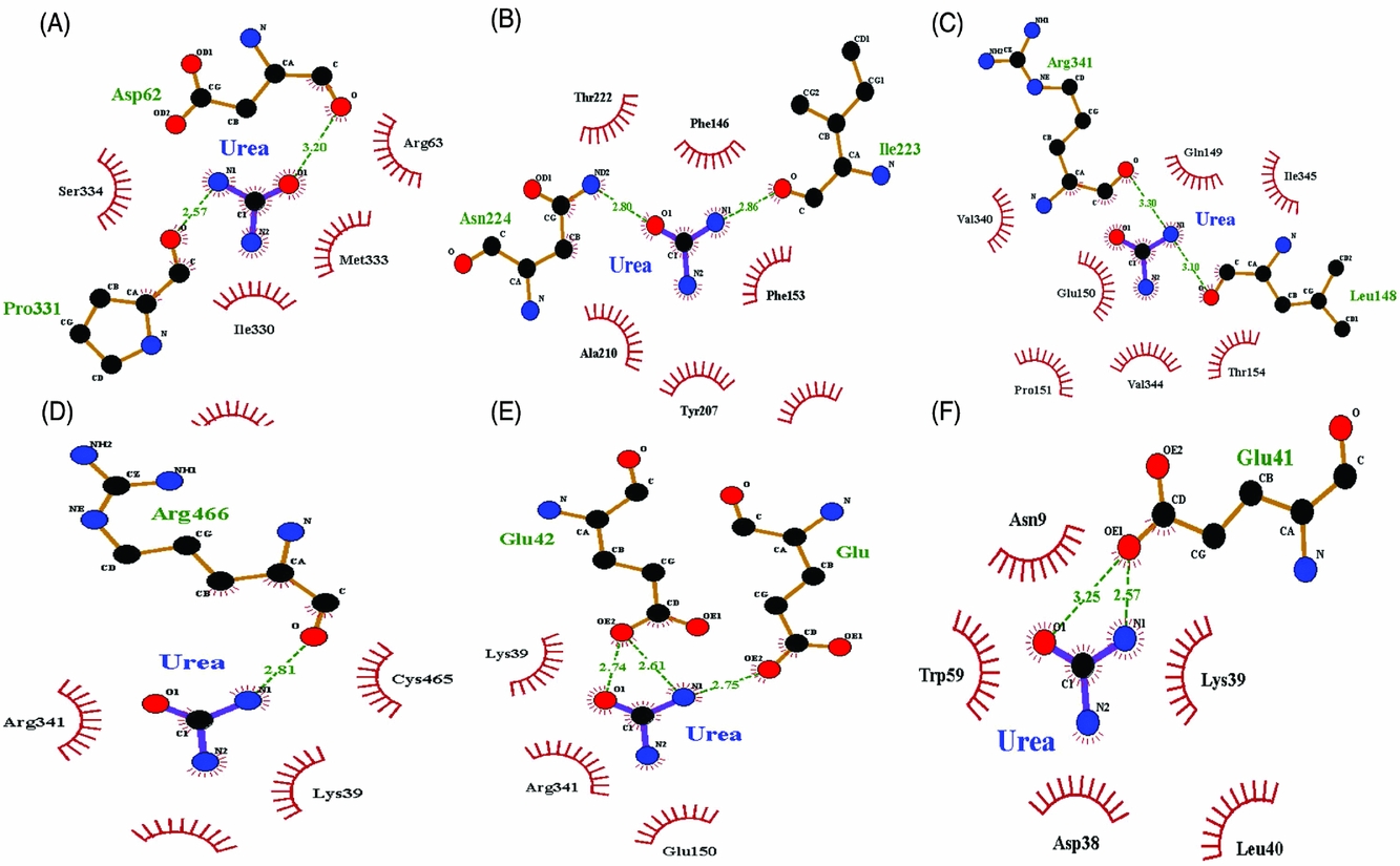
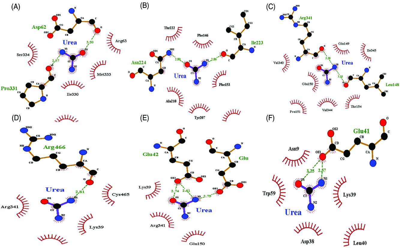
As LIGPLOT software showed, there were hydrophobic interactions between each urea and identified residues (Fig. 2A–F). The nitrogen atoms of the urea molecules mainly contributed to the hydrophobic interactions with the residues as shown in Fig. 2A–F (red arcs). The results showed the hydrogen bonds between urea molecules and some residues of ZP-N domain of ZP3 (green dashes) as shown in Fig. 2A Asp62 and Pro331, Fig. 2B Asn224 and Ile223, Fig. 2C Arg341and Leu148, Fig. 2D Arg466, Fig. 2E Glu42, and in Fig. 2F Glu41.
Two-dimension urea/ZP-N domain of ZP3 interaction diagrams. To investigate the free energy and behaviour of urea binding to ZP-N domain of ZP3 (PDB code: 3D4C), we ran docking procedure six consecutive times (A–F), such that we used the previous docked urea/ZP-N complex as the template macromolecule for the next docking run. LIGPLOT software was used to generate images and to visualize hydrophilic interactions, hydrogen bonding, and hydrophobic contacts between urea and ZP-N domain of ZP3 protein. The LIGPLOT shows the hydrogen bonds between urea and ZP-N domain of ZP3 at (A) Asp62 and Pro331; (B) Asn224 and Ile223; (C) Arg341and Leu148; (D) Arg466; (E) Glu42; and (F) Glu41 which are shown by dashed green lines. Also, the distance between acceptor and donor atoms is shown in the middle. Arcs that radiate spokes towards the urea molecule indicate hydrophobic interactions.
Scanning electron microscopy shows urea-induced shrinkage in oocyte
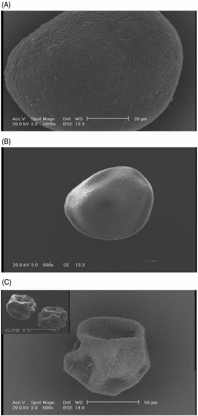
Scanning electron microscopy was used to investigate the effect of urea on the oocyte/zona pellucid a structure. As shown in Fig. 3, compared with the control group or urea-exposed group (20 mg/dl) (Fig. 3A, B, respectively), urea-exposed counterparts (40 mg/dl) showed a shrinkage in their structure (Fig. 3C).
Scanning electron microscopy observation of bovine oocytes that were matured in the maturation medium supplemented with the different concentrations of (A) 0 mg/dl urea, the control group; (B) 20 mg/dl urea; and (C) 40 mg/dl urea for 24 h. Upon incubation of oocytes, 40 mg/dl urea induced shrinkage in oocytes.

Urea-exposed oocytes negatively show reduced developmental competence
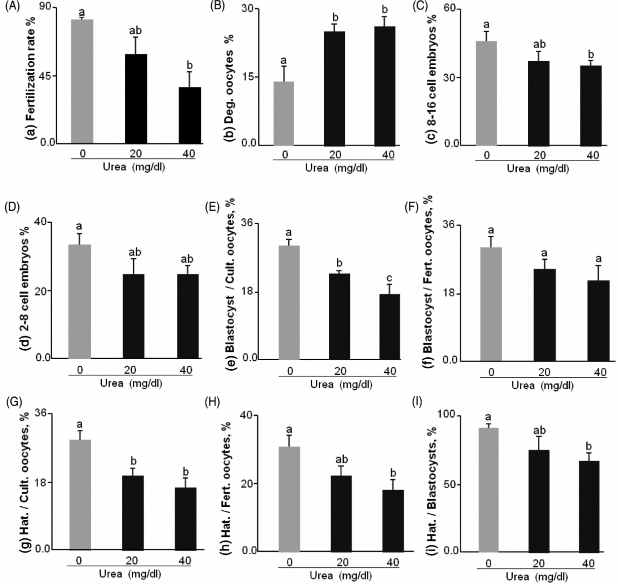
Compared with the control group, the addition of 20 or 40 mg/dl urea to the oocyte maturation medium dose-dependently decreased fertilization rate (87.6, 81.1, and 75.1%, respectively) (Fig. 4A). Urea dose-dependently increased (P < 0.01) the rate of degenerated oocytes, by day 3 post-insemination (Fig. 4B). The presence of urea in the oocyte maturation medium dose-dependently reduced (P < 0.01) cleavage rate on day 3 (at the 6–18-cell stage but not the 2–8-cell stage) (Fig. 4C, D), blastocyst formation on day 7 (as a percentage of cultured or fertilized oocytes) (Fig. 4E, F), and rate of hatching on day 9 (as a percentage of cultured or fertilized oocytes or number of blastocyst on day 7) (Fig. 4G–I, respectively).
The effect of urea supplementation (0, 20, or 40 mg/dl) in maturation medium on oocyte competence. (A) Fertilization rate (%). (B) Degenerated oocytes (%). (C) 8–16-cell embryos (%). (D) 2–8-cell embryos (%). (E) Blastocysts/cultured oocytes (%). (F) Blastocysts/fertilized oocytes (%). (G) Hatching/cultured oocytes (%). (H) Hatching/fertilized oocytes (%). (I) Hatching/blastocysts (%). Blast, blastocyst; Deg, degenerated; Cult, cultured; Fert, fertilized; Hat, hatching. n = 143–213 oocytes per treatment across five replicates. Numerical values are presented as the mean ± standard error of the mean (SEM). a,b,cDifferent letters indicate significant differences between the treatments at P < 0.05.

Urea-exposed oocytes alter the expression of marker genes in resultant embryos
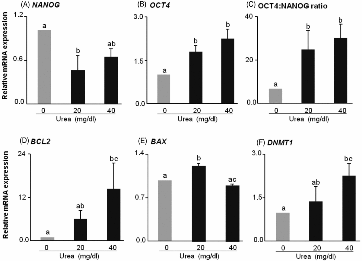
Urea at 20 mg/dl downregulated (P < 0.01) the expression of NANOG in blastocysts produced from urea-exposed oocytes. Blastocysts resulting from high urea-exposed oocytes (40 mg/dl) tended to reduce NANOG expression (Fig. 5A). Urea dose-dependently upregulated (P < 0.01) the expression of OCT4 in blastocysts (Fig. 5B). Because of this altered expression pattern, the OCT4:NANOG ratio was significantly increased (P < 0.01) with increasing urea concentrations (Fig. 5C). mRNA expression of OCT4 was over two-fold higher in blastocysts derived from urea-exposed oocytes (40 mg/dl) compared with the control group. Urea dose-dependently upregulated (P < 0.01) the expression of anti-apoptotic marker, BCL2 in blastocysts produced from urea-exposed oocytes (Fig. 5D). Urea at 20 mg/dl upregulated (P < 0.01) and at 40 mg/dl down-regulated the expression of BAX compared with 20 mg/dl urea group (Fig. 5E). Urea dose-dependently upregulated (P < 0.01) the expression of DNMT1, an epigenetic marker, in blastocysts (Fig. 5F).
Relative mRNA expression of some marker genes in bovine blastocysts resulted from urea-exposed oocytes. (A) NANOG. (B) OCT4. (C) OCT4:NANOG. (D) BCL2. (E) BAX. (F) DNMT1. n ≥ 7 blastocysts per treatment across five replicates. Each replicate of qRT-PCR was repeated three times to minimize the technical errors. Numerical values are presented as the mean ± standard error of the mean (SEM). a,b,cDifferent letters indicate significant differences between the treatments at P < 0.05.

Sensitivity of developmental parameters to the presence of urea in maturation medium
Interestingly, statistical analysis predicted that the hatching rate on day 9 and blastocyst rate on day 7 had the highest negative sensitivity (−0.26) to urea (Table 2). Also, the rate of hatching or blastocyst per cultured oocytes (−0.26 and −0.25, respectively) showed a greater negative sensitivity than hatching or blastocyst rate per fertilized oocytes to urea (−0.25 and −0.18, respectively).
The sensitivity of the different developmental parameters to urea. The negative or positive values mean negative or positive sensitivity of different developmental parameters to urea. Negative sensitivity means that the parameter is decreased as urea concentration increases. Positive sensitivity means that the parameter is increased as urea concentration increases

Discussion
To show urea contribution in reduced fertility, we recently reported that urea had the ability to disrupt in vitro the immunological and secretory response of oviduct epithelial cells (Kowsar et al., Reference Kowsar, Marey, Shimizu and Miyamoto2016), implying a role for urea in disrupting the local environment of fertilization and early embryo development.
Importantly, urea has been able to induce protein denaturation and conformation by making hydrogen bonds with polar or charged groups (Caflisch & Karplus, Reference Caflisch and Karplus1999). AutoDock4 is widely used in the fields of biology and biomedicine and provides a quick estimation of bound conformations and free energies of binding small ligands i.e. urea with the target macromolecules, the ZP-N domain of ZP3 or ZP2 and full-length chicken ZP3, and predicted full-length mammalian ZP3 (Morris et al., Reference Morris, Huey, Lindstrom, Sanner, Belew, Goodsell and Olson2009; Barzegar et al., Reference Barzegar, Mansouri and Azamat2016). Using AutoDock4 via the Lamarckian GA, molecular docking showed that urea had affinity for the ZP-N domain of both ZP3 and ZP2 proteins and for full-length chicken ZP3 and predicted full-length mammalian ZP3. These docking analyses suggested that urea, in general, favoured affinity with the ZP proteins.
The binding energy values for several molecular dockings of urea with the ZP-N domain of both ZP2 and ZP3 proteins, full-length chicken ZP3, and predicted full-length mammalian ZP3 were negative and showed no substantial changes. This result implies that the addition of urea did not decrease the total free energy, suggesting that ZP proteins especially the ZP-N domain of both ZP2 and ZP3 still had enough space and affinity to be docked by greater numbers of urea molecules. Regarding the important role of both ZP2 and ZP3 proteins for the general assembly of complete ZP (Callebaut et al., Reference Callebaut, Mornon and Monget2007; Wassarman, Reference Wassarman2008), the binding of urea with ZP-N domains of both ZP2 and ZP3 might interfere with the normal assembly of these ZP proteins. LIGPLOT analysis also showed that urea formed stable interactions with non-polar, acidic and basic residues of the ZP-N domain of ZP3 through hydrophobic and hydrogen bonding interactions, implying a change in the normal conformation of ZP3 sperm receptor. However, we could not confirm changes in ZP conformation under urea effect using the present molecular docking. Further studies such as molecular dynamic simulation are needed to confirm these changes in ZP protein conformation.
To evaluate the possible effect of urea on bovine ZP, using scanning electron microscopy, we found that urea induced shrinkage in oocyte structure. Similarly, Zeuthen & MacAulay (Reference Zeuthen and MacAulay2012) showed that urea may shrink Xenopus oocytes. Interestingly, Wagner et al. (Reference Wagner, Huber, Wärntges, Zempel, Kaba, Fux, Orth, Busch, Waldegger, Lambert, Nilius, Heinle and Lang2000) reported that urea induced a sustained shrinkage in vascular smooth muscle cells via inhibition of voltage-gated Ca2+ channels, which resulted in a decrease in cellular Ca2+. Importantly, calcium channels have been shown to play a critical role in Ca2+ increase in the oocyte which is a pivotal process for meiosis re-initiation and consequently oocyte maturation (Tosti, Reference Tosti2006). It would be of interest to investigate the hypothesis that urea may negatively affect oocyte maturation via inhibition of Ca2+ channels and reduction of Ca2+ concentration in bovine oocytes.
In the present in vitro study, urea dose-dependently reduced oocyte fertilizability, cleavage rate on day 3, blastocyst rate on day 7, and hatching rate on day 9. De Wit et al. (Reference De Wit, Cesar and Kruip2001) reported that 36 mg/dl (6 mM) of urea in the maturation medium reduced embryonic development as a percentage of cultured but not cleaved oocytes. Similarly, when the sensitivity of fertilizability or developmental competence of oocytes to the presence of urea in maturation medium was analyzed, the results showed that the hatching rate per cultured oocyte had the highest negative sensitivity to urea. Also, the blastocyst rate per cultured oocytes showed a greater negative sensitivity to urea than that per fertilized oocytes. These findings meant that as urea concentration increased, the rate of hatching or blastocyst per cultured oocytes was greatly decreased. In addition, the rate of degenerated oocytes by day 3 drastically showed a positive sensitivity to urea, meaning that the presence of urea in maturation medium may increase the rate of oocyte degeneration. These findings together suggested that dietary protein through elevation of BUN and consequent increase in urea concentration in the FRT may contribute to reduce the rate of cleaved oocytes and resultant embryo development in healthy dairy cows. However, further studies are needed to reveal the unknown interaction among urea, early embryo, and oviduct secretions as early embryonic development can be easily affected by the oviductal secretions that have also been influenced by urea (Kowsar et al., Reference Kowsar, Marey, Shimizu and Miyamoto2016).
The expression of OCT4 showing an important role in self-renewal, pluripotency and lineage commitment was dose-dependently up-regulated in blastocysts produced from urea-exposed oocytes. Conversely, NANOG expression, which maintains pluripotency in epiblast progenitors, was downregulated (Arnold & Robertson, Reference Arnold and Robertson2009; Kalmar et al., Reference Kalmar, Lim, Hayward, Muñoz-Descalzo, Nichols, Garcia-Ojalvo and Martinez Arias2009; Madeja et al., Reference Madeja, Sosnowski, Hryniewicz, Warzych, Pawlak, Rozwadowska, Plusa and Lechniak2013). Consequently, the OCT4/NANOG ratio was elevated as urea concentration increased [over four-fold higher in blastocysts produced from urea-exposed oocytes (40 mg/dl) compared with their untreated counterparts]. It has been shown that cells co-expressing NANOG and OCT4 are more stable in the pluripotent state than those that over-express these genes individually (Theunissen et al., Reference Theunissen, van Oosten, Castelo-Branco, Hall, Smith and Silva2011). Indeed, the disruption of the OCT4/NANOG ratio leads to the inappropriate differentiation of ICM and embryonic stem cells to trophectoderm and extra-embryonic endoderm, respectively (Mitsui et al., Reference Mitsui, Tokuzawa, Itoh, Segawa, Murakami, Takahashi, Maruyama, Maeda and Yamanaka2003; Boyer et al., Reference Boyer, Lee, Cole, Johnstone, Levine, Zucker, Guenther, Kumar, Murray, Jenner, Gifford, Melton, Jaenisch and Young2005). For example, from a toxicology point of view, ethanol has been shown to disturb the balance of these lineage specifiers (Sánchez-Alvarez et al., Reference Sánchez-Alvarez, Gayen, Vadigepalli and Anni2013). Thus any imbalances may result in the loss of pluripotency or differentiation of embryonic stem cells (Boyer et al., Reference Boyer, Lee, Cole, Johnstone, Levine, Zucker, Guenther, Kumar, Murray, Jenner, Gifford, Melton, Jaenisch and Young2005). The higher OCT4/NANOG transcript ratio observed in the present study might also have resulted from OCT4 expression in both ICM and trophectoderm while NANOG is poorly expressed in ICM in bovines (Berg et al., Reference Berg, Smith, Pearton, Wells, Broadhurst, Donnison and Pfeffer2011; Khan et al., Reference Khan, Dubé, Gall, Peynot, Ruffini, Laffont, Le Bourhis, Degrelle, Jouneau and Duranthon2012). It seems in healthy dairy cows that have been fed high dietary protein diets to support their milk production, high urea concentrations in the follicular fluid may negatively affect oocyte competence and in turn early embryonic development. This idea was manifested by disrupting the normal and fine-tuned expression of key transcription factors.
Urea as a stress factor has cytotoxic effects on the growth and viability of cell populations, implying that cells exposed to increasing doses of urea may die via apoptosis (Glinos et al., Reference Glinos, Bardi, Dermitzaki, Perez and Talieri1983; Kowsar et al., Reference Kowsar, Marey, Shimizu and Miyamoto2016). Recently, we showed that high levels of urea may disrupt the oviduct epithelial monolayer, damaging the oviduct epithelium and disrupting sperm reservoir and fertilization site (Kowsar et al., Reference Kowsar, Marey, Shimizu and Miyamoto2016). In the present study, the expression of BCL2, an anti-apoptotic gene, was dose-dependently elevated in blastocysts produced from urea-exposed oocytes. However, the BAX gene, a pro-apoptotic gene, was downregulated in blastocysts produced from urea-exposed oocytes (40 mg/dl). Importantly, OCT4 can actively repress apoptotic pathways (Sánchez-Alvarez et al., Reference Sánchez-Alvarez, Gayen, Vadigepalli and Anni2013; Kim et al., Reference Kim, Koo, Park, Moon, da Torre, Javaregowda, Kang, Park, Saadeldin, Choi, Lee and Jang2015). Kim et al. (Reference Kim, Koo, Park, Moon, da Torre, Javaregowda, Kang, Park, Saadeldin, Choi, Lee and Jang2015) reported that in porcine embryos, BCL2 was up-regulated in OCT4-overexpressed fibroblast cells. Ogony et al. (Reference Ogony, Malahias, Vadigepalli and Anni2013) showed that ethanol as a toxic factor dose-dependently induced OCT4 and downregulated apoptosis genes. These findings together may imply that, as a possible autonomous control system, blastocysts upregulate and downregulate anti-apoptotic and pro-apoptotic genes, respectively in response to urea, as a stressor.
DNMT1, one of the major DNA methyltransferase enzymes, regulates methylation status during DNA replication, genomic methylation and gametic imprinting (Grohmann et al., Reference Grohmann, Spada, Schermelleh, Alenina, Bader, Cardoso and Leonhardt2005). Genomic hypo-methylation and consequently early embryonic lethality can be expected due to a deficiency in DNMT1 (Li et al., Reference Li, Beard and Jaenisch1993). DNMT1 over-expression leads to the activation of the silent maternal allele and increased embryonic lethality (Biniszkiewicz et al., Reference Biniszkiewicz, Gribnau, Ramsahoye, Gaudet, Eggan, Humpherys, Mastrangelo, Jun, Walter and Jaenisch2002; Giraldo et al., Reference Giraldo, DeCourcy, Ball, Hylan and Ayares2013). Importantly, during early embryonic development, the methylation level is drastically reduced, especially at the later pre-implantation stages (Messerschmidt et al., Reference Messerschmidt, Knowles and Solter2014). In this study, in addition to an increase in oocyte degeneration by day 3 post-insemination, DNMT1 expression was also increased in blastocysts produced from urea-exposed oocytes. Urea seems to interfere with the early embryonic development via alterations in the normal level of DNMT1.
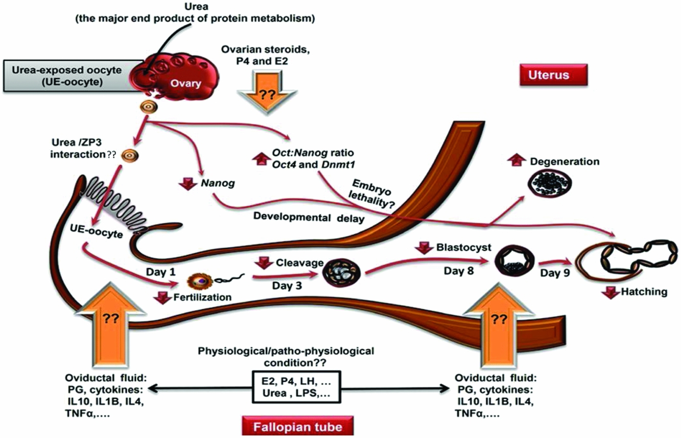
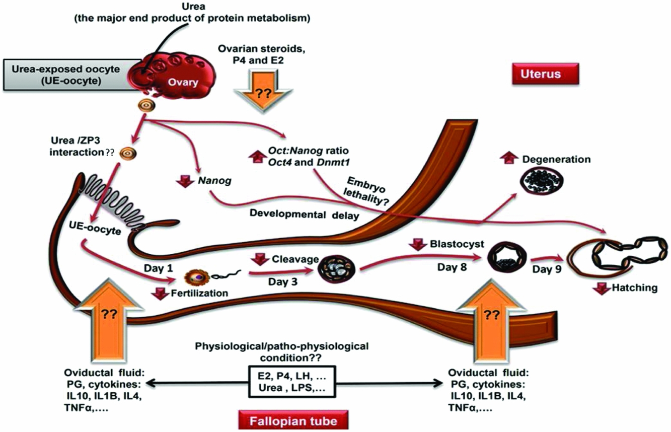
Taken together, urea negatively interacts with the ZP-N domain of ZP3 and ZP2, and full length sperm receptor ZP3, induced oocyte shrinkage, reduced oocyte competence, and interferes with the normal expression of marker genes involved in the early embryonic development. Of importance, as proposed in Fig. 6, regarding the possible interactions between embryo, oviduct secretions and urea, these findings can be easily affected under physiological and patho-physiological conditions (Cebrian-Serrano et al., Reference Cebrian-Serrano, Salvador, García-Roselló, Pericuesta, Pérez-Cerezales, Gutierrez-Adán, Coy and Silvestre2013; Ibrahim et al., Reference Ibrahim, Salilew-Wondim, Rings, Hoelker, Neuhoff, Tholen, Looft, Schellander and Tesfaye2015). Thus, further investigations are needed to unveil how these interactions regulate oocyte and embryo responses to the negative effect of urea, in particular during a high-protein diet.
Schematic illustration of a proposal implying how urea may contribute to low fertility. Urea as a major end product of protein metabolism in dairy cows may contribute to the impaired early embryonic development via affecting oocytes before fertilization. Of great importance, regarding possible interactions between embryo, oviduct secretions conditions (Cebrian-Serrano et al., Reference Cebrian-Serrano, Salvador, García-Roselló, Pericuesta, Pérez-Cerezales, Gutierrez-Adán, Coy and Silvestre2013; Ibrahim et al., Reference Ibrahim, Salilew-Wondim, Rings, Hoelker, Neuhoff, Tholen, Looft, Schellander and Tesfaye2015) and urea (Kowsar et al., Reference Kowsar, Marey, Shimizu and Miyamoto2016), these in vitro findings physiologically and patho-physiologically can be easily affected. Thus further investigations are needed to unveil how oviductal secretions regulate the negative effect of urea, in particular during a high-protein diet in dairy cows. E2, estradiol; IL, interleukin; LH, luteinizing hormone; LPS, lipopolysaccharide; P4, progesterone; PG, prostaglandins.
Acknowledgements
The authors thank the FKA, Animal Husbandry and Agriculture Co, for their help in conducting this research. Also, authors would like to thank Mr Alireza Mansouri for his technical assistance in molecular docking.
Financial support
This study was supported by the Isfahan University of Technology and a Grant-in-Aid for Scientific Research (No. 16H05013) of the Japan Society for the Promotion of Science (JSPS).
Ethical standards
Animal experiments were carried out according to the Guiding Principles for the Care and Use of Research Animals promulgated by the Isfahan University of Technology, Iran. The protocol and methods were approved by the Committee on the Ethics of Animal Experiments of the Isfahan University of Technology.
Conflict of interest
The authors have declared no conflict of interest.