Introduction
Strongyloides is a genus including some 50 species of obligate gastrointestinal parasites of vertebrates. These species infect mammals, birds, reptiles and amphibians (Speare, Reference Speare and Grove1989). For most nematodes that parasitize the vertebrate gastrointestinal tract, the male and female worms live in the host gut and females lay eggs that pass out of the host in faeces. Depending on the species, the eggs, or infective larvae that develop from them, are ingested (eggs or larvae) or penetrate the host skin (larvae) to achieve transmission to a new host. A period of larval migration follows within the new host before new adults establish in the gut. Strongyloides follows this basic strategy of parasitic infection; however, there are some significant differences. First, female-only adult worms form the parasitic stages of Strongyloides. In addition, there is a dioecious adult generation that occurs outside of the host (Schad, Reference Schad and Grove1989). This free-living adult generation is rather unique among nematode parasites of vertebrates. Originally, the parasitic and free-living adult generations of Strongyloides were described as distinct species of Anguillula (Bavay, Reference Bavay1876, Reference Bavay1877) until they were discovered to be separate stages of one life cycle (Grassi, Reference Grassi1879).
The cytoskeleton provides the basic scaffold for the maintenance of cell shape and motility. There are three types of cytoskeletal proteins: microtubules, intermediate filaments and actin. Actin is a globular protein that contains a nucleotide (either ATP or ADP) associated with the cleft located between the two lobes of the protein. Under physiological conditions, actin monomers (G-actin) assemble into polar, helical filaments (F-actin). Because actin filaments are polar structures, they have two structurally and biochemically distinct ends, which are called the ‘barbed end’ and the ‘pointed end’. Under steady-state conditions, the net addition of subunits to actin filaments occurs at the barbed end. Irreversible hydrolysis of the bound ATP subsequently destabilizes the filament and results in net dissociation of actin subunits at the pointed end. It is the actin cytoskeleton that regulates dynamic cell motility, morphology, intracellular trafficking and bundle formation in plant and animal cells, including those of parasites (Pollard & Borisy, Reference Pollard and Borisy2003; Bahia et al., Reference Bahia, Avelar, Vigorosi, Cioli, Oliveira and Mortara2006).
Little is known about the distribution of cytoskeletal components in Strongyloides species. In the late 1990s ultrastructural studies were undertaken on a discrete peri-vulval area of cuticular modification in the parasitic female of Strongyloides cebus. Ciliary-like arrangements of microtubules were observed, suggesting that this region was sensory in nature (Viney & Ashford, Reference Viney and Ashford1990). The aim of the present study was to verify whether or not F-actin accumulates in contractile structures along the female body of S. venezuelensis.
Materials and methods
Maintenance of the life cycle of Strongyloides venezuelensis
Strongyloides venezuelensis was maintained in Wistar rats (Rattus norvegicus), routinely infected in the Laboratory of Parasitology of the Universidade Federal de Uberlândia (UFU), MG, Brazil. All animals used for the experiments were male and 4–6 weeks old. Experimental procedures were approved by the Animal Ethics Committee of the Universidade Federal de Uberlândia.
Rats were inoculated via a subcutaneous route with 1500 S. venezuelensis L3 (third-stage larvae), then at 8 days post-infection (p.i.), they were sacrificed by cervical dislocation. To recover adult worms, intestines were removed, placed in Petri dishes containing saline, longitudinally sectioned and incubated for 2 h at 37°C. Adult worms from the intestine were recovered under light stereomicroscopy.
Detection of filamentous actin
After fixation with 4% formaldehyde for 1 h, each parasite suspension was washed by centrifugation three times (5 min, 5000 rpm) with phosphate-bauffered saline (PBS) and maintained in PGN (PBS, 0.2% gelatin, 0.1% NaN3) until use. Parasite suspensions were centrifuged for 5 min at 2000 rpm and the supernatant was carefully drawn off with a micropipette. Samples were resuspended and incubated with 250 μl of blocking/permeabilizing solution (PBS, 0.2% gelatin, 0.5% NP40, 0.1% NaN3, solution; henceforth referred to as PGN-NP40) for 1 h. Samples were incubated directly in PGN-NP40 solution for 1 h with phalloidin-rhodamine 1 μg/ml (Molecular Probes, Eugene, Oregon, USA) and DAPI 1:25 (4′,6-diamidino-2-phenylindole dihydrochloride; Molecular Probes) to label filamentous actin and nuclei, respectively. Glass multi-well microscope slides were coated with 30 μl of poly-l-lysine (100 μg/ml in water) and allowed to air dry. Parasite suspensions (30 μl) were added to pre-coated wells, and parasites were allowed to attach and air dry. Then the slides were mounted in glycerol buffered with 0.1 m Tris pH 8.6 and 0.1% p-phenylenediamine to reduce bleaching.
Slides were examined on a Bio-Rad 1024UV confocal system, attached to a Zeiss Axiovert 100 microscope using either a 40 × 1.2 numerical aperture (NA) water immersion or a 63 × 1.4 NA differential interference contrast (DIC) objective (DeMarco et al., Reference DeMarco, Kowaltowski, Mortara and Verjovski-Almeida2003; Bahia et al., Reference Bahia, Avelar, Vigorosi, Cioli, Oliveira and Mortara2006). Images were acquired at 0.9 μm intervals, with 2.7 μm intervals and the z-series rendered using Volume-J. Images were processed using NIH-Image J (http://rsb.info.nih.gov/ij/) or Adobe Photoshop 7.0 software.
Results and discussion
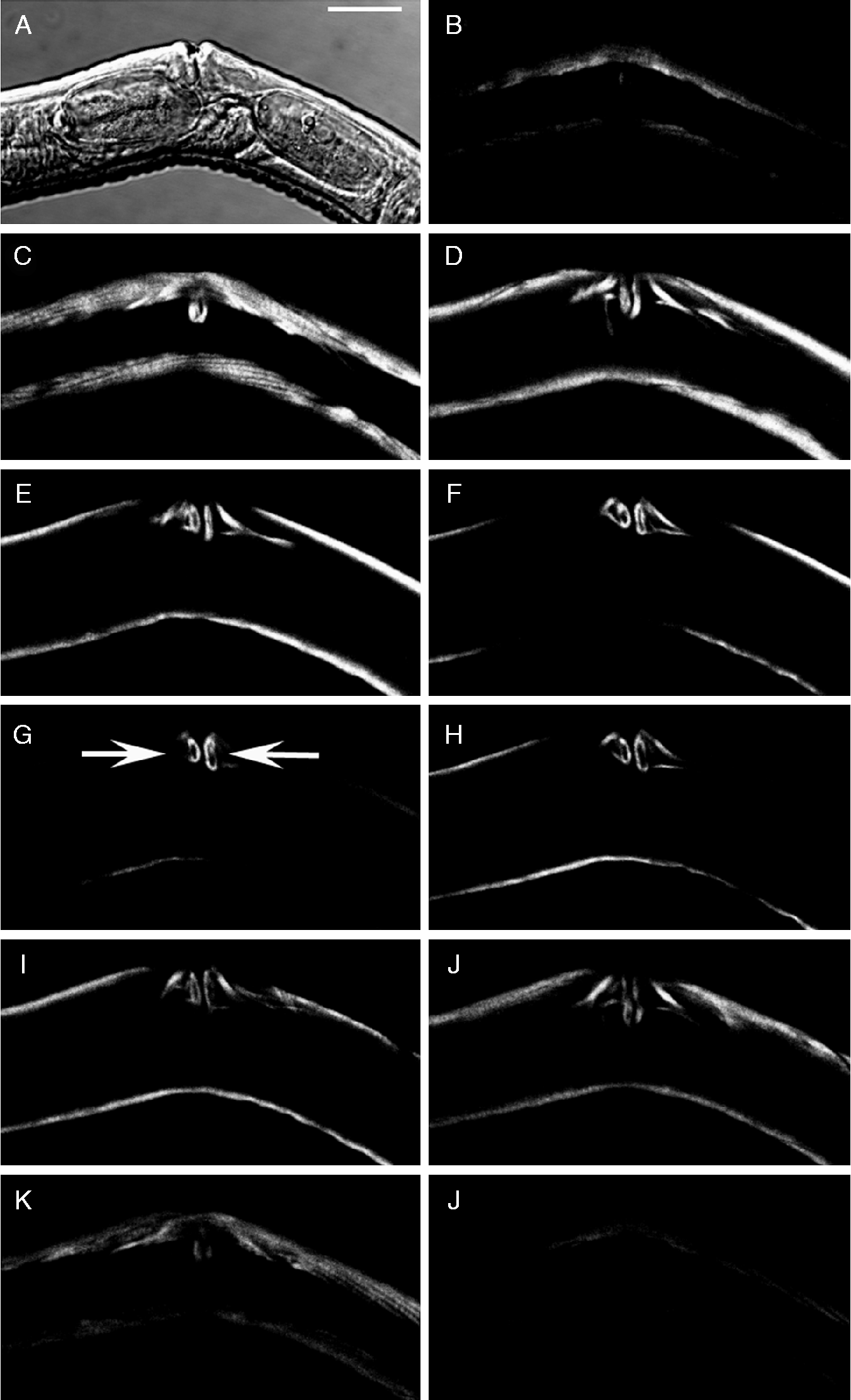
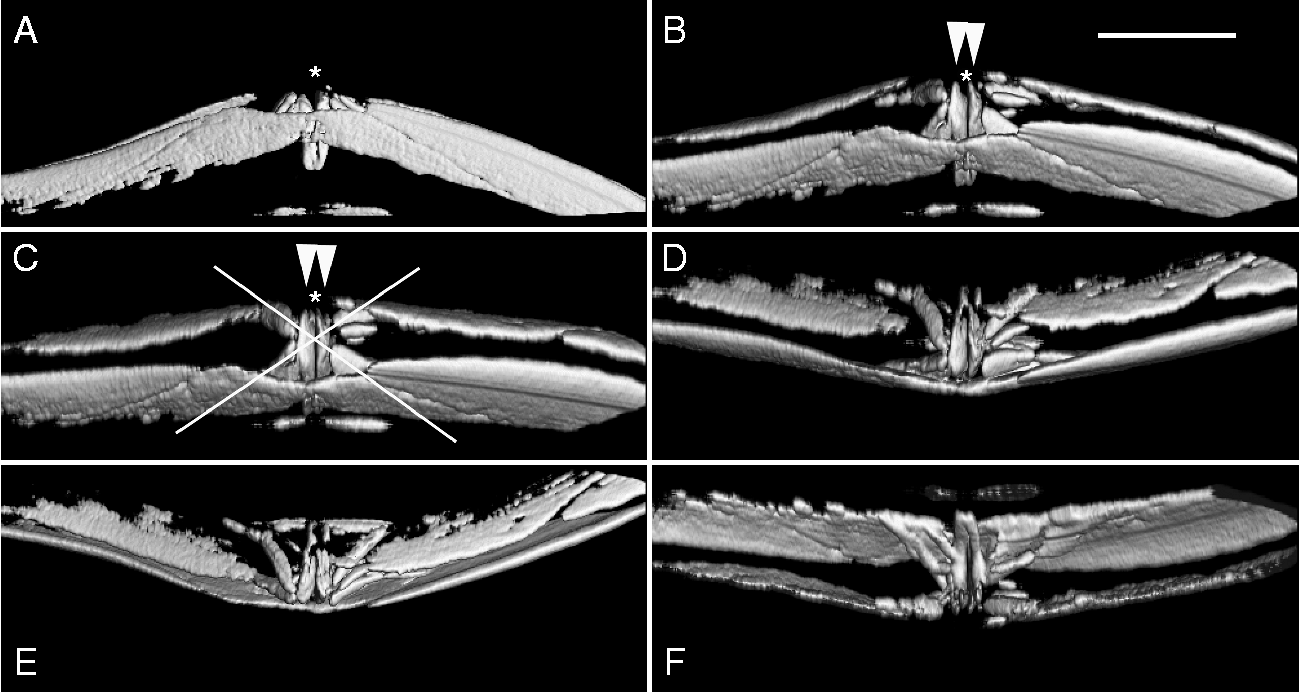
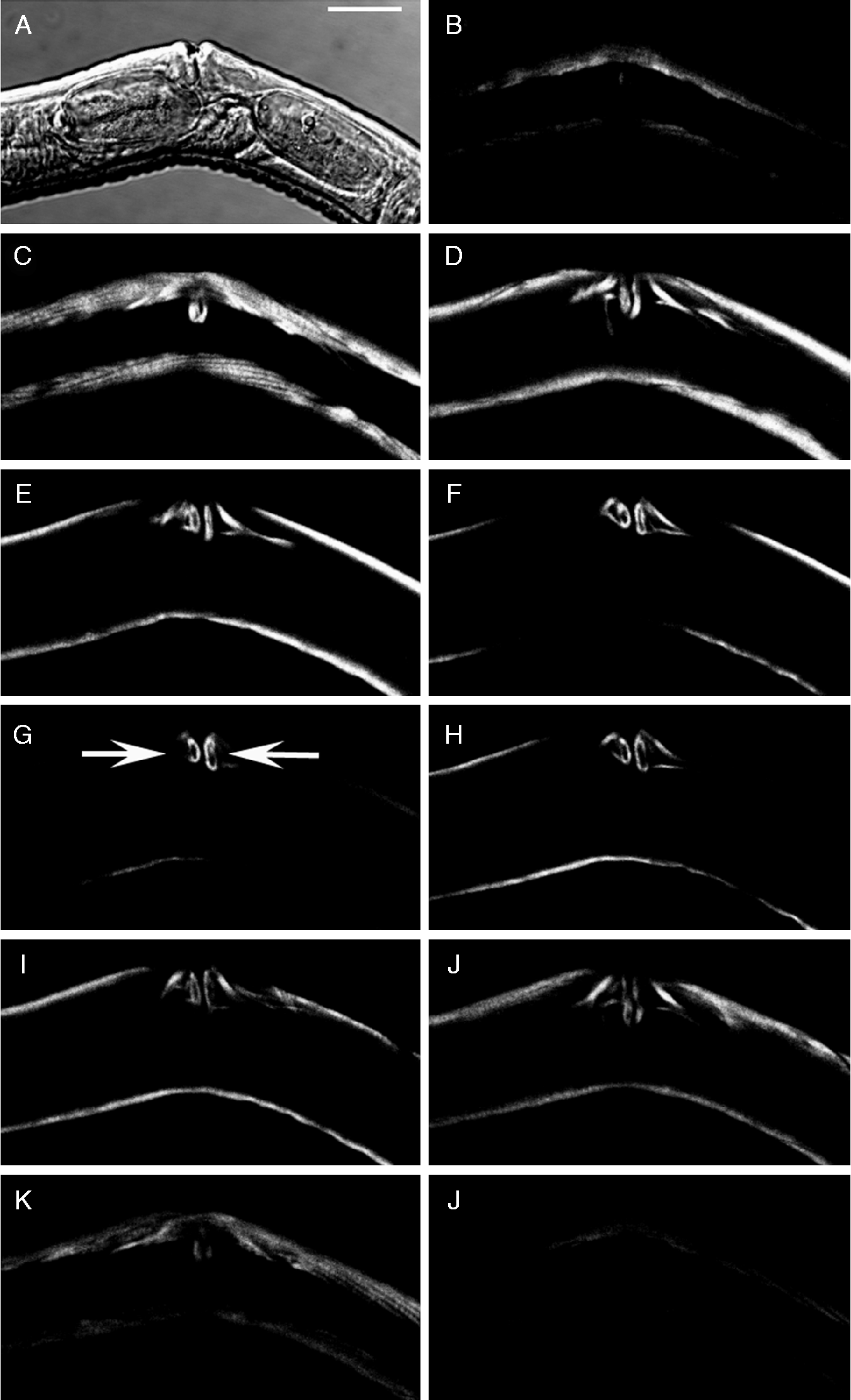
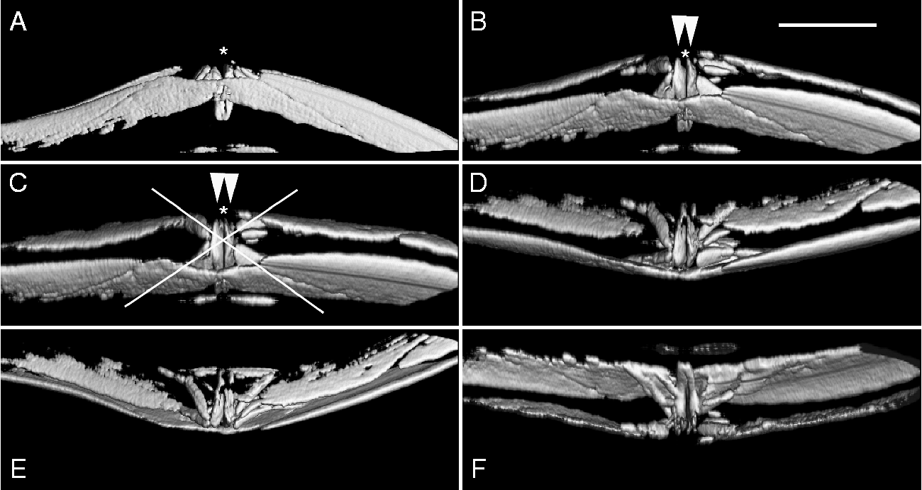
Adult worms from the intestine were labelled with phalloidin-rhodamine as described and imaged under confocal microscopy. Our results showed that F-actin accumulated in the vulva of female S. venezuelensis (figs 1 and 2). In the vulva actin forms an intricate and symmetrical X-shaped (fig. 2C) array of fibres lining the vulva aperture (marked * in fig. 2), that deepens to about half of the parasite's width in an elongated hollow structure (arrows in fig. 1G and arrowheads in fig. 2B and C).
F-actin stained with phalloidin-rhodamine accumulates in a distinctive manner in the vulva of female S. venezulensis. Images were acquired at intervals of 0.9 μm and are represented at 2.7 μm (B–L). The image in (A) is a differential interference contrast (DIC) image. Hollow structures line the vulval aperture (arrows in G). Scale bar = 20 μm.
The muscle structure of the vulva (arrowheads) of female Strongyloides venezuelensis (A–F) using selected angles of the x-axis rotation. *, Lining of the vulval aperture. Scale bar = 20 μm.
Actin accumulation in particular structures has been associated with specific functions. Actin labelling of whole parasites and larvae with fluorescent phalloidin has provided insights into the general muscle distribution (Mair et al., Reference Mair, Maule, Day and Halton2000) and has revealed known contractile structures such as the Schistosoma mansoni cercaria oral sucker (Mair et al., Reference Mair, Maule, Fried, Day and Halton2003). In addition, the observation of protein in the flame cells of S. mansoni larvae has been interpreted as displaying contractile activity of these excretory structures (Bahia et al., Reference Bahia, Avelar, Vigorosi, Cioli, Oliveira and Mortara2006).
We have provided the first demonstration of F-actin accumulation in the vulva of S. venezuelensis. Actin gathering at this site may represent an important role during oviposition, probably providing the contractile basis for egg elimination. It is yet to be determined whether such a pattern is a common feature among Strongyloides species. There is sparse literature describing aspects of actin dynamics in Strongyloides species. Indeed, we have a great deal of research to undertake in order to better comprehend the dynamics of actin polymerization during Strongyloides infection, motility and oviposition. Moreover, to date, the molecules that regulate actin polymerization have not been described in Strongyloides. Our study is the first demonstration of F-actin accumulation in a specific site important to the maintenance of the S. venezuelensis life cycle. In addition, these results raise an important issue concerning the manipulation of F-actin integrity and the fate of Strongyloides infections.
Acknowledgements
We thank Fundaçáo de Amparo a Pesquisa do Estado de Minas Gerais (FAPEMIG), Coordenaçáo de Aperfeiçoamento de Pessoal de Nível Superior (CAPES), Conselho Nacional de Desenvolvimento Científico e Tecnológico (CNPq) for grants and fellowships. We also thank Renato Arruda Mortara PhD for confocal microscopy assistance and image acquisition. Also, we would like to thank BioMed Proofreading for correcting the language.