Introduction
Tympanic membrane perforations are a common finding in ENT practice. Acute traumatic perforations often heal spontaneously, but in cases of chronic otitis media, surgery is generally necessary.Reference Gladstone, Jackler and Varav1 Type 1 tympanoplasty, described as closure of the perforation without ossiculoplasty, is the most commonly performed procedure.Reference Wullstein2 Multiple techniques have been developed with numerous differences in graft positioning, graft material and approach. Anterior perforations often remain challenging with consequent worse surgical outcome.Reference Schraff, Dash and Strasnick3,Reference Applebaum and Deutsch4
We believe insufficient stabilisation of the graft and inadequate visualisation are key aspects in poor outcomes for anterior membrane reconstruction and have been using the ‘en hamac’ technique as a possible solution. We also believe that routinely performed complete canalplasty improves visualisation during surgery and can enhance ear canal aeration and self-cleaning post-operatively leading to improved success rates. Despite the multiple advantages of canalplasty, it has not been implemented as a routine step in type 1 tympanoplasty. We therefore present our experience with type 1 tympanoplasty using these techniques to improve surgical outcome.
Materials and methods
A retrospective review was performed using the prospective Otology-Neurotology Database® (‘ONDB’) tool for otological surgery. Data from a single surgeon (BL) were analysed for all patients who underwent a primary type 1 tympanoplasty for chronic otitis media without cholesteatoma from 2010 to 2016. Minimal clinical and audiometric follow up was 18 months. Cases were excluded where there was a loss at follow up (one case), if there were incomplete audiometric records, or if there was combined ossiculoplasty or mastoid surgery and underlying immunodeficiency (one case).
Audiometry was performed pre- and post-operatively by certified clinical audiologists using pure tone audiometry to test bone conduction and air conduction thresholds. The average air conduction, bone conduction and air–bone gap (ABG) were calculated using the four frequencies of 0.5, 1, 2 and 4 kHz. Although the Committee on Hearing and Equilibrium from the American Academy of Otolaryngology-Head and Neck Surgery recommends choosing 3 kHz instead of 4 kHz, this showed similar results in the ABG.Reference Berliner, Doyle and Goldenberg5 The pre-operative results were compared with the most recent audiogram. All statistical analyses (t-test) were performed using SPSS® statistical software (version 25). Results were reported complying with level 1 of the guidelines.6
Surgical technique
The surgery was performed under general anaesthesia without routine use of facial nerve monitoring. A retroauricular approach was used in most cases. We prefer ‘question mark’ incisions over C-shaped incisions in children for better cosmetic outcomes. An anterior based skin flap and anterior based periosteal flap were elevated for access.Reference Fisch7 An endaural approach without full canalplasty and en hamac fixation was used for small posterior perforations (10 cases). We chose to use a spiral-shaped total meatal skin flap to fully expose the bony ear canal.Reference Fisch7 This facilitates a complete canalplasty, which we systematically performed in all cases to ensure adequate visualisation, access and post-operative ear canal ventilation. Canalplasty was complete when all overhanging bone was removed and the entire tympanic annulus was visible with one position of the microscope. The shape of the external ear canal becomes an inverted cone where the outer diameter is about twice the size of the tympanic membrane.Reference Fisch7 The residual external ear canal skin was opened as two swinging doors by a vertical incision. Grafting was done using a combination of temporalis fascia and cartilage. Cartilage was harvested at the cymba and overlying soft tissue and perichondrium was removed. The cartilage was sliced using a Storz® cartilage cutter and placed as underlay at the perforation site, supported by Gelfoam® pledgets in the middle ear. Next, temporalis fascia was placed over the cartilage in anterior underlay and posterior overlay. No composite grafts were used. Endaural cases were performed using tragal cartilage which is placed similarly as in retroauricular cases. The perichondrium was removed but kept as graft and placed in anterior underlay and posterior overlay, similar to the temporalis fascia. Graft support was ensured by placing Gelfoam pledgets in the middle ear and ear canal.
In cases of anterior perforations, defined as a perforation extending beyond the umbo, we used the en hamac technique to improve graft support.Reference Fisch7 After placement of the fascia graft, a 2–3 mm wide tunnel between the fibrous annulus and the bony sulcus was made using the 45° Fisch® needle. Careful elevation prevented damage to the fibrous annulus. This tunnel was placed anterosuperiorly to the short process of the malleus, at the 2 o'clock (right ear) or 10 o'clock (left ear) position. Afterwards, a slip of the fascia graft was pulled through this tunnel using the same 45° needle (Figure 1). This anchors the graft anteriorly and prevents medialisation. The slip of fascia was then covered by the meatal skin and Gelfoam was placed in the ear canal.
(a) Image showing anterior slip and pull-through for graft stabilisation ‘en hamac’. (b) Diagram showing anchoring points for fibrin adhesive glue marked with ҂.

Results
A total of 64 cases were selected for analysis, of which 30 were female and 34 were male (female:male ratio = 0.88 per cent). The average patient age at time of procedure was 20 years (median 10; range 5–63). Perforation sites are shown in Table 1. Ten cases were performed endaurally, with a retro-auricular approach being used in 53. The en hamac technique was performed in 25 (retro-auricular) cases.
Perforation site

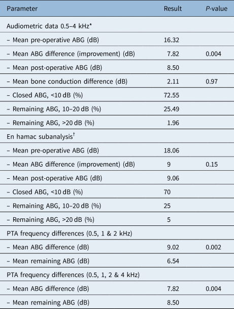
Tympanic membrane perforation closure was seen in 62 of all patients (96.88 per cent). Two non-hamac cases had persistent perforation after surgery (95 per cent tympanic membrane perforation closure) whereas none of the en hamac cases had residual or recurrent perforation (p = 0.02). Bone conduction data was not available for 13 cases, resulting in 51 cases available for audiometric analysis. Audiometric data is shown in Table 2. Subgroup analysis of the en hamac cases and the impact of frequency selection in reporting pure tone audiometry results were calculated and are shown in Table 2.
Audiometric data

*n = 51; †n = 20. ABG = air–bone gap
Complications were rare and minor. Necrosis of the external ear canal skin flap was seen in three patients (5.88 per cent) but healed spontaneously. Epidermal inclusion cyst formation was seen in three patients (5.88 per cent). In one patient (1.96 per cent), we observed a small dehiscent temporomandibular joint after canalplasty. The patient temporarily suffered from temporomandibular joint dysfunction which resolved spontaneously.
Discussion
Numerous techniques have been described to improve type 1 tympanoplasty outcomes in otological surgery. Other studies mainly focus on approach, graft type and placement, whereas other surgical steps are often neglected. We believe these results can be optimised in three simple ways.
First, we systematically perform a complete canalplasty in each case. This provides optimal visualisation during surgery and facilitates optimal graft placement.Reference Vijayendra, Ittop and Sangeetha8 The level of tympanic sulcus can easily be identified, and lateralisation of the graft can be prevented. Canalplasty improves healing by enhancing the self-cleaning capacity and ventilation of the reconstructed ear canal. We believe this helps to prevent myringitis post-operatively and provides a wide endaural access for revisions if needed. Although a thorough canalplasty may seem unnecessary or time-consuming, time spent on canalplasty is easily compensated by the time gained by improved exposure during grafting and post-operative care. Even though presently no clinical trials are available on this topic, we believe these advantages are key to improving tympanoplasty outcomes.
Secondly, we prefer to use a combined grafting technique using temporalis fascia as well as cartilage. Our technique, as described above, can be classified as a cartilage tympanoplasty group B according to Tos et al. Reference Tos9 Temporalis fascia has ideal properties for grafting (i.e. a low rejection rate, good tensile strength and conductive properties) but is at risk for atrophy and weakening, which can cause reperforation or retraction.Reference Buckingham10,Reference Lyons, Su, Vissers, Peters, Smit and Grolman11 The increased stiffness of cartilage makes it less prone to retraction and reperforation.Reference Dornhoffer12,Reference Jesic, Dimitrijevic, Nesic, Jotic and Slijepcevic13 Initial claims about worse ABG closure using cartilage were not confirmed in a recent large meta-analysis, which showed similar audiometric outcomes compared to fascia tympanoplasty.Reference Jalali, Motasaddi, Kouhi, Dabiri and Soleimani14 Studies have also shown the advantages of higher graft integration rate in cartilage tympanoplasty compared to temporalis fascia grafting alone.Reference Jalali, Motasaddi, Kouhi, Dabiri and Soleimani14 By combining the two grafts we can provide optimal graft integration as well as a sturdy tympanic membrane to prevent reperforation.
Lastly, anterior marginal perforations have a worse outcome because of reduced vascular supply, limited anterior margin, poor visualisation and inadequate graft stabilisation.Reference Schraff, Dash and Strasnick3,Reference Applebaum and Deutsch4 By using the en hamac technique we can stabilise the graft by creating an anterior ‘hammock-like’ fixation of the graft.Reference Sauvage, Heurtebise and Puyraud15 This French hammock technique is different from the hammock technique described by Peng and Lalwani, which utilises a large tympanomeatal flap incorporating anterior ear canal skin and places the graft like a hammock extending from the anterior to the posterior external auditory canal.Reference Peng and Lalwani16 However, it is similar to other techniques of anterior stabilisation (anterior hitch and anterior pull-through) and can be considered largely the same.Reference Harris, Wong, Yang and Miller17,Reference Primrose and Kerr18 A uniform vocabulary is needed, as this heterogeneity interferes with direct comparison and pooling of results.
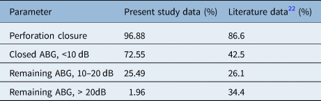
When comparing our results with previously published data we see an overall higher tympanic membrane perforation closure, as shown in Table 3. Because of our study design we were unable to compare our data to matched control cases. The only study to date comparing an anterior stabilisation technique to the standard underlay technique shows a tympanic membrane perforation closure of 93.2 per cent versus 84.6 per cent.Reference D'Eredità and Lens19 However, these results were not statistically significant (odds ratio = 0.4; 95 per cent confidence interval = 0.12–1.41) so further comparative studies are needed.
Data comparison between the literature and this study

ABG = air–bone gap
Audiometric analysis of our data shows overall better results than literature data, even with the use of cartilage as graft (Table 3). However, it needs to be stressed that our data is retrospective and direct comparison with other studies remains difficult. Bone conduction changes in our cohort were minimal and the difference was not statistically significant.
• Anterior tympanic membrane perforations are often a challenging aspect in type 1 tympanoplasty because of inadequate visualisation and insufficient stabilisation of the graft
• The ‘en hamac’ anterior fixation provides improved graft stabilisation for anterior perforations with a high tympanic membrane closure rate and excellent audiometric outcomes
• Routinely performed canalplasty can safely lead to high success rates because of the improved visualisation during surgery and enhanced post-operative aeration and self-cleaning capacity of the ear canal
Still, it was not possible to close the ABG in all cases. Subclinical subtle ossicular chain rigidity and middle-ear volume could play a role in these cases. Indeed, it has been shown that middle ear and mastoid volumes can cause substantially different conductive hearing loss in identical tympanic membrane perforations.Reference Lerut, Pfammatter, Moons and Linder20,Reference Voss, Rosowski, Merchant and Peake21 In our study, computed tomography volume assessment and tympanometry volume recordings were not adequately available to assess this hypothesis.
It has been shown that canalplasty does not affect the audiometric results of tympanoplasty, but preliminary study results show that it can result in a small deterioration in perceived quality of sound. To what extent these changes are clinically relevant still needs to be determined.Reference Van Spronsen, Brienesse, Ebbens and Dreschler22
The impact of frequency selection in reporting audiometric results (Table 2) confirms previously published data that shows a worse outcome for 4 kHz than lower frequencies.Reference Goldenberg and Berliner23 Consequently our data shows less ABG closure when including 4 kHz in the results. Indeed, we often see that the typical audiometric inverted V-shape changes to a small isolated high frequency ABG after surgery.Reference Tan, Santa Maria, Eikelboom, Anandacoomaraswamy and Atlas24 Unfortunately statistical analysis (t-test) of these ABG differences was not statistically significant (p = 0.76), presumably because of our limited sample size.
Conclusion
Type 1 tympanoplasty using fascia and cartilage with complete canalplasty and the en hamac (anterior hitch) technique is a safe and efficient procedure with optimal results. We present a cohort with long-term clinical and audiological follow-up, showing a high perforation closure rate (96.88 per cent) as well as optimal hearing results (mean remaining ABG 8.50 dB; 72.55 per cent ABG closure less than 10 dB). Complications are rare and minor and although procedure time is generally longer, we believe time spent on canalplasty is easily compensated by the time gained post-operatively. Therefore, the principles and modifications suggested in this article can be regarded as useful techniques for the otologist to improve surgical results in type 1 tympanoplasty.
Competing interests
None declared