Implications
Until now, research on prebiotic substances has been mainly carried out on laboratory animals such as rats. These studies mainly concentrated on intestinal bacterial communities, but in recent years some research on immunology and blood traits has also been carried out. This study now aims to give insights in effects of prebiotic substances on intestinal and systemic health in preruminant calves. To achieve this, hematology and expression of relevant genes in young cattle fed two different kinds of prebiotics have been investigated.
Introduction
Calf diseases, mainly diarrhea, are a severe problem during the early period of animal life and are often prevented or treated with antibiotics (Svensson et al., Reference Svensson, Lundborg, Emanuelson and Olsson2003). However, antimicrobials in sub-therapeutic doses are also used to promote growth for meat production and this led to problems with resistant bacteria and antibiotic residues in meat (McEwen and Fedorka-Cray, Reference McEwen and Fedorka-Cray2002). Thus, antimicrobial growth promoters have been banned in the European Union (EG 183/2003). This caused adverse effects on animal health and thus increased the use of therapeutic antibiotics (Casewell et al., Reference Casewell, Friis, Marco, McMullin and Phillips2003). Now, alternatives have to be found to stabilize gut health and to promote growth. These may include changes in nutrition, for example the inclusion of prebiotics (Lallès et al., Reference Lallès, Bosi, Smidt and Stokes2007). A prebiotic like inulin or lactulose has been defined as a non-digestible substance that beneficially affects the host by modulation of the intestinal flora (Gibson and Roberfroid, Reference Gibson and Roberfroid1995).
Inulin is a natural β-(2-1)-linked fructo-oligosaccharide with up to 60 units common in plants used in the Western diet (Van Loo et al., Reference Van Loo, Coussement, de Leenheer, Hoebregs and Smits1995). It has been shown to lead to a shift in the intestinal bacterial flora toward more beneficial bifidobacteria (Gibson et al., Reference Gibson, Beatty, Wang and Cummings1995), to impede carcinogenesis (Femia et al., Reference Femia, Luceri, Dolara, Giannini, Biggeri, Salvadori, Clune, Collins, Paglierani and Caderni2002) or to stimulate the immune system (Schley and Field, Reference Schley and Field2002). In addition, it was shown to enhance growth performance of livestock (Van Loo, Reference Van Loo2007).
Lactulose is a semi-synthetic disaccharide (Schumann, Reference Schumann2002) that is mainly fermented by beneficial bacteria like lactobacilli or bifidobacteria (Mitsuoka et al., Reference Mitsuoka, Hidaka and Eida1987). It has been commonly used to treat constipation (Attar et al., Reference Attar, Lémann, Ferguson, Halphen, Boutron, Flourié, Alix, Salmeron, Guillemot, Chaussade, Ménard, Moreau, Naudin and Barthet1999) or hepatic encephalopathy (Bircher et al., Reference Bircher, Müller, Guggenheim and Haemmerli1966), and has been reported to have effects similar to inulin on carcinogenesis, immunology (Schumann, Reference Schumann2002) or animal growth performance (Fleige et al., Reference Fleige, Preißinger, Meyer and Pfaffl2007a).
This study investigated long-term treatment of calves with the prebiotics inulin and lactulose and their potentially beneficial effects on hematology and intestinal health. Inulin has been chosen since it is commonly accepted to have prebiotic characteristics, but only little is known about its effects on calves. Lactulose has been used in previous studies on calves in combination with the probiotic strain Enterococcus faecium (Fleige et al., Reference Fleige, Preißinger, Meyer and Pfaffl2007a, Reference Fleige, Preißinger, Meyer and Pfaffl2007b and Reference Fleige, Preißinger, Meyer and Pfaffl2009). In this study, its effects should be verified in the absence of additional effectors.
Material and methods
Animal husbandry and feeding
The animal housing and slaughtering procedures followed the actual German law on animal production and veterinary inspection (Bayerische Landesanstalt für Landwirtschaft (LfL), Grub, Germany).
Forty-two Holstein–Friesian bull calves were purchased from the Viehzentrum Waldkraiburg GmbH. Animals were transported to the experimental station Karolinenfeld (LfL, Institut für Tierernährung und Futterwirtschaft) and subdivided into three experimental groups (n = 14 per group). Weight (52.9 ± 6.2 kg) and age (22 ± 5 days) were balanced between groups.
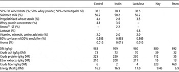
The composition of diets is given in Table 1. Basic to all diets was the milk replacer Milkibeef Top (Milkivit, Trouw Nutrition, Burgheim, Germany). Control groups were fed the pure replacer, and the other groups were fed the same milk replacer iso-energetically and iso-nitrogenically enriched with 2% (dry matter) of either inulin (Beneo ST, Orafti, Tienen, Belgium) or lactulose (Lactusat, Milei GmbH, Leutkirch, Germany). This concentration is approximately equivalent to suggestions on the consumption of dietary fiber in human diets (Van Loo et al., Reference Van Loo, Coussement, de Leenheer, Hoebregs and Smits1995). In a previous study, lactulose has been used in similar concentrations (Fleige et al., Reference Fleige, Preißinger, Meyer and Pfaffl2009). Individual feeding was achieved by transponder automatic feeders (Förster Technik, Engen, Germany). During the experimental period of 20 weeks, the milk replacer concentration was rising from 125 to 200 g/l with daily intake volumes rising from 6 to 16 l. Calves had free group access to fresh drinking water and up to 300 g hay per day and animal. Since the calves were housed on straw, a further uptake of roughage could not be excluded. The animals were slaughtered after 20 weeks.
Ingredients and analysis of nutrient and energy content of the diets (energy content of the milk replacer was estimated with the program Zifo; LfL, 2005)

DM = dry matter.
Tissue and blood sampling
Ten weeks after the beginning of the feeding experiment, and during the slaughtering process after 20 weeks of experimental diet, blood samples were taken from the jugular vein by venipuncture or during bleeding for examination by a veterinary laboratory (Vetmed Labor, Unterhaching, Germany). Venipuncture was included into the daily routine so that animals had free access to the milk replacer before sampling. Owing to transportation and a waiting period before slaughtering, animals were fasted during the second blood sampling. From each of the blood samples, 10 ml were also transferred to paxGene Blood RNA tubes (BD, Heidelberg, Germany) for RNA extraction. Tubes were stored at −20°C until extraction. Immediately after slaughter, organs were removed from the carcass. Small pieces were cut from central parts of the jejunum, ileum and colon and from the tip of the spleen. In addition, mesenteric lymph nodes were sampled. Intestinal samples were washed twice in physiological salt solution to remove digesta. All tissues were immediately flash frozen in liquid nitrogen. Samples were taken in duplicate and stored at −80°C until extraction to prevent RNA degradation.
RNA extraction and quality control
RNA extraction of whole blood stabilized in paxGene blood RNA tubes was performed using the paxGene blood RNA kit (Qiagen, Hilden, Germany) according to the manufacturer's instructions. RNA extraction of tissue samples was performed with TriFast reagent (Peqlab, Erlangen, Germany) according to the manufacturer's instructions. RNA was extracted from small pieces (∼50 μg) of tissue. Samples were not allowed to thaw until pieces were immersed in TriFast to avoid RNA degradation.
RNA quantity and purity of all samples (260 nm/280 nm absorption ratio) were measured with a NanoDrop spectrophotometer (Peqlab, Erlangen, Germany). RNA quality was assessed using the Bioanalyzer 2100 with RNA Nano Chips (Agilent Technologies, Palo Alto, CA, USA).
Real-time quantitative reverse transcription PCR (qRT-PCR)
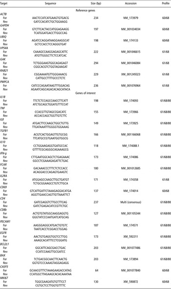
qRT-PCR was performed using a Rotor-Gene 6000 (Corbett Life Science, Sydney, Australia) and the SuperScript III Platinum SYBR Green One-Step qRT-PCR kit (Invitrogen, Carlsbad, CA). Specific primer sets were either adapted from Fleige et al. (Reference Fleige, Preißinger, Meyer and Pfaffl2007b and Reference Fleige, Preißinger, Meyer and Pfaffl2009) or designed using primer3 v. 0.4.0 (http://frodo.wi.mit.edu/) and purchased from Eurofins MWG Operon (Ebersberg, Germany). The one-step qRT-PCR profile consisted of a reverse transcription (10 min at 50°C), a denaturation step (5 min at 95°C) and 40 cycles of amplification and quantification (with 15 s at 95°C, 30 s annealing at appropriate temperature, 20 s elongation at 68°C and a 15 s measurement step at 80°C). Primer sequences and PCR profile information are given in Table 2.
Primer pairs, product sizes, gene identities and appropriate profiles for qRT-PCR. For every tissue one pair of reference genes was chosen to normalize crossing point values of target genes. All genes were measured in each tissue

Data evaluation
Data of hematology examination are presented as mean ± s.e.m. with n = 14.
For qRT-PCR, the crossing point values were obtained with Rotor-Gene 6000 software version 1.7 (Corbett Life Science). For relative quantification of mRNA expression, a number of potential reference genes were measured and for each tissue the optimal pairs of reference genes were chosen using GenEx Pro Ver 4.3.4 (MultiD Analyses AB, Gothenburg, Sweden). The arithmetic mean of their crossing point values was used to normalize target gene values. The calculation of relative expression values was carried out using the 2-ΔΔCT method (Livak and Schmittgen, Reference Livak and Schmittgen2001). For each group, results are given in geometric means of expression ratios relative to the control group and r.e.m. Statistical analysis of gene expression data was carried out using the one-way ANOVA method of SigmaStat 3.0 (SPSS Inc., Chicago, IL, USA). In case of significant differences, a post-hoc Tukey–Kramer test was performed to further investigate group differences.
Results
Hematology
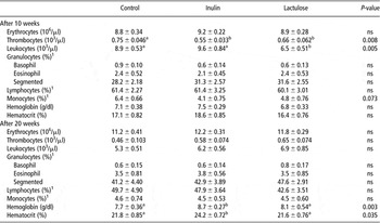
The results are listed in Table 3. Ten weeks after the start of the feeding regimes, animals of the lactulose group showed significantly reduced concentrations of peripheral blood leukocytes (P = 0.005) compared with the inulin and control group. Thrombocytes were significantly decreased by both experimental feedings compared with the control (P = 0.008). For the proportion of monocytes of total leukocytes, a trend for a decrease in both prebiotic groups could be found (P = 0.073).
Results of hematological examination of blood samples taken 10 weeks after beginning or during slaughtering

1Of total leukocytes.
All values are presented as mean ± s.e.m. (n = 14). P-values are results of ANOVA analysis. Within rows, values without a common superscript additionally have been found significantly different by post-hoc Tukey–Kramer test (P < 0.05).
At slaughter, all these effects were no longer visible, but instead both hemoglobin concentration (P = 0.003) and hematocrit (P = 0.035) were significantly increased in inulin-treated animals compared with control and lactulose-treated animals. At 10 weeks, a similar pattern was visible, which could not be statistically verified (P = 0.286 or P = 0.158, respectively).
RNA quality
RNA purity was regarded as good with an average A260/A280 ratio of 2.0 ± 0.01 for all tissues. Values >1.80 are considered as an indicator for high purity. RNA quality for all tissues was investigated with a Bioanalyzer 2100 (Agilent Technologies) and yielded an average RNA integrity number (RIN) of 6.9 ± 0.13. RIN is a measure for RNA quality ranging from totally degraded RNA (RIN = 1) to intact RNA (RIN = 10) (Fleige and Pfaffl, Reference Fleige and Pfaffl2006; Bustin et al., Reference Bustin, Benes, Garson, Hellemans, Huggett, Kubista, Mueller, Nolan, Pfaffl, Shipley, Vandesompele and Wittwer2009). A RIN of 5 was regarded as a minimal requirement for qRT-PCR. Single samples not sufficient for qRT-PCR were re-extracted, and when values did not reach adequate levels were omitted from analysis.
Reference gene pairs
Reference genes are regarded as acceptable if their mRNA expression is stable between treatment groups. GenEx Pro software (MultiD Analyses AB) is one possibility to choose ideal reference gene pairs to normalize mRNA expression data. Here, for each tissue, the best reference gene pair was chosen from all genes measured (Bustin et al., Reference Bustin, Benes, Garson, Hellemans, Huggett, Kubista, Mueller, Nolan, Pfaffl, Shipley, Vandesompele and Wittwer2009). Thus, normalization was carried out with actin β (ACTB) and G protein-associated kinase (GAK) in the jejunum, vacuolar protein sorting 4 homolog A and GAK in the ileum, ubiquitin (UBIQ) and glyceraldehyde-3-phosphate dehydrogenase in the colon, Ras-related protein Rab-21 and peptidase (mitochondrial processing) alpha in the spleen and ACTB and UBIQ in blood.
mRNA expression changes of inflammation and immune modulating factors
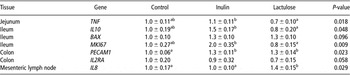
An overview of all significant gene expression changes found in this study is given in Table 4. Genes or tissues without significant changes are not shown.
Changes in relative mRNA expression levels found in the in vivo study

All values are presented as geometric mean ± s.e.m. (n = 14) of relative expression values with respect to the control levels. P-values are results of ANOVA analysis. Within a row means without a common superscript have been found significantly different after post-hoc Tukey–Kramer test (P < 0.05).
In the jejunum, relative expression of the pro-inflammatory tumor necrosis factor-α (TNF) was influenced by the feeding group (P = 0.018). Lactulose induced a downregulation of its expression compared with the control as well as the inulin group (Tukey–Kramer test: P = 0.025 or P = 0.017, respectively).
In the ileum, the relative mRNA amount of anti-inflammatory interleukin 10 (IL10) was significantly influenced by the diet (P = 0.048). Specifically, it was upregulated by inulin and downregulated by lactulose, both relative to the control. Post-hoc test showed that expression levels were significantly different between the treatment groups (P = 0.017) but not between each of these groups and the control.
In the colon, the expression of platelet endothelial cell adhesion molecule-1 (PECAM1) mRNA was significantly increased by both dietary treatments (P = 0.023). The Tukey–Kramer test revealed significant differences in its expression between control and lactulose (P = 0.017) as well as between control and inulin (P = 0.016). Simultaneously, expression of the lymphocyte activation marker interleukin 2 receptor alpha chain (IL2RA) tended to decrease in lactulose-treated animals (P = 0.058).
In mesenteric lymph nodes, expression of the chemo-attractant interleukin 8 (IL8) was significantly upregulated by lactulose (P = 0.029). The upregulation was significant when compared with control (P = 0.027) as well as to inulin (P = 0.016).
Interleukin 1β, transforming growth factor-β1, interferon-γ (IFN-g), receptor for Fc fragment of IgA, CD69, CD4 and CD8b were not influenced by the diet in any tissue analyzed. In the spleen and blood, no regulation of investigated genes were found (data not shown).
mRNA expression changes of proliferation and apoptosis-related genes
Changes in expression of proliferation or apoptosis-related genes were found only in the ileum (Table 4). There, the proliferation marker MKI67 was significantly influenced by both prebiotics (P = 0.009). Inulin led to an upregulation of its expression relative to the control (P = 0.028), whereas lactulose decreased it slightly. Such expression was significantly different between the inulin and the lactulose group (P = 0.003) but not between both and the control. Simultaneously, lactulose and inulin showed a weak tendency to increase the relative expression of pro-apoptotic BCL2-associated X protein (BAX; P = 0.096).
BCL2-like-1, epidermal growth factor receptor and Caspase 3 were not influenced by the diet (data not shown).
Discussion
Until now, only a few studies on prebiotics have been completed with calves, and to the authors’ knowledge, only one other long-time study was performed where changes in gene expression and hematological traits during feeding of lactulose have been investigated. There, the authors described a decrease in thrombocyte numbers in 3% lactulose-fed calves (Fleige et al., Reference Fleige, Preißinger, Meyer and Pfaffl2009) that could be supported for both prebiotics in this study after 10 weeks of feeding. A high rate of thrombocyte aggregation is considered as one of many risk factors for coronary heart disease and has been shown to be negatively correlated with intakes of higher amounts of dietary fiber in humans (Pietinen et al., Reference Pietinen, Rimm, Korhonen, Hartman, Willett, Albanes and Virtamo1996) and rats (Bagger et al., Reference Bagger, Andersen, Nielsen and Ryttig1996).
At the end of the experimental period, only hemoglobin concentrations and hematocrit were affected by inulin feeding. Ohta et al. (Reference Ohta, Ohtsuki, Uehara, Hosono, Hirayama, Adachi and Hara1998) reported that these two parameters were significantly reduced after gastrectomy and subsequent malabsorption of iron in rats, but the levels could be recovered by 0.75% oligofructose feeding. Iron deficiency is a common problem in veal calves fed only milk replacer and no solid feed or fiber-rich supplementation (Cozzi et al., Reference Cozzi, Gottardo, Mattiello, Canali, Scanziani, Verga and Andrighetto2002). Inulin feeding could have had improving effects on iron absorption capabilities as was shown previously in rats (Ohta et al., Reference Ohta, Ohtsuki, Baba, Takizawa, Adachi and Kimura1995).
Immune- and inflammation-modulating effects of prebiotic substances – especially for inulin and oligofructose – have been reported repeatedly (e.g. Field et al., Reference Field, McBurney, Massimino, Hayek and Sunvold1999; Schley and Field, Reference Schley and Field2002; Hosono et al., Reference Hosono, Ozawa, Kato, Ohnishi, Nakanishi, Kimura and Nakamura2003). In this study, leukocyte counts in peripheral blood were decreased in lactulose-treated calves and monocyte numbers were decreased in both treatment groups. A similar effect has been found when mannanoligosaccharides were fed to dogs (Middelbos et al., Reference Middelbos, Godoy, Fastinger and Fahey2007). In addition, the expression of IL2RA – an activation marker on the surface of lymphocytes – tended to be lowered in the colon of lactulose-treated calves compared with the control. Fleige et al. (Reference Fleige, Preißinger, Meyer and Pfaffl2009) found a similar lowering of its expression in the mesenteric lymph nodes of lactulose-treated calves. Middelbos et al. (Reference Middelbos, Godoy, Fastinger and Fahey2007) suggested that a lowering of the infectious load in the intestine due to the feeding of mannan moieties may decrease the necessity of activated immune cells in the vicinity. This may also be reflected in peripheral blood. A similar decrease in pathogen load in the intestine is also attributed to prebiotics such as inulin or lactulose (Bovee-Oudenhoven et al., Reference Bovee-Oudenhoven, Termont, Heidt and Van der Meer1997; Flickinger et al., Reference Flickinger, Van Loo and Fahey2003).
A similar pattern as for leukocyte counts and IL2RA expression could be found for TNF expression in the jejunum. Lactulose significantly reduced its expression confirming again a decreased activation of an inflammatory or immune response in the intestine. A similar effect on TNF has been found before in a rat colitis model after treatment with lactulose (Camuesco et al., Reference Camuesco, Peran, Comalada, Nieto, Di Stasi, Rodriguez-Cabezas, Concha, Zarzuelo and Galvez2005). In contrast, pro-inflammatory IL8 was significantly enhanced in the mesenteric lymph nodes of animals in the lactulose group. Fleige et al. (Reference Fleige, Preißinger, Meyer and Pfaffl2009) also reported increases of IL8 expression in the spleen and mesenteric lymph nodes of calves after feeding of lactulose, but these results were not statistically significant. IL8 expression can be increased by lipoteichoic acid of gram-positive pathogens (Standiford et al., Reference Standiford, Arenberg, Danforth, Kunkel, VanOtteren and Strieter1994) that may have been transported to the mesenteric lymph nodes by antigen-presenting cells. But since no induction of IL8 was found in gut tissues, where these bacteria should show a first effect, it has to be considered whether other factors possibly independent of gut flora may be responsible for the significant increase in IL8 expression in the mesenteric lymph nodes of lactulose-fed calves.
Anti-inflammatory IL10 showed an increased expression in the ileum of inulin-treated calves, especially when compared with lactulose-treated animals. The ileum wall is interstratified with Peyer's patches, parts of the gut-associated lymphoid tissue. Cells from these patches have been shown to release anti-inflammatory cytokines such as IL10 after stimulation with probiotics, prebiotics or fermentation products thereof (Säemann et al., Reference Säemann, Böhmig, Österreicher, Burtscher, Parolini, Diakos, Stöckl, Hörl and Zlabinger2000; Hosono et al., Reference Hosono, Ozawa, Kato, Ohnishi, Nakanishi, Kimura and Nakamura2003; Roller et al., Reference Roller, Rechkemmer and Watzl2004). Therefore, it seems probable that fermentation products deriving from inulin degradation or secretory molecules from bacteria stimulated by inulin feeding are causative agents in this increase of IL10 expression.
PECAM1 is a cell-surface molecule of leukocytes and endothelial cells and is crucial for the transmigration of leukocytes through the capillary wall into inflamed tissue (Wakelin et al., Reference Wakelin, Sanz, Dewar, Albelda, Larkin, Boughton-Smith, Williams and Nourshargh1996), which rather stands for pro-inflammatory action. Nevertheless, it has been found to be downregulated on endothelial cells in typical pro-inflammatory situations such as stimulation with TNF or IFN-g without effect on leukocyte transmigration (Shaw et al., Reference Shaw, Perkins, Lim, Liu, Nusrat, Schnell, Parkos and Luscinskas2001). In addition, this molecule also plays a role in angiogenesis (DeLisser et al., Reference DeLisser, Christofidou-Solomidou, Strieter, Burdick, Robinson, Wexler, Kerr, Garlanda, Merwin, Madri and Abelda1997), and therefore an upregulation could also mean an increase in perfusion of the surrounding tissue. A stimulation of angiogenesis in gastric ulcer healing by probiotic bacteria – targets of prebiotics – has been found previously (Lam et al., Reference Lam, Yu, Wong, Wu, Shin, Tai, So, Woo and Cho2007) and may be related to the present finding.
Expression of the proliferation marker MKI67 in the ileum was increased in the inulin group. Interestingly, morphological investigations during the same study rather showed a slight decrease in the villus length and number of proliferative cells in the same group (Masanetz et al., Reference Masanetz, Wimmer, Plitzner, Limbeck, Preißinger and Pfaffl2010). Maybe the main MKI67 expression was not located in the epithelial layer, which was investigated by microscopy, but in other parts of the gut wall. Nevertheless, both enhancement (Brunsgaard and Eggum, Reference Brunsgaard and Eggum1995) and decline (Femia et al., Reference Femia, Luceri, Dolara, Giannini, Biggeri, Salvadori, Clune, Collins, Paglierani and Caderni2002) of gut epithelial proliferation have been described after treatment with different prebiotic substances in animal models. Simultaneously, a trend for an increased expression of BAX mRNA has been found in the ileum in both prebiotic groups. It has been reported before that butyrate – a fermentation product of prebiotic substances – is able to increase apoptosis via upregulation of BAX expression (Mandal et al., Reference Mandal, Olson, Sharma, Vadlamudi and Kumar2001). This is fitting to the proposed anti-carcinogenic effects described for inulin (Femia et al., Reference Femia, Luceri, Dolara, Giannini, Biggeri, Salvadori, Clune, Collins, Paglierani and Caderni2002).
Differences between effects of both prebiotic substances or different gut segments may be due to fermentation properties of the substances themselves (Branner et al., Reference Branner, Böhmer, Erhardt, Henke and Roth-Maier2004). In addition, the density and composition of calf intestinal flora also vary greatly within the gut with decreasing anaerobic bacteria counts and a shift toward bifidobacteria from the rumen to the colon (Collado and Sanz, Reference Collado and Sanz2007). In addition, diverse stimulatory effects on beneficial bacterial subpopulations and fermentation product profiles were reported for inulin and lactulose (Rycroft et al., Reference Rycroft, Jones, Gibson and Rastall2001).
Changes in hematological traits between the samplings at 10 and 20 weeks may be correlated to age-related changes that have been demonstrated previously (Mohri et al., Reference Mohri, Sharifi and Eidi2007). For example, similar to this study, a significantly higher platelet count was found in young calves that decreased to adult control levels over time (Brun-Hansen et al., Reference Brun-Hansen, Kampen and Lund2006). Differences in regulations of blood characteristics may also play a tremendous role in the effectivity of prebiotic substances. In future studies, changes of hematology in growing cattle fed different diets should be investigated in detail.
In conclusion, both prebiotic substances used in this study showed effects on immune regulation and inflammation both systemically and locally in the gut of calves. Inulin and lactulose decreased signs of immune activation and increased anti- or lowered pro-inflammatory signals, respectively. Presumably, these effects were generated by a decline in pathogen load in the intestine commonly attributed to prebiotic treatment. Effects of both substances on proliferation and apoptosis are rather diverse and remain to be studied in detail. In addition, positive effects on hemoglobin and hematocrit have only been shown for inulin. These presumed effects on iron absorption are highly interesting and remain to be studied in future experiments, especially with a look at other mineral absorption capabilities. On the whole, a beneficial effect of prebiotic feeding on veal calf performance may be expected for both inulin or lactulose regarding the results of this study.
Acknowledgments
The current research work was supported by grants from the ‘Vereinigung zur Förderung der Milchwissenschaftlichen Forschung’ (Germany). Special thanks go to Milei GmbH for providing the lactulose preparation, to Milkivit, Trouw Nutrition (Burgheim) for the preparation of the milk replacer and to Orafti for the Beneo® ST.