I. INTRODUCTION
Visible light-driven photocatalysts have attracted significant interests in recent years because visible light is an important clean energy and can be easily utilized. To use solar energy efficiently, the key step is to explore new visible light-driven photocatalysts.Reference Bell1,Reference Mann2 The various types of inorganic compounds that are active photocatalyst, including TiO2, WO3, ZnO, and Bi2WO6 have been synthesized by various kinds of methods.Reference Fei, Cui, Yan, Qi, Yang, Wang, He and Li3–Reference Liu, Guo, Wang, Huang, Zhu and Chen6 It is highly attractive and desirable to synthesize new type of materials with fine structures, although it is still a big challenge.
Bismuth silicate (Bi2SiO5) was first reported in 1996.Reference Koiwa, Kanehara, Mita, Iwabuchi, Osaka, Ono and Maeda7 Bi2SiO5 has an orthorhombic crystal structure and belongs to Cmc21 space group. It is composed of (SiO3)2− pyroxene file layers inserted between (Bi2O2)2+ layers.Reference Geoges, Goutenoire and Lacorre8 After the first report, nanostructured Bi2SiO5 has attracted great interests owing to the interesting functional performance during the past several years. It has been reported as a material with relatively good dielectric propertiesReference Koiwa, Kanehara, Mita, Iwabuchi, Osaka, Ono and Maeda7 and pyroelectric and nonlinear optical effects.Reference Geoges, Goutenoire and Lacorre8 Until recently, synthetic microstructure/nanostructure photocatalysts are found to display interesting photocatalytic activities in environmental application.Reference Chen, Bi, Wu, Wang, Li and Fu9–Reference Zhang, Wang, Sun, Xu, Shang and Ren11 Chen et al.Reference Chen, Bi, Wu, Wang, Li and Fu9 prepared pure Bi2SiO5 nanoplates by a hydrothermal method, which first showed photocatalytic activities. Zhang et al.Reference Zhang, Hu and Li10 synthesized Bi2SiO5 microspheres that exhibited good photoactivitity in the degradation of phenol in water. Zhang et al.Reference Zhang, Wang, Sun, Xu, Shang and Ren11 presented a facile self-assembly approach for mass production of Bi2SiO5 nanobelts/nanoplates, as photocatalysts could degrade aqueous dye and phenol. Although some advances have been achieved in the synthesis and application of such materials,Reference Sato, Saito, Nishiyama and Inoue12–Reference Hou, Wu, Wang, Ding, Li and Fu16 however, current chemical knowledge on Bi2SiO5 as a photocatalytic material is very limited. Therefore, it is still a challenge to prepare pure Bi2SiO5 through a facile synthetic process that is cost-effective and energy efficient. Furthermore, the growth mechanism of the Bi2SiO5 via a facile process, as an important fundamental issue, needs to be investigated.
In this paper, we report a novel and simple hydrothermal method for the preparation of Bi2SiO5 nanoplates/nanoflowers structures using silica aerogelsReference Zhu, Xie, Lü and Jiang17–Reference Zhu, Xie, Chen and Jiang19 as precursors. A new precursor system containing bismuth ion and silica aerogels in ethanol–H2O mixed solvent was adopted. Phase structure, chemical composition, and morphology were investigated systematically with various techniques. Results show that the Bi2SiO5 nanoplates/nanoflowers structures have novel or enhanced properties. Furthermore, the band structure (BS) of Bi2SiO5 was elucidated by the experimental and density functional theoretical (DFT) calculation methods. Moreover, as a novel photocatalyst, Bi2SiO5 nanostructures were found to exhibit enhanced photocatalytic activity in aqueous dye removal.
II. EXPERIMENTS
A. Catalyst preparation
All chemicals were of analytical grade and were used without further purification. In a typical synthesis, silica aerogels were prepared as reported previously.Reference Zhu, Xie, Lü and Jiang17–Reference Zhu, Xie, Chen and Jiang19 For the Bi2SiO5 nanoflowers samples, 1 mmol silica aerogel was dissolved in a 20 mL ethanol–H2O mixed solvent (volume ratio 3:1) followed by the addition of polyvinylpyrrolidone (PVP, K-30). Then a solution of 2 mmol Bi(NO3)3·5H2O was dissolved into 2 mL HNO3 solution (4 mol/L). The obtained mixture was added into the above, under stirring for about 20 min until a clear solution formed. Finally, the pH value of the solution was adjusted to 9 through the dripping of 25 wt% NH3·3H2O. After stirring for 30 min, the mixture was transferred into a teflon-lined autoclave with a capacity of 25 mL and maintained at 180 °C for 12 h. The product with flower-like nanostructures was isolated by centrifugation, washed with deionized water and ethanol for several times, respectively, and finally dried in a vacuum at 80 °C for 12 h. For the Bi2SiO5 nanoplates sample, it was obtained without PVP with the other conditions unchanged.
B. Characterizations
The phase of the as-synthesized products was characterized using x-ray diffraction (XRD) (D8 Advance diffractometer, Bruker, Germany) with Cu Kα radiation (λ = 1.5406 Å) at a scanning rate of 10° min−1; scanning electron microscopy (SEM) (JSM-7001F, JEOL, Japan) was used to investigate the microstructures of samples; energy-dispersive x-ray (EDX) microanalysis was also performed during the SEM observations; the Brunauer–Emmett–Teller (BET)-specific surface area was obtained from the N2 adsorption/desorption analyses at 77 K (NOVA 2000, Quantachrome, Boynton Beach, FL); and ultraviolet-visible diffuse reflectance spectra (UV-VIS DRS) were recorded on a UV-2450 spectrophotometer (Shimadzu, Kyoto, Japan) equipped with spherical diffuse reflectance accessory in the range of 190–800 nm. Surface topography of the Bi2SiO5 nanoplates was also characterized by atomic force microscope (AFM, MicroNanoD3000, Zhuolun, Shanghai, China) under ambient conditions.
C. Photocatalytic experiment
The photocatalytic efficiency of as prepared samples were investigated by the extent of photodecolorization of Rhodamine B (Rh B) under irradiation of a 100 W xenon lamp as light source (luminous flux = 15 lm/W, the xenon lamp is used for solar simulation, ultraviolet radiation below 242 nm). The samples (100 mg) were added to 100 mL of Rh B (10 mg/L) solution, which was kept in the dark for 30 min before irradiation to ensure adsorption/desorption equilibrium. At given time intervals of 30 min, an aliquot of the reaction solution was collected for UV-spectroscopy analysis. Decolorization rate was calculated as (A 0 − A)/A 0, where A 0 shows initial dye solution of maximum absorbance at 554 nm and A shows final solution of maximum absorbance.
D. Detection of hydroxyl radical
The detection of the formed hydroxyl radicals (·OH) on the surface of Bi2SiO5 was measured by a Gary eclipse fluorescence spectrophotometer (Agilent Technologies, Santa Clara, CA). Terephthalic acid, which reacts readily with ·OH to produce a fluorescence product, 2-hydroxyterephthalic acid, was used as a probe molecule. It is known that the fluorescence intensity around 425 nm, which can be ascribed to the characteristic of 2-hydroxyterephthalic acid, is proportional to the amount of hydroxyl radicals formed in solution.Reference Ishibashi, Fujishima, Watanabe and Hashimoto20,Reference Xiao, Si, Zhang, Xiao and Tan21 Under irradiation of a 100 W xenon lamp, the detection experiment process is similar to the photodegradation process. The concentration of terephthalic acid was set at 5 × 10⁻⁴ mol/L in dilute NaOH (2 × 10⁻³ mol/L) solution. The Bi2SiO5 (200 mg) was added to 50 mL above solution, which was kept in the dark for 20 min before irradiation to ensure adsorption/desorption equilibrium. The sampling was carried out every 10 min; the solution was measured on a Gary eclipse fluorescence spectrophotometer after centrifugation. The 2-hydroxyterephthalic acid can give a fluorescence peak at about 425 nm by excitation with the wave length of 315 nm.
E. Density functional theory calculation
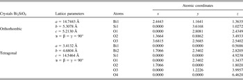
The combination of theory calculation and experimental method has offered great opportunities for establishing the link between the catalytic properties and the catalyst structure.Reference Zheng, Liu and Deng22 Therefore, it is necessary to explore the structure properties of Bi2SiO5 by the theoretical method. The lattice parameters of the models, the orthorhombic structure, and the tetragonal structure of Bi2SiO5 were optimized, the lattice parameters and the atomic coordinates, which were optimized and used in the calculations, are listed in Table I. During the calculations of the BS and density of state (DOS) parameters of Bi2SiO5,Reference Koiwa, Kanehara, Mita, Iwabuchi, Osaka, Ono and Maeda7 the electron correlation effects were modeled using the local density approximation level and Ceperley-Alder equivalent to Perdew-Zunger (CA-PZ), the combination of exchange and correlation functional.Reference Bulgac23 In the calculations, the ultra soft pseudopotentials were used. In addition, plane wave cutoff energy of 300 eV and a 5 × 1 × 5 k-point grid were adopted to sample the Brillouin zone.Reference Segall, Lindan, Probert, Pickard, Hasnip, Clark and Payne24 All calculations were performed using the Cambridge Sequential Total Energy Package (CASTEP) module in Materials Studio package.Reference San25
The lattice parameters and the atomic coordinates in orthorhombic (a) and tetragonal (b) Bi2SiO5 used in calculation.

III. RESULTS AND DISCUSSION
A. Structural characterization of the as-prepared samples
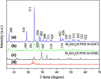
Figure 1(a) shows the XRD patterns of as-prepared flower-like Bi2SiO5 samples. All the diffraction peaks of the pattern can be indexed to a pure orthorhombic Bi2SiO5 phase (JPCDS no. 36-0287, a = 1.5217 nm, b = 0.5477 nm, c = 0.5325 nm, space group Cmc21). No other peaks are found, indicating the high purity and crystallinity of the samples. Figure 1(b) shows the XRD pattern of Bi2SiO5 nanoplates. All the peaks can be readily indexed to the tetragonal phase of Bi2SiO5 (JPCDS no. 36-0288) with lattice constants a = 3.802 Å, b = 3.802 Å and c = 15.194 Å. Figures 1(c) and 1(d) show the XRD patterns of as-prepared samples using commercial SiO2 as precursors (c with PVP, d without PVP). No peaks of Bi2SiO5 phase are found, suggesting none of the Bi2SiO5 was obtained using commercial SiO2 as precursors. The possible reason of this phenomenon is that silica aerogels have very high-specific surface area and irregular pore structure led to more reaction sites than that of commercial SiO2 in the hydrothermal process.
XRD patterns of as-prepared samples: (a) Bi2SiO5 nanoflowers, (b) Bi2SiO5 nanoplates, (c) sample using commercial SiO2 as precursors via PVP, (d) sample using commercial SiO2 as precursors without surfactant.

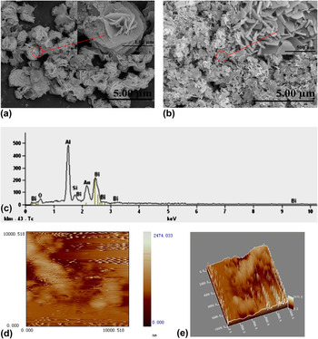
The morphologies of the synthesized samples were studied by SEM, which are shown in Figs. 2(a)–2(c). The SEM images of the Bi2SiO5 product were shown at low and high magnification, respectively. As shown in Fig. 2(a), flower-like Bi2SiO5 was prepared via a template-PVP hydrothermal route in which the silica aerogels was first applied to reaction as a precursor. For comparison, template-free hydrothermal route and keeping other conditions unchanged, only irregular nanoplates were obtained as shown in Fig. 2(b). The nonionic surfactant (PVP) presents a certain degree of viscosity that plays the role of decreasing the spontaneous interaction of (Bi2O2)2+ and (SiO3)2− ions even under the hydrothermal conditions. The precursor solution can absorb to the surface of these vesicles layer upon layer overlay by the electrostatic interaction. This process provides nucleation domains for the hydrothermal reaction between (SiO3)n2− and (Bi2O2)n2+, the hydrolysis of (Bi2O2)n2+, and decrease the crystal growth rate of Bi2SiO5, so the fast nucleation and aggregation rates of Bi2SiO5 result in the three-dimensional flower-like aggregation originate from the reduction of the interfacial energy surface.Reference Zheng, Duan, Chen and Xie26Figure 2(c) is the EDX spectrum obtained from flower-like Bi2SiO5 samples. The result from EDX displays that the products are composed of the elements of Bi, Si, O, and Au. The Au element is generated from the Au nanoparticles spattered for the SEM test. The EDX analysis shows that the atom ratio of Bi and Si is nearly 2:1, indicating that the composition of the products is Bi2SiO5. Flower structure is made up of many thin nanoplates with a thickness of about 20 nm and an average length of 400 nm. These nanoplates are aligned perpendicularly to the flower surface with clearly oriented layers, pointing toward a common center. All these results suggest that the PVP can affect the morphology of nanoflowers.
(a) Low-magnification SEM image of the Bi2SiO5 nanoflowers, (b) Low-magnification SEM image of the Bi2SiO5 nanoplates, (c) EDX pattern of Bi2SiO5 nanoflowers, (d, e) AFM micrographs of the Bi2SiO5 nanoplates.

Figures 2(d) and 2(e) show the AFM images of Bi2SiO5 nanoplates. As shown in Fig. 2(d), Bi2SiO5 nanoplates displays a flat surface, which is in accordance with the result of SEM. Figure 2(e) is the topographic AFM image, which brings a clear show of the flat surface. The lateral dimension of the obtained numerous overlapping Bi2SiO5 nanoplates is about 2–3 µm.
B. Possible growth pattern and formation mechanism
The crystal growth pattern and formation mechanism in solution are so complicated that the actual crystallization mechanism remains an open question. The Ostwald ripening, selective polymer adsorption, and oriented attachment, etc. were adopted to account for the process of crystal growth.Reference Pacholski, Kornowski and Weller27
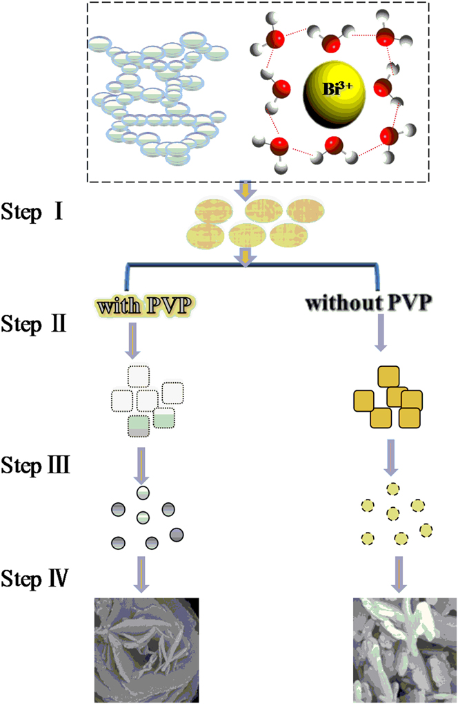
From Fig. 3, it can be concluded that Bi2SiO5 nanostructures can be obtained via a nucleation–self-assembly–dissolution–crystallization growth process. It is well known that usage of a polymer-assisted reaction to control the nucleation and growth is a simple but effective way.Reference Dai, Luo, Fu, Chen and Lu28 In our approach, the Bi2SiO5 should be formed from the combination of (Bi2O2)2+ ion from the dehydration of Bi(NO3)2 and silicate ion from the dissolved SiO2, which was decomposed from silica aerogels. The formation of the Bi2SiO5 sol played an important role in the controlled releasing of (Bi2O2)2+ and (SiO3)2−. With the temperature increased to 180 °C, the silica aerogels were gradually decomposed to generate the (SiO3)2−, which was then combined with (Bi2O2)2+ generated from the hydrolysis of the Bi3+.Reference Zheng, Duan, Chen and Xie26 The reaction for the formation mechanism is shown in Eqs. (1)–(3) as follows
 $${\rm{2Bi}}^{{\rm{3 + }}} \,{\rm{ + \,\,2H}}_{\rm{2}} {\rm{O}} \,\leftrightharpoons\, {\rm{(Bi}}_{\rm{2}} {\rm{O}}_{\rm{2}} {\rm{)}}^{{\rm{2 + }}} \,{\rm{ + \,\,4H}}^{\rm{ + }} \quad,$$
 $$\!\!\!\!\!{\rm{Silica\; aerogels}} \,\leftrightharpoons\,{\rm{SiO}}_{\rm{2}} \,{\rm{ + }}\,{\rm{H}}_{\rm{2}} {\rm{O}}\, \,\leftrightharpoons\,\,{\rm{\rm{SiO_3} ^{{\rm{2}} - } \,{\rm{ + }}\,{\rm{2H}}^{\rm{ + }} \,\,\,\,,$$
 $${\rm{(Bi}}_{\rm{2}} {\rm{O}}_{\rm{2}} {\rm{)}}^{{\rm{2 + }}} \,{\rm{ + }}\,{\rm{SiO_3}} ^{{\rm{2}} - } \,\leftrightharpoons\,\,{\rm{Bi}}_{\rm{2}} {\rm{SiO}}_{\rm{5}}\quad .$$
The possible growth pattern and formation mechanism of Bi2SiO5 nanostructures.
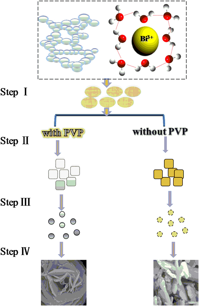
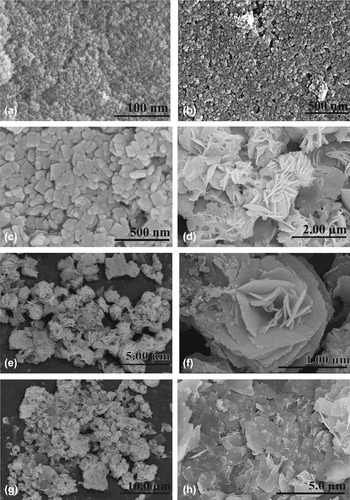
The growth mechanism is also based on the SEM micrographs (Fig. 4). Firstly, silica aerogels were porous in nature with continuous network structure. The particle distribution of aerogels was uniform and is as shown in Fig. 4(a). The free energy of dissolution related to the H2O molecules causes a fast dissociation of the chemical salts in solution,Reference Cavalcate, Sczancoski, Li, Longo and Varela29 so that the (Bi2O2)2+ and silica aerogels were rapidly solvated by these molecules. Secondly, silica aerogels precursors with surfactant were irregular particles [Fig. 4(b)]. After stirring for about 20 min, large amounts of particles appeared due to the subsequent reaction [Fig. 4(c)]. In the case of PVP (nonionic surfactant), this polymer easily dissolved in H2O and formed a long polymeric chain structure by uncountable N-vinylpyrrolidone monomers.Reference Zeng30 After hydrothermal treatment for 6 h, the irregular amorphous particles were stacked together tend to change or direct the growth of the nanoflowers [Fig. 4(d)]. After hydrothermal treatment for 12 h, the nanoflowers were generated. This fact implied that the nanoflowers were grown by the nanoplates. The production of novel structure in the hydrothermal process is in accordance with the well-known “Ostwald ripening mechanism.”Reference Zeng30Figures 4(g) and 4(h) represent the samples obtained at longer reaction time, respectively. The images reveal that nanoflowers with some microparticles were stacked together (24 h), while stable microparticles agglomeration was observed at 180 °C for 36 h.
SEM images of Bi2SiO5 nanoflowers synthesized under different conditions: (a) the silica aerogels, (b) the silica aerogels with the PVP, (c) the precursor before the hydrothermal treatment, (d) the precursor at the temperature of 180 °C for 6 h; (e, f) the precursor at the temperature of 180 °C for 12 h, (g) the precursor at the temperature of 180 °C for 24 h, (h) the precursor at the temperature of 180 °C for 36 h.

In the previous investigation, PVP, a water-soluble polymer, was used as capping molecules and served as surfactant-templating in the synthesis of novel structure.Reference Saravanan, Diwakar, Mohankumar, Pandurangan and Jayavel31,Reference Shi, Chen, Chen, Zhou, Lou, Wang and Yuan32 Saravanan et al.Reference Saravanan, Diwakar, Mohankumar, Pandurangan and Jayavel31 found PVP can modify the Ostwald ripening kinetics in such a way that the growth rate decreases with the size of the CdS nanoparticles and effectively narrow the size distribution. Shi et al.Reference Shi, Chen, Chen, Zhou, Lou, Wang and Yuan32 attributed the concentration of PVP in the reaction mixture plays a key role for controllable structures and morphologies because PVP can attach on the (012) plane of the BiOBr selectively and insulate the growth of this plane.
To sum up, the role of the PVP is 2-fold: (i) it can control the growth of the particles by forming passivation layers around the nanoparticles via co-ordination bond formation between the nitrogen atom of the PVP and the metal ion, (ii) it can prevent agglomeration by steric effect due to the repulsive force acting among the polyvinyl groups (tail part). Therefore, the PVP encapsulation creates a restricted environment around the nanocrystals.Reference Saravanan, Diwakar, Mohankumar, Pandurangan and Jayavel31 Moreover, PVP may attach on some planes of the Bi2SiO5 nanoflowers selectively and suppress the growth of these planes, which conforms very well to the results of XRD.Reference Shi, Chen, Chen, Zhou, Lou, Wang and Yuan32 We speculate that PVP can regulate the morphology of samples and control the crystal structure. Based on the discussion above, it is rational to conclude that the orthorhombic structure is the equilibrium phase.
C. BET surface areas of the samples
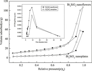
To investigate the surface area and pore structure of Bi2SiO5 samples, nitrogen adsorption–desorption measurements were carried out. The physisorption of N2 has been studied to characterize the porosity of Bi2SiO5 nanoflowers/nanoplates. A typical nitrogen adsorption–desorption isotherm of Bi2SiO5 nanoflowers/nanoplates is presented in Fig. 5. Both samples exhibit type-IV adsorption isotherms, which are considered to indicate the presence of mesopores. Figure 5(inset) shows the pore size distributions measured by the Barret–Joyner–Hatenda model. The BET surface area for Bi2SiO5 nanoflowers is 44.95 m2/g and Bi2SiO5 nanoplates is 12.34 m2/g, respectively. The average diameter of the pores obtained by BET analysis is about 9.0 and 8.8 nm, which would benefit the adsorption of organic molecules in the photocatalysis.Reference Zheng, Duan, Chen and Xie26
N2 absorption–desorpotion isotherms and pore size distribution plot (inset) of samples: (a) Bi2SiO5 nanoflowers, (b) Bi2SiO5 nanoplates.

D. UV-vis DRS patterns of the samples
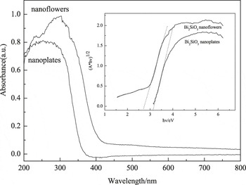
Figure 6 shows the typical UV-visible diffuse reflectance spectra of Bi2SiO5 nanoflowers and Bi2SiO5 nanoplates photocatalysts. As is vividly depicted in Fig. 6, the as-prepared photocatalyst Bi2SiO5 nanoflowers has a much stronger absorption in the visible light area than nanoplates.Reference Li and Zhu33 Following the equation αhν = A(hν − E g)n/2,Reference Fu, Pan, Yao and Zhu34 the energy of the band gap of as-prepared photocatalyst estimated from the absorption edges were 2.71 and 3.15 eV, respectively. These results point out that these samples have a band gap suitable for photocatalytic degradation of organic pollutants under visible light irradiation.
UV-vis DRS patterns of as-prepared samples and graph of samples replotted as (A × hν)1/2 versus (hν) (inset): (a) Bi2SiO5 nanoflowers, (b) Bi2SiO5 nanoplates.

E. Theoretical evaluation
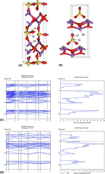
The BSs of two Bi2SiO5 structures were investigated by DFT, as shown in Fig. 7. It can be seen that the minimum of conduction band and the maximum of valence band (VB) were located at the X-point and the S-point, respectively, which indicates that Bi2SiO5 is an indirect gap semiconductor. The theoretical band gap value of orthorhombic and tetragonal Bi2SiO5 structures were determined to be 2.75 and 0.93 eV, respectively. The large disagreement between the theory and the experiment for the nanoplates is not only due to the non-perfect experimental samples (the experimental nanoplates might therefore show a non-perfect stoichiometry or a non-high-quality crystallinity) considered in the UV-vis/DRS experiment but also due to our simulated perfect tetragonal crystal of the nanoplates by DFT. This can be interpreted as follows: on the other hand, the slight disagreement between experimental and theoretical values for the nanoflowers might be due to the equilibrium phase of experimental samples conforming to the simulated perfect orthorhombic crystal. The highest occupied and lowest unoccupied molecular orbital levels were composed of the hybrid orbital of O 2p and Bi 6s and Si 3p orbitals, respectively. The BS indicates that charge transfer upon photoexcitation occurs from the O 2p + Bi 6s hybrid orbitals to the empty Si 3p orbitals. This transition could be due to the 6s electrons which usually occurs at a lower energy than the charge transfer transition in (SiO3)2−. The VB of Bi2SiO5 was composed of O 2p and Bi 6s, which is similar to that of Bi2WO6.Reference Fu, Pan, Yao and Zhu34 These results suggest Bi2SiO5 as a photocatalyst is energetically possible under irradiation.
Unit-cell structure of orthorhombic (a) and tetragonal (b) Bi2SiO5, showing ball and stick (Red, purple, and yellow spheres represent O, Bi, and Si atoms, respectively). Energy band diagram and DOSs for orthorhombic (c) tetragonal (d) Bi2SiO5 structures calculated by DFT.

F. Trapping photoluminescence spectra of ·OH
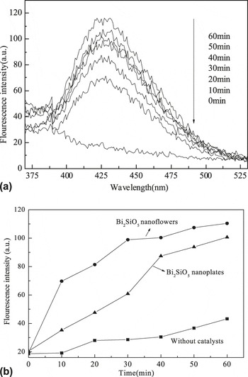
The amount of ·OH radical could be estimated from the fluorescence intensity. Figure 8(a) shows the ·OH-trapping photoluminescence spectra of Bi2SiO5 nanoflowers in a terephthalic acid solution under light irradiation. The fluorescence intensity at 426 nm increases when light is irradiated continuously.Reference Ishibashi, Fujishima, Watanabe and Hashimoto20,Reference Xiao, Si, Zhang, Xiao and Tan21 The stronger fluorescence intensity means that more ·OH is produced. The generated rate of ·OH can be calculated by the slope of straight lines in Fig. 8(b). This exhibits that the change in fluorescence intensity for sample Bi2SiO5 nanoflowers is steeper than nanoplates, suggesting that a greater formation rate of ·OH radicals occurred over Bi2SiO5 nanoflowers. The greater the formation rate of ·OH radicals is, the higher the separation efficiency of electron–hole pairs is achieved. Therefore, higher photocatalytic activity can be predicted.Reference Chen, Bi, Wu, Wang, Li and Fu9
(a) Fluorescence spectral changes observed during illumination of Bi2SiO5 nanoflowers in the solution of terephthalic acid (excitation at 312 nm, emission at 426 nm); (b) plot of the induced fluorescence intensity at 426 nm against irradiation time for terephthalic acid on the sample (A) Bi2SiO5 nanoflowers, (B) Bi2SiO5 nanoplates.

G. Photocatalytic oxidative degradation
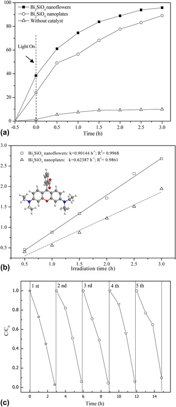
RhB, a widely used dye, was selected as a representative pollutant to evaluate the photocatalytic activity of the prepared samples. As shown in Fig. 9(a), the photocatalytic test demonstrates that nanoflowers display higher activity than the nanoplates. This is because nanoflowers had higher surface area to absorb RhB molecules more efficiently and hence their higher photocatalytic activity under solar light irradiation. However, degradation of RhB was very slow in the absence of photocatalyst.
(a) Decolorization rate of RhB over different samples, (b) ln (C 0/C)–T for photocatalytic degradation of RhB (the structures of RhB in the inset); (c) UV-vis spectral changes observed during illumination of Bi2SiO5 nanoflowers.

Moreover, the plot of ln(C 0/C) versus irradiation time were investigated in Fig. 9(b), where C was the concentration of RhB at a given irradiation time and C 0 concentration of RhB before irradiation (i.e., when adsorption equilibrium was reached). According to the Langmuir-Hinshelwood modelReference Ollis35: r = −dC t/dt = kC t, the photocatalytic degradation reaction of RhB on the samples followed the pseudo-first-order linear relationship with reaction rate constants (k). The k of nanoflowers was 0.90144 h−1 and was also 1.45 times higher than that of nanoplates. This demonstrates that the hierarchical nanostructures exhibited excellent in situ photocatalytic performance.Reference Yu, Shu and Xie36
As shown in Fig. 9(c), after five cycles of run of photodegradation of RhB,Reference Huang, Chen, Xia, Tao, Gan, Weng and Zhang37 the activity of the Bi2SiO5 nanoflowers did not show obvious loss. It indicates that the photocatalytic activity of Bi2SiO5 nanoflowers was stable.
IV. CONCLUSIONS
In summary, Bi2SiO5 nanoflowers/nanoplates were successfully synthesized with silica aerogel as precursors via a hydrothermal method for the first time. Photocatalytic evaluation via decomposition of dye under solar light irradiation revealed that Bi2SiO5 nanoflowers exhibited higher activity than nanoplates due to its suitable morphology, smaller particle size, and higher BET surface area. And the band gap structure of Bi2SiO5 was determined by UV-vis/DRS and DFT calculation. With a band gap of 2.75 eV, Bi2SiO5 had strong absorption in visible light. Our research results could provide new insights into design and preparation of materials with novel properties. The study provided new insight into design and synthesis of nanomaterials and self-assembly architectures, which exhibited better photocatalytic activity.
ACKNOWLEDGMENTS
This work was partly supported by the National Natural Science Foundation of China (Grant No. 21003065), Natural Science Foundation of Jiangsu Province (Grant No. BK2010166), Natural Science Foundation of Jiangsu Provincial Department of Education (Grant No. 11KJB430004), Scientific Innovation Research of College Graduate in Jangsu Province (Grant No. CXZZ12 0682), Social Development Foundation of Zhenjiang (Grant Nos. SH2011005 and SH2012011), Industry Technology Foundation of Zhenjiang, China (Grant No. GY2012017) and Research Foundation for Talented Scholars of Jiangsu University (Grant No. 10JDG133).