Geleophysic dysplasia is an extremely rare skeletal dysplasia resembling lysosomal storage disease. Aside from typical facial features and skeletal abnormalities, it is also associated with valvular involvement of different degrees.Reference Scott, Yeung, Dickinson, Karbani and Crow 1
Worldwide, this association has been described only in very few cases.
Herein we report this abnormality in three sisters from one family in whom this association was noted. In our report, we focused on the cardiac involvement.
Materials and methods
The medical charts of the three girls were reviewed, including the demographic data, growth parameters, history, and presenting symptoms at first presentation. Photographs of the face and extremities of the three girls were taken after a written consent was obtained from the parents. Electrocardiography and X-ray of the chest and skeletal survey were assessed. The echocardiographic images at the time of presentation and on successive follow-up were reviewed in detail, as reported by an independent reviewer using Phillips iE33 (Philips Ultrasound, Bothell-Everett Highway, Bothell, WA, United States of America) echocardiography machine. The pressure gradients across the valves, both peak and mean, when applicable, were documented. The valve annuli were measured in centimetre and Detroit Z-score value was used. The data were presented as initial and follow-up for 2 years for the first two patients and 1 year for the third patient.
Case 1
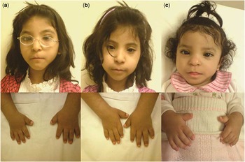
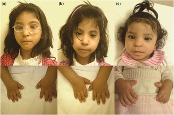
The first patient was a 7-year-old girl born to first cousin parents. She was the oldest of four children, with two affected sisters and one healthy 3-year-old brother. She had a history of repeated respiratory disease requiring hospital admissions during infancy. She was first seen at the age of 41/2 years. At that time, her height was 96 cm and her weight was 12 kg; both were less than the third centile for her age. Her heart rate was 96 beats per minute, and blood pressure was 98/55 mmHg. For the last 3 years, her weight and height grew parallel but considerably below the third centile. She was a happy and friendly person with normal intelligence. She had a distinctive facial appearance with a thin upper lip, smooth and wide philtrum, anteverted nares, hypertelorism, and a broad nasal bridge. She had thick hairy skin. Her hands and feet were small and broad (Fig 1a). She had limitations of flexion at small joints of her hands with bilateral contractures of both elbow and knee joints. She walked on the tips of her toes. She had no hepatomegaly. Auscultation revealed normal first and second heart sound with a grade 3/6 ejection systolic murmur maximally heard at the pulmonary area.
Photograph of the face and hands; ( a ) the eldest girl, ( b ) the second, and ( c ) the third sister. Note the typical facial features and small broad hands.
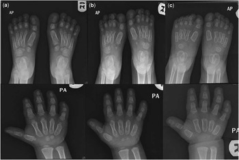
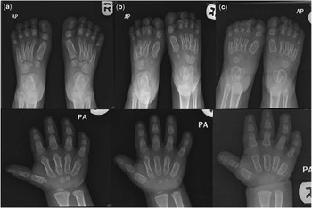
Chest X-ray showed normal heart size and normal pulmonary vascular markings. Skeletal survey showed short tubular bones of the hands and feet (Fig 2a), small irregular capital femoral epiphyses, and ovoid vertebral bodies. Electrocardiography demonstrated normal sinus rhythm and a sign of left ventricular hypertrophy.
X-ray of the feet and hands; ( a ) the eldest girl, ( b ) the second, and C the third sister. Note the short broad and proximal tapering of metacarpals and metatarsal bones.
The molecular testing ADAMTSL2 gene – a disintegrin and metalloproteinase with thrombospondin repeats like 2 – showed compound heterozygous mutations (IVS5-89G>A transition and c.338G>T). The c.338G>T has been reported in geleophysic patients; however, the IVS5-89G>A transition is a novel mutation that has not been published.
Echocardiography
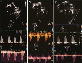
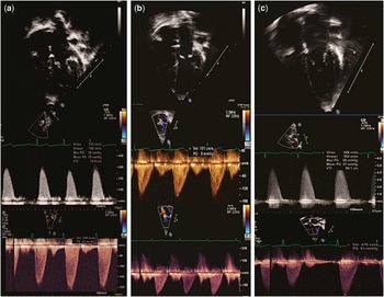
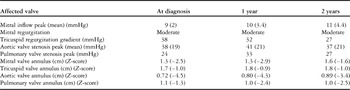
Echocardiography revealed a small secundum atrial septal defect. Both atrioventicular valves were abnormally thickened with redundant leaflets. There was mild tricuspid valve regurgitation with a peak systolic gradient of 38 mmHg. The mitral subvalvular apparatus was generally normal; the mitral valve was moderately regurgitant at the coaptation site; and the peak inflow gradient was 9 mmHg, with a mean of 2 mmHg. There were two separate papillary muscles, and mild hypertrophy of the left venticular posterior wall and interventicular septum. The patient had thickened semilunar valves, mild pulmonary valve stenosis, and a maximum systolic gradient of 24 mmHg. She had normal main pulmonary artery and branches. There was evidence of mild aortic valve stenosis, with a maximum systolic gradient of 38 mmHg and a mean of 19 mmHg (Fig 3a). The left ventricular systolic dimension and function were within normal limits.
Two dimensional and Doppler echocardiography of associated valve involvement: ( a ) the eldest girl, ( b ) the second, and ( c ) the third sister. The first upper row showing the atrioventricular valves in diastole; note the thickening of the leaflets. The second row showing the systolic aortic valve gradient at the time of diagnosis. After 2 years in ( a ) and ( b ) and 1 year in ( c ), there is no significant progression seen.
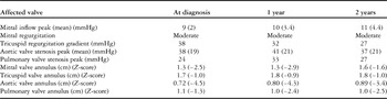
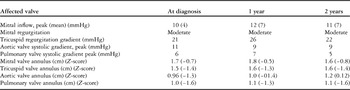
The patient has been followed up for 2 years with repeated echocardiography, which revealed no significant progression of all cardiac lesions (Table 1).
Showing the echocardiographic findings of the first patient at first examination, 1 and 2 years later. There was no progression of the underlying valvular lesion.
Case 2
The second sister was 6 years old, and was observed to have the same features as her older sister (Fig 1b); however, there was no history of respiratory problems and she was asymptomatic. The hands and feet were short with limited movement at all small and large joints. She was also a tip-toe walker and had the same facial features as her older sister. She was first seen when she was 3 1/2 years old; her height was 87 cm and weight was 11.2 kg, both below the third centile for her age. Her heart rate was 91 beats per minute and blood pressure was 80/66 mmHg. There was no hepatomegaly. Auscultation revealed a systolic murmur of grade 3/6 at the apex. Chest X-ray showed normal heart size and normal pulmonary vascular markings. Skeletal survey showed similar skeletal features as her sister (Fig 2b). Her electrocardiography showed normal sinus rhythm and normal tracing for her age.
Echocardiography
Echocardiography showed mildly thickened atrioventricular valves and mild tricuspid valve regurgitation with a peak systolic gradient of 22 mmHg. She had moderate mitral valve regurgitation and a peak mitral inflow gradient of 10 mmHg and a mean of 4 mmHg. She had normal left ventricular size, normal semilunar valves, and no pulmonary or aortic valves stenoses (Fig 3b). The left ventricular wall was normal.
The patient has been followed up for 2 years with repeated echocardiography, which revealed no significant progression of all cardiac lesions (Table 2).
Showing the echocardiographic findings of the second patient at first examination, 1 and 2 years later.
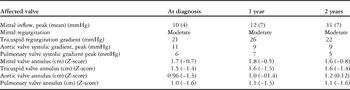
There was no progression of the underlying valvular lesion
Case 3
The youngest sister was born normally with no maternal or postnatal problems; however, she was noted by the parents to have the same facial profile. She had episodes of mild chest infection in early infancy, which required hospital admission once. Evaluation at 15 months of age revealed retarded weight and height – 8 kg and 73 cm, respectively – both of which were less than the 10th centile for her age. She had short hands and feet and contracture at the wrist and elbow joints. She had the same facial features as her older sisters (Fig 1c). Her heart rate was 108 beats per minute and blood pressure was 92/57 mmHg. There was no heptomegaly and there was an ejection systolic murmur grade 3/6 at the right upper sternal border.
Chest X-ray showed normal heart size and pulmonary vascular markings. Skeletal survey demonstrated similar features as her sisters (Fig 2c). Her electrocardiography showed normal sinus rhythm and signs of biventricular hypertrophy; there was no ST segment changes.
Echocardiography
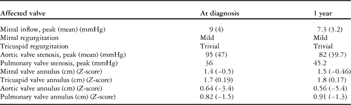
Echocardiography revealed an intact interatrial septum, normal tricuspid leaflets, trivial tricuspid regurgitation, and normal mitral valve leaflets with mild to moderate mitral regurgitation. There were no signs of mitral stenosis. She had a normal right ventricular size. She had hypertrophied interventricular septum and left ventricular posterior wall. She had a normal left ventricular systolic function. She had thickened pulmonary valve leaflets with mild pulmonary valve stenosis, with a peak systolic gradient of 36 mmHg. She had mild post-stenotic dilatation of the main pulmonary artery and normal pulmonary artery branches. The small aortic annulus measures 7 mm (Z-score of −3.0). She had thickened aortic valve leaflets and severe aortic valve stenosis with a peak systolic gradient of 95 mmHg and a mean of 47 mmHg (Fig 3c). There was no post-stenotic dilatation of the ascending aorta. The patient had a small patent arterial duct with left-to-right shunt and no coarctation of the aorta.
The patient has been followed up for 1 year with repeated echocardiography every 2 to 4 months, which revealed no significant progression of all cardiac lesions over this period (Table 3).
Showing the echocardiographic findings of the third patient at first examination, 1 year later.
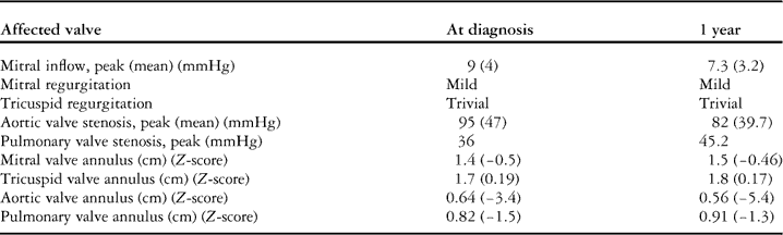
There was no progression of the underlying valvular lesion
Discussion
Geleophysic dysplasia has been described in 1960 as a form of atypical gargoylism by Vanace et al.Reference Vanace, Friedman and Wagner 2 Spranger et alReference Spranger, Gilbert, Tuffli, Rossiter and Opitz 3 presented it as a focal mucopolysacchriodosis. The disease is considered as a severe form of acromelic skeletal dysplasia, which is a rare form of dysplasia that includes three disorders: geleophysic dysplasia, Weill–Marchesani syndrome, and acromicric dysplasia; however, evidence of lysosomal-like PAS-positive vacuoles in liver, skin, and trachea have been documented in several reports.Reference Le Goff and Cormier-Daire 4 , Reference Lipson, Kan and Kozlowski 5
Le Goff et al identified ADAMTSL2 gene – a disintegrin and metalloproteinase with thrombospondin repeats like 2 – as being responsible for the autosomal type of geleophysic dysplasia, which is located in chromosome 9p34.2, and demonstrated its high expression in the cardiac myocytes, epidermis, and tracheal wall. ADAMTSL2 gene bound to latent transforming growth factor β binding protein-1, which is responsible for the availability and storage of transforming growth factor β in extracellular matrix. Misfolding of ADAMTSL2 protein that interacts with transforming growth factor β binding protein-l might be the underlying mechanism of geleophysic dysplasia.Reference Le Goff, Morice-Picard and Dagoneau 6 The genetic heterogeneity of geleophysic dysplasia has been observed in a relatively large collection of cases of geleophysic and acromiric dysplasia.Reference Allali, Le Goff and Pressac–Diebold 7
Up to date, <40 cases have been published in different population with geleophysic dysplasia. Almost all of those patients have a combination of short stature, peculiar dysmorphic feature with joint contracture, and cardiac valve involvement, similar to our patients.Reference Le Goff and Cormier-Daire 4 The most frequent cardiac lesions described are the mitral valve, then the aortic valve followed by pulmonary valve and the least is the tricuspid valve.Reference Scott, Yeung, Dickinson, Karbani and Crow 1 Spranger et alReference Spranger, Gilbert and Arya 8 described one girl with atrial septal defect and patent arterial duct without valvular involvement. Pontz and SantolayaReference Santolaya, Groninga and Delgado 9 , Reference Pontz, Stöss and Henschke 10 seperately described five cases with normal cardiac structure. Reports regarding progression were controversial, although most authors suggested slow progression over a long period of time and postulated that a severe and intermediate form of the disease could explain the severity of cardiac involvement.Reference Lipson, Kan and Kozlowski 5
In our series, three sisters had the same dysmorphic feature but showed different cardiac involvement, with the youngest expressing the most severe aortic valve stenosis and the middle one having the least valvular involvement. Titomanlio et alReference Titomanlio, Della Casa and Lecora 11 reported a case of geleophysic dysplasia diagnosed at the age of 8 months with mild pulmonary valve stenosis and atrioventricular valve regurgitation, which showed initial progression and then complete resolution at the age of 8 years.
From follow-up of the first two cases, we observed that the valvular involvement remained static over 2 years. This observation along with the case presented by Titomanlio helped us to pursue more conservative management strategy for the third case despite severe aortic valve stenosis. Subsequent follow-up at 12 months revealed unchanged aortic valve gradient and preserved left ventricular function.
Conclusion
There are very few case reports describing the cardiac involvement in geleophysic dysplasia; however, this family represents the first family reported from the Arab region. The cardiac involvement in most cases affects one or more cardiac valves in the form of thickening leading to stenosis. The published reports suggest progression of the disease. We could not confirm this in our series. In our three cases, the youngest sister had the most severe form of valve involvement, whereas the middle girl had the least involvement.
This might suggest that the expression of the gene could determine the severity of the disease, rather than the proposed progression with age.
Acknowledgements
The authors would like to thank Mr. Mohammed Alenazi, ARDMS, senior echocardiography technologist, Prince Salman Heart Center, King Fahad Medical City, for his assistance in preparing the images and photos for publication.
Conflicts of Interest
None.
Financial Support
This research received no specific grant from any funding agency, commercial or not-for-profit sectors.