INTRODUCTION
The taxonomic knowledge of benthic organisms in Antarctica is important (Winston, Reference Winston and Eldredge1992; Arntz et al., Reference Arntz, Brey and Gallardo1994) even if some groups of the deep fauna are still poorly known given the high level of apparent endemism for the majority of taxa (Arntz et al., Reference Arntz, Gutt, Klages, Battaglia, Valencia and Walton1997) due to speciation processes favoured by the origin and isolation of the continent (Sarà et al., Reference Sarà, Balduzzi, Barbieri, Bavestrello and Burlando1992).
Sponges are important elements of the benthos in Antarctica and may dominate particular benthic communities (see McClintock et al., Reference McClintock, Amsler, Baker and Van Soest2005). Sponge species richness (352 species) is remarkably high and comparable to diversity values reported for temperate and tropical waters (e.g. van Soest, Reference van Soest, van Soest, van Kempen and Braekman1994; McClintock et al., Reference McClintock, Amsler, Baker and Van Soest2005).
Antarctic sponges have been studied since the 19th Century and are relatively well known compared to other world faunas (Calcinai & Pansini, Reference Calcinai and Pansini2000). Moreover, due to the peculiar characteristics of the Antarctic environment, their spatial distribution is certainly more homogeneous than for faunas at lower latitudes (Sarà et al., Reference Sarà, Balduzzi, Barbieri, Bavestrello and Burlando1992). Knowledge about them, however, is still far from complete, as evidenced by continual new discoveries from the region (Campos et al., Reference Campos, Mothes and Veitenheimer Mendes2007; Plotkin & Janussen, Reference Plotkin and Janussen2007; Ríos & Cristobo, Reference Ríos and Cristobo2007).
Specimens were collected during the XX Italian Expedition in 2004 and 2005 by the Department of Animal Biology and Marine Ecology of the University of Messina and entrusted to our laboratories for identification. A total of 33 Demosponge species, already known for the Ross Sea, were identified, with an additional two species described here as new.
MATERIALS AND METHODS
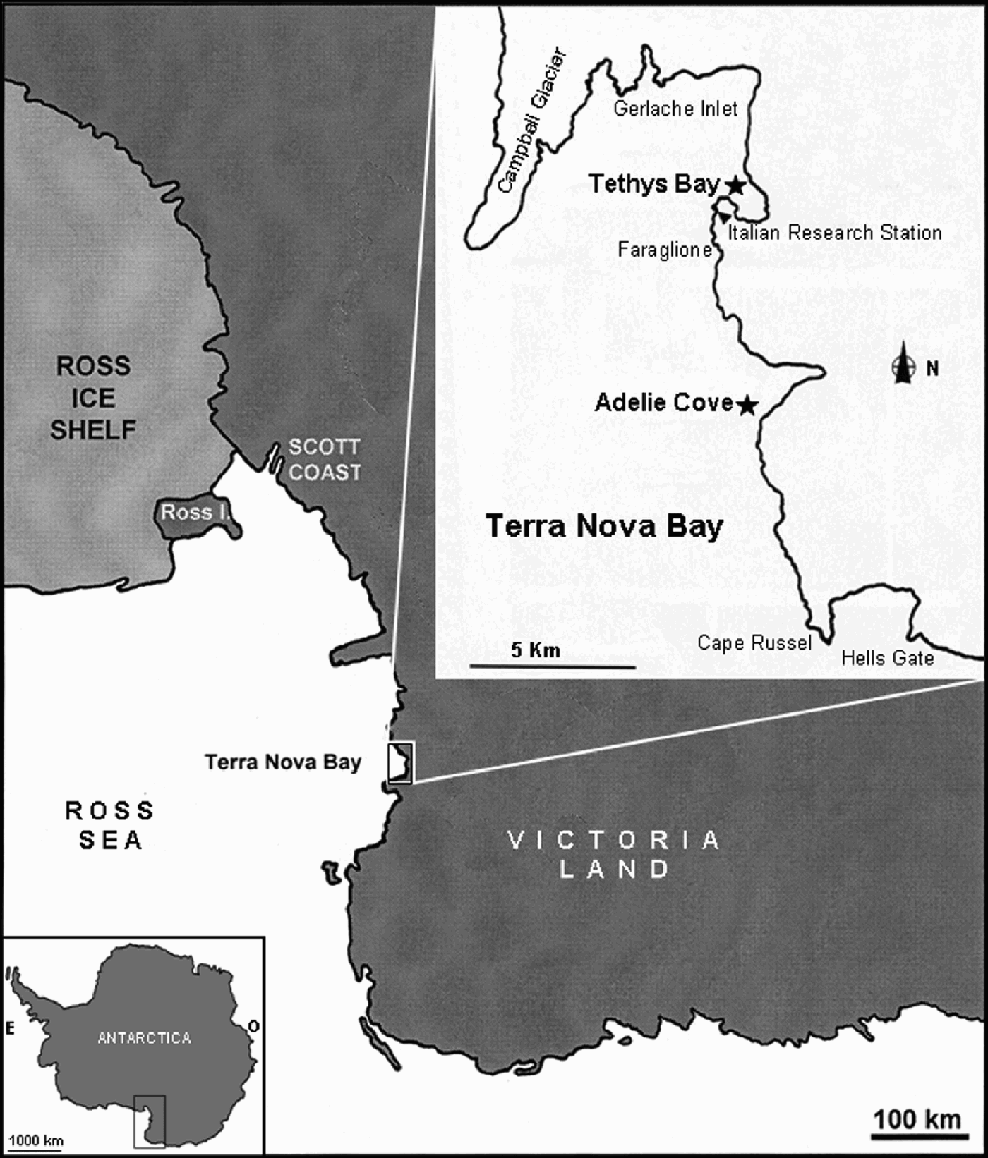
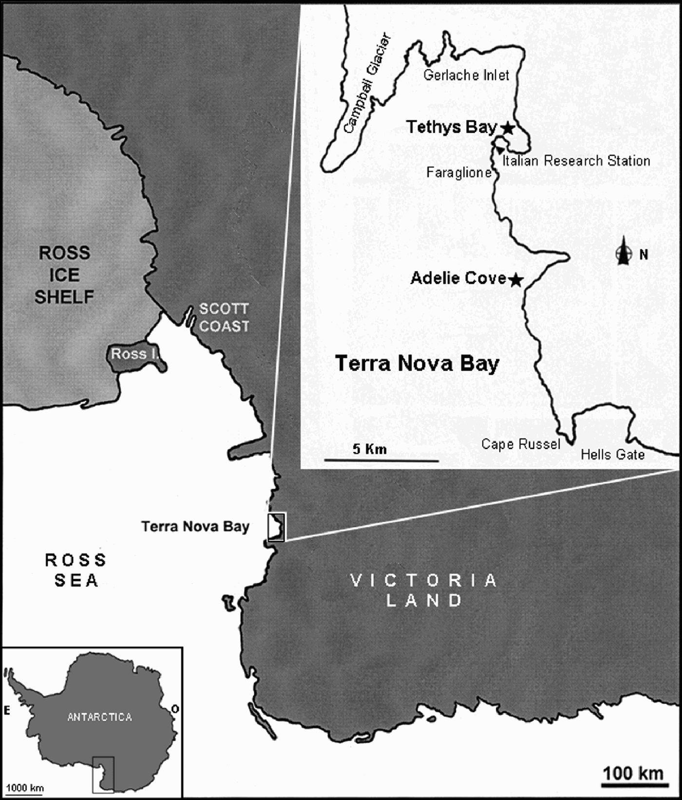
Specimens were collected by dredge and grab at a depth of 120 m at Adelie Cove and at 200 m at Tethys Bay (Figure 1) in Terra Nova Bay (Ross Sea). Specimens were frozen on board for shipping and, after defrosting at the laboratory they were preserved in alcohol 70% or dried. Since only portions of the specimens were collected the original shape of the sponges is unknown.
Map of Terra Nova Bay (Ross Sea), with the location of the Italian Research Station (arrow) and of the sampling stations (Adelie Cove and Tethys Bay).
Spicule complement was analysed according to Rützler (Reference Rützler, Stoddart and Johannes1978). Skeletal architecture was examined with light and scanning electron microscope (SEM); hand-cut sections of the ectosome and choanosome were made following Hooper (Reference Hooper2000).
From 30 measurements for each spicule type, size-range, mean and standard deviation (in parentheses) were obtained. For SEM analyses dissociated spicules were transferred onto stubs and sputtered with gold. The SEM preparations were observed by a Philips XL 20 scanning electron microscope.
The holotypes are deposited at the Museo Civico di Storia Naturale ‘G. Doria’ of Genova (MSNG).
RESULTS
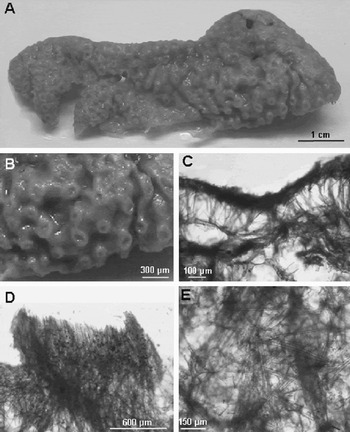
Crella (Crella) aurantiaca sp. nov. (A) Holotype; (B) detail of the sponge surface with grooves and slightly elevated areolae; (C) cross-section showing the diverging bundles of anisotornotes supporting the crust of acanthoxeas and the low density choanosome; (D) surface areolae walls are reinforced by parallel arranged tornotes; (E) thick bundles of tornotes and acanthostyles in the inner part of the choanosome with echinating acanthostyles.

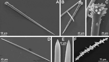
Crella (Crella ) aurantiaca sp. nov. (A) Large acanthostyle with a few spines on the shaft; (B) entirely spined smaller acanthostyles with blunt spines on the head; (C) magnification of the acanthostyle head; (D) anisotornote; (E) magnifications of the extremities of an anisotornote; (F) acanthoxea.

TYPE MATERIAL
Holotype: [MSNG 54588]. AC-c5; dry specimen, Terra Nova Bay, Adelie Cove, 74°46′32″S 164°03′01″E; water depth 120 m, Collected 20 January 2005.
DESCRIPTION
The studied material, most probably coming from a massive specimen, consists of a sponge fragment about 7 cm long and 3 cm thick (Figure 2A). The surface is highly grooved, provided with areolated areas which are slightly raised in the fresh sponge (Figure 2B). The consistency is firm. The dull orange original colour rapidly faded after preservation.
Skeleton
Ectosome: dense, 100–130 µm thick, composed of tangential, disorderly arranged acanthoxeas, forming a superficial crust supported by the choanosomal skeleton (Figure 2C). The elevated pore fields protrude from this crust. Their walls are built by anisotornotes arranged in palisade (Figure 2D).
Choanosome: plumose skeleton of ascending tracts of megascleres rather irregularly arranged. In the peripheral part of the sponge body spicule tracts are formed by anisotornotes slightly diverging in bouquets towards the surface to support the crust. In the inner part of the choanosome the spicule bundles, made of acanthostyles and tornotes, are seldom connected by echinating acanthostyles (Figure 2E). An unequivocal distinction between the two categories of acanthostyles cannot be ascertained. The scarcity of interstitial spicules in the mesohyl and the abundance of empty spaces produce a low density choanosomal skeleton.
Spicules
Megascleres: acanthostyles in two size-categories; the larger ones are slightly curved with spines more concentrated on the head and on the distal part of the spicule, but rare along the shaft (Figure 3A). The spines are relatively long and generally blunt on the spicule head. They measure 377 (468) 550 × 15 (18.7) 20 µm. The smaller acanthostyles are similar in shape, but the spines are in general numerous along the shaft with respect to the larger acanthostyles (Figure 3B & C). They measure 194 (210.4) 265 × 15 (19) 20 µm. Anisotornotes generally straight with conical and bluntly pointed extremities (Figure 3D & E). They measure 326 (357) 387 × 10 µm.
Microscleres: acanthoxeas slightly curved or straight, uniformly covered by long, triangular spines perpendicular to the shaft (Figure 3F). They measure 56 (65) 76.5 × 7 (9.5) 11.5 µm.
ETYMOLOGY
The species is named after its live orange coloration.
REMARKS
This new species is assigned to the subgenus Crella due to the presence of true echinating acanthostyles, a tangential ectosomal crust of acanthoxeas and to the absence of chelae. However, its peculiar and unusual characteristic is the presence of acanthostyles together with tornotes in the deeper choanosomal tracts.
In Antarctica, three species of the genus Crella were recorded (Sarà et al., Reference Sarà, Balduzzi, Barbieri, Bavestrello and Burlando1992): C. (Crella) tubifex (Hentschel, 1914), C. (Pytheas) crassa Hentschel, 1914 and C. stylifera Hentschel, 1914. Crella (C.) tubifex is the only member of the subgenus Crella recorded for this area. It differs from C. (Crella) aurantiaca sp. nov. having only megascleres. They consist in a single category of acantostyles (176–392 × 10–11 µm), amphistrongyles (472–650 × 7–8 µm) and large acanthostrongyles (440–560 × 12.5–15 µm) instead of acanthoxeas. Crella (Pytheas) crassa has amphistrongyles and isochelae as microscleres. Crella stylifera, not yet assigned to any subgenus (van Soest et al., Reference van Soest, Boury-Esnault, Hooper, Rützler, de Voogd, Alvarez, Hajdu, Pisera, Vacelet, Manconi, Schoenberg, Janussen, Tabachnick and Klautau2008), has polytylote styles (280–320 × 5–7 µm), a single type of acanthostyles (120–192 × 9–13 µm), slightly curved acanthoxeas (144–168 × 5 µm) and arcuate isochelae (27–30 × 10–13 µm). Other species attributed to the subgenus Crella such as C. (Crella) elegans (Schmidt, 1862), C. (Crella) mollior Topsent, 1925 and C. (Crella) papillosa (Schmidt, 1870) differ in gross morphology and skeletal structure from the present species, and have temperate or tropical distributions.
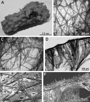
Mycale (Aegogropila) denticulata sp. nov. (A) Holotype; (B) ectosomal reticulation of paucispicular (2–6 spicules) tracts of mycalostyles forming triangular or polygonal meshes; (C) plumose skeleton of stout multispicular tracts of mycalostyles; (D) multispicular tracts diverging in proximity of the sponge surface, support the ectosome; (E) ectosomal reticulation; (F) the mycalostyle extremities slightly protrude outside making the surface bristly.

Mycale (Aegogropila) denticulata sp. nov. (A) A thick bundle of mycalostyles with abundant dispersed spicules; (B) mycalostyle; (C) head of a mycalostyle; (D, E) front and lateral view of palmate anisochelae with the characteristic tooth.

TYPE MATERIAL
Holotype: [MSNG 54589].TB-a1; dry specimen, Terra Nova Bay, Tethys Bay, 74°41′17.9″S 164°03′27″E; water depth 200 m. Collected 18 January 2005.
DESCRIPTION
The studied fragment is cylindrical, 10 cm long and 4 cm thick (Figure 4A), presumably belonging to an erect, possibly branching sponge. The surface may be considered as mammillate, having round, slightly elevated knobs, often merging into one another scattered over the surface. It appears hispid at closest examination. Oscules were not detectable. Consistency is firm but compressible, friable in the dried state. Colour in life is dark yellow and becomes white after alcohol preservation or drying.
Skeleton
The skeleton is typical of the subgenus Aegogropila Gray, 1867.
Ectosome: a paucispicular (2–6 spicules) reticulation of mycalostyles forming triangular or polygonal meshes (100–230 µm across) with a thin membrane perforated by ostia 60–80 µm in diameter (Figure 4B & E).
Choanosome: a plumose skeleton of stout multispicular tracts of mycalostyles (50–75 µm thick) (Figure 4C). These tracts, diverging in proximity of the sponge surface, support the ectosome (Figure 4D). The mycalostyle extremities slightly protrude outside the sponge, making the surface hispid (Figure 4E & F).
The slightly elevated knobs are supported by thick tracts of mycalostyles 500–1000 µm (Figure 5A).
Abundant megascleres and rare anisochelae are scattered in the choanosome. Anisochelae are not arranged in rosettes.
Spicules
Megascleres: mycalostyles straight, slightly curved and often faintly sinuous (Figure 5B). They measure 490 (591.6) 652 × 10 (12.5) 15 µm. They are not isodiametric and taper into a bluntly pointed extremity. Tyles are regular, faintly swollen (Figure 5C). Malformed spicules are occasionally found.
Microscleres: a single category of anisochelae with a large palmate frontal ala and lateral alae clearly separated from the shaft; the distal extremities of the alae are slightly curved outwards. The smaller end of the spicule is characterized by a long pointed tooth on the small frontal ala (Figure 5D & E). The anisochelae are 41 (44.4) 51 µm long.
ETYMOLOGY
The species is named after the tooth that characterizes the smaller extremity of the anisochelae.
REMARKS
According to the characteristics of the ectosomal skeleton the new species is assigned to the subgenus Aegogropila. Mycale (Aegogropila) denticulata sp. nov. is characterized by anisochelae in a single size-category as the only microscleres present.
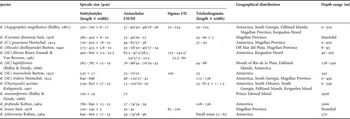
Thirteen species of the genus Mycale have been recorded from Antarctica and sub-Antarctic areas (Sarà et al., Reference Sarà, Balduzzi, Barbieri, Bavestrello and Burlando1992; van Soest et al., Reference van Soest, Boury-Esnault, Hooper, Rützler, de Voogd, Alvarez, Hajdu, Pisera, Vacelet, Manconi, Schoenberg, Janussen, Tabachnick and Klautau2008). They are listed in Table 1 with their spicule size, geographical distribution and depth-range. Among them M. (Aegogropila) magellanica (Ridley, 1881) is the only species assigned to the subgenus Aegogropila. Specimens of M. (Aegogropila) magellanica present in the author's collection (Pansini & Sarà, Reference Pansini, Sarà, Arntz and Rios1999) were examined. It clearly differs from Mycale (Aegogropila) denticulata sp. nov. in having three size-categories of anisochelae (57–90 µm, 30–46 µm and 18–26 µm), two other types of microscleres (sigmas 10–224 µm and raphides 20–105 µm) and slightly longer mycalostyles (320–700 × 6–17 µm). Remarkable skeleton and spicule differences separate the other Antarctic and sub-Antarctic Mycale species from M. (Aegogropila) denticulata sp. nov.
Species of the genus Mycale recorded in Antarctica and sub-Antarctic areas with spicule size, geographical distribution and depth-range.

The closest species appears to be M. mammiformis (Ridley & Dendy, 1886), showing the same spicule complement. However, the mycalostyles of M. mammiformis are different in shape and dimension from those of the new species, being much longer (1000 × 19 µm), straight and slightly polytylote. The anisochelae also differ in shape, lacking the long tooth and having a large frontal ala and wide lateral alae, and in size, being 72 µm long.
Other Mycale species have been recorded from southern adjacent biogeographical regions (South Africa, Vema Seamount, Tasmania, South Australia, New Zealand) by Lévi (Reference Lévi1963, Reference Lévi1969), Uriz (Reference Uriz1988), Wiedenmayer (Reference Wiedenmayer1989), Bergquist & Fromont (Reference Bergquist and Fromont1988), but they strongly differ from M. denticulata sp. nov. Even Mycale (Aegogropila) flagelliformis Bergquist & Fromont, Reference Bergquist and Fromont1988, the only representative of the subgenus Aegogropila at these latitudes differs in having three size-categories of anisochelae and two types of sigmas. The present species is clearly new to science.
ACKNOWLEDGEMENTS
The authors thank Professor V. Bruni and Dr L. Michaud of the Department of Animal Biology and Marine Ecology of the University of Messina (Italy) for the collection of the specimens.