Introduction
The study of parthenogenesis by means of artificial oocyte activation (AOA) is important to investigate the roles of paternal and maternal genomes in controlling early embryonic development. In addition, artificial activation of oocytes is a key procedure for a successful somatic cell nuclear transfer (SNT) and intracytoplasmic sperm injection (ICSI) (Kim et al., Reference Kim, Simerly, Funahashi, Schatten and Day1996). Oocyte activation includes many well defined morphological and biochemical changes, essential for normal development. Some of them occur within seconds or minutes after sperm–oocyte plasma membrane interaction, while others occur over the course of several hours (Yanagimachi, Reference Yanagimachi, Knobil and Neil1994).
In all species studied so far, the efficiency of blastocyst production by SNT is impaired by several biological and technical problems (Vajta et al., Reference Vajta, Lewis, Trounson, Purup, Maddox-Hyttel, Schmidt, Petersen, Greve and Callesen2003) and only 1–2% of the embryos reconstructed by SNT were able to develop to term (Prather et al., Reference Prather, Hawley, Carter, Lai and Greenstein2003). In horses, although the use of ICSI and SNT resulted in the production of live foals, the development of embryos and live birth remains low, unstable, and far from satisfactory, despite numerous efforts (Oikawa et al., Reference Oikawa, Takada, Kikuchi, Numabe, Takenaka and Horiuchi2005). Besides the technical difficulties caused by the low number of slaughterhouses, the darkness of the ooplasm, and the toughness and elasticity of the oolemma, plus the inability to respond to mechanical activation may be also responsible for the low developmental rate.
It has been shown previously that a soluble sperm factor triggers calcium oscillations and egg activation in mammals. This sperm factor has been identified as PLCζ (Saunders et al., Reference Saunders, Larman, Parrington, Cox, Royse and Blayney2002). It has been suggested that after the fusion of sperm and egg plasma membrane, the sperm-derived PLCζ protein diffuses into the egg cytoplasm, resulting in the hydrolysis of PIP2 (phosphatidylinositol-4,5-bisphosphate) from an unknown source to generate IP3 (Saunder et al., Reference Saunders, Swann and Lai2007). The increase in free intracellular calcium also inactivates the cytostatic factor (CSF), promoting the destruction of cyclin B1 and resulting in the inactivation of the maturation promoting factor (MPF; White & Yue, Reference White and Yue1996), a dimmer formed by the protein cyclin B1 (regulatory subunit) and protein P34CDC2 (catalytic subunit) (Homa, Reference Homa1995). The blockage of the activity of MPF allows the occurrence of the early events of oocyte activation, such as the resumption of meiosis (transition for anaphase/telophase), extrusion of the second polar body, and exocytosis of cortical granules.
Different procedures for AOA have been established and are commonly divided into three subtypes (mechanical, electrical, and chemical stimuli) that elicit one or several calcium transients. Chemical compounds can induce calcium increase, initiating oocyte activation. Some artificial activation treatments, such as strontium chloride, promote an increase in intracellular free calcium concentrations by the release of calcium from cytoplasmic stores (Cuthbertson et al., Reference Cuthbertson, Whittingham and Cobbold1981), others such as electrical stimulus and calcium ionophores promote the influx of calcium from extracellular medium, and treatments such as ethanol and ionomycin promote both of these effects (Loi et al., Reference Loi, Ledda, Fulka, Cappai and Moor1998). These treatments are commonly combined with protein synthesis inhibitors such as cycloheximide, which prevent cyclin synthesis, and phosphorylation inhibitors, such as 6-dimethylaminopurine (6-DAMP; Liu & Yang, Reference Liu and Yang1999), which prevent MPF activation. The major disadvantage of using the protein kinase inhibitors or the protein synthesis inhibitors is that these drugs do not specifically inhibit the activity of a particular kinase or the synthesis of a specific protein that control cell-cycle progression. The purine analogue roscovitine, which inhibits the adhesion of ATP to the MPF subunit Cdk1 (P34CDC2) and effectively suppresses meiosis in cattle, swine, and equines (Mermillod et al., Reference Mermillod, Tomanek, Marchal and Meijer2000; Marchal et al., Reference Marchal, Tomanek, Terqui and Mermillod2001; Franz et al., Reference Franz, Choi, Squires, Seidel and Hinrichs2003; Choi et al., Reference Choi, Love, Varner and Hinrichs2006), may significantly affect AOA.
Treatments in which only calcium ionophore and ethanol are used have failed to induce significant rates of artificial activation if compared with the combination of these drugs with cycloheximide, suggesting that the initial peak of calcium is not alone sufficient to inhibit CSF (Hinrichs et al., Reference Hinrichs, Schmidt and Selgrath1995).
Literature data concerning the role of calcium in artificial fertilization are controversial. An increase in embryonic development in media containing calcium concentrations three to six times higher than the usual concentration was observed in a study of nuclear transfer (NT) in mules (Woods et al., Reference Woods, White, Vanderwall, Aston, Bunch and Campbell2002). Moreover, the calcium oscillations triggered by the sperm cells not only induce resumption of meiosis, but seem to be also associated with many other events essential for activation of the zygote genome (Aoki et al., Reference Aoki, Hara and Schultz2003). On the other hand, it has been shown that the addition of calcium to the culture medium decreases the formation of equine blastocysts produced by ICSI (13% versus 23% for control) and that 80% of embryos cultured in high calcium media show signs of degeneration (Hinrichs et al., Reference Hinrichs, Choi, Walckenaer, Varner and Hartman2007).
Here, a combination of ionomycin with cycloheximide, roscovitine, strontium, or 6-dimethylaminopurine (6-DMAP) was tested in the presence of low and high calcium concentrations in an attempt to establish the best protocol for artificial activation of bovine and equine oocytes.
Material and methods
The present experiments comply with all relevant institutional and national animal welfare guidelines and policies, and were approved by the Ethics Committee of São Paulo State University.
All chemicals were purchased from Sigma Co (St Louis, USA), unless specified otherwise.
Collection, selection, and in vitro maturation of bovine and equine oocytes
For collection of bovine oocytes, ovarian follicles with diameters between 2 and 8 mm were aspirated and the oocyte was selected according to the presence of three or more layers of compact cumulus cells with homogeneous cytoplasm.
For in vitro maturation, selected bovine oocytes were cultured in four-well dishes (NUNC®, Roskilde, Denmark) containing 400μl maturation medium (TCM199; Invitrogen, California, USA), 10% fetal calf serum (FCS), 16 μg/ml pyruvate, 10 g/ml luteinising hormone (LH; Lutropin®, Tecnopec, São Paulo, Brazil), 10 g/ml follicle stimulating hormone (FSH; Follitropin®, Tecnopec, São Paulo, Brazil), 1 μg/ml estradiol, and 75 μg/ml gentamicin, at 38.5°C in an atmosphere of 5% CO2 in air and high humidity for 24 h.
Equine oocytes were collected through dissection and curettage of individual follicles from 10 to 30 mm in diameter. Selected oocytes showed at least one layer of cumulus cells, and spherical appearance. The selected oocytes were classified according to presence or absence of groups of expanded cumulus and/or granulosa cells and divided into two groups: expanded and compact.
For in vitro maturation, equine oocytes were also cultured in four-well dishes (NUNC®) containing 400 μl maturation medium (human tubal fluid (HTF) medium; Irvine Scientific–California, USA):Basal Medium Eagle (BME; Invitrogen) (1:1), 0.3% bovine serum albumin (BSA; 0.3 mg/ml), 0.32 mm pyruvate, 1 mM l-glutamine, 0.4 mm cysteine, 0.1 mm taurine, 0.4 mm glycine, 3.3 mm sodium lactate, 100 ng/ml insulin-like growth factor I (IGF-1), 50 ng/ml epidermal growth factor (EGF), 100 ng/ml equine growth hormone (EGH; Equigen®, BrasaGen, Adelaide, Australia), 0.03 UI/ml equine FSH (eFSH; Bioniche Life Science, Ontario, Canada), 500 ng/ml estradiol, and 75 μg/ml gentamicin, at 39°C in an atmosphere of 5% CO2 in air and high humidity for 30 h.
Artificial activation of bovine oocytes
Each experiment included at least five replicates. Following the period of in vitro maturation, the oocytes were incubated in minimum essential medium with HEPES (MEM-H)/1.8 mM calcium concentration (Invitrogen, California, USA) with 5 mg/ml hyaluronidase type V. Granulosa cells were extracted with a fine glass pipette and the oocytes with homogeneous cytoplasm were selected and divided among the groups of parthenogenetic activation.
Group Ionomycin + Cycloheximide in Low Calcium (n = 86)
Incubated in MEM-H + 10% FCS + 5 μM ionomycin for 8 min, washed in MEM-H + 10% FCS and incubated in MEM + 10% FCS + 10 g/ml cycloheximide + 5 μg/ml cytochalasin B (Calbiochem, California, USA) for 4 h.
Group Ionomycin + Roscovitine in Low Calcium (n = 128)
Incubated in MEM-H + 10% FCS + 5 μM ionomycin for 8 min, washed with MEM-H + 10% FCS and incubated in MEM + 10% FCS + 66 μM roscovitine + 5 μg/ml cytochalasin B for 5 h.
Group Ionomycin + Strontium in Low Calcium (n = 98)
Incubated in MEM-H + 10% FCS + 5 μM ionomycin for 8 min, washed with MEM-H + 10% FCS and incubated in MEM + 10% FCS + 20 mM strontium chloride (JT Baker, New Jersey, USA) + 5 μg/ml cytochalasin B for 5 h.
Group Ionomycin + 6-DMAP in Low Calcium (n = 45)
Incubated in MEM-H + 10% FCS + 5 μM ionomycin for 8 min, washed with MEM-H + 10% FCS and incubated in MEM + 10% FCS + 2 mM 6-DMAP + 5 μg/ml cytochalasin B for 4 h.
Group Ionomycin + Cycloheximide with High Calcium (n = 46)
Incubated in MEM-H with 0.84 mg/ml CaCl2 + 10% FCS + 5 μM ionomycin for 8 min, washed with MEM-H + 10% FCS and incubated in MEM + 0.42 mg/ml CaCl2 + 10% FCS + 10 g/ml cycloheximide + 5 μg/ml cytochalasin B for 4 h.
Group Ionomycin + Roscovitine in High Calcium (n = 129)
Incubated in MEM-H with 0.84 mg/ml CaCl2 + 10% FCS + 5 μM ionomycin for 8 min, washed with MEM-H + 10% FCS and incubated in MEM + 0.42 mg/ml CaCl2 + 10% FCS + 66 μM roscovitine + 5 μg/ml cytochalasin B for 5 h.
Group Ionomycin + Strontium in High Calcium (n = 84)
Incubated in MEM-H with 0.84 mg/ml CaCl2 + 10% FCS + 5 μM ionomycin for 8 min, washed with MEM-H + 10% FCS and incubated in MEM + 0.42 mg/ml CaCl2 + 10% FCS + 20 mM strontium chloride + 5 μg/l cytochalasin B for 5 h.
Group Ionomycin + 6-DMAP in High Calcium (n = 49)
Incubated in MEM-H with 0.84 mg/ml CaCl2 + 10% FCS + 5 μM ionomycin for 8 min, washed with MEM-H + 10% FCS and incubated in MEM + 0.42 mg/ml CaCl2 + 10% FCS + 2 mM 6-DMAP + 5 μg/ml cytochalasin B for 4 h.
Following the activation period, the oocytes were cultivated in HTF:BME (1:1), 0.6% BSA + 10% FCS + 0.01% inositol + 75 μg/ml gentamicin at 39°C in an atmosphere of 5% CO2, 5 % O2, and 90% N2 for 24 h.
Artificial activation of equine oocytes
Each experiment included at least five replicates. After the in vitro maturation period, equine oocytes were denuded as described above and divided among the following groups of activation, following the initial separation between expanded and compact cumulus complex oocytes:
Group Ionomycin + Cycloheximide in Low Calcium (expanded n = 87; compact n = 49)
Incubated in MEM-H + 10% FCS + 5 μM ionomycin for 8 min, washed with MEM-H + 10% FCS and incubated in MEM + 10% FCS + 10 g/ml cycloheximide + 5 μg/ml cytochalasin B for 4 h.
Group Ionomycin + Roscovitine in Low Calcium (expanded n = 97; compact n = 63)
Incubated in MEM-H + 10% FCS + 5 μM ionomycin for 8 min, washed with MEM-H + 10% FCS and incubated in MEM + 10% FCS + 66 μM roscovitine + 5 μg/ml cytochalasin B for 5 h.
Group Ionomycin + 6-DMAP in Low Calcium (expanded n = 77; compact n = 88)
Incubated in MEM-H + 10% FCS + 5 μM ionomycin for 8 min, washed with MEM-H + 10% FCS and incubated in MEM + 10% FCS + 2 mM 6-DMAP + 5 μg/ml cytochalasin B for 4 h.
Group Ionomycin + Cycloheximide in High Calcium (expanded n = 89; compact n = 51)
Incubated in MEM-H with 0.84 mg/ml CaCl2 + 10% FCS + 5 μM ionomycin for 8 min, washed with MEM-H + 10% FCS and incubated in MEM + 0.42 mg/ml CaCl2 + 10% FCS + 10 g/ml cycloheximide + 5 μg/ml cytochalasin B for 4 h.
Group RH: Ionomycin + Roscovitine in High Calcium (expanded n = 84; compact n = 59)
Incubated in MEM-H with 0.84 mg/ml CaCl2 + 10% FCS + 5 μM ionomycin for 8 min, washed with MEM-H + 10% FCS and incubated in MEM + 0.42 mg/ml CaCl2 + 10% FCS + 66 μM roscovitine + 5 μg/ml cytochalasin B for 5 h.
Group Ionomycin + 6-DMAP in High Calcium (expanded n = 73; compact n = 88)
Incubated in MEM-H 0.84 mg/ml CaCl2 + 10% FCS + 5 μM ionomycin for 8 min, washed with MEM-H + 10% FCS and incubated in MEM + 0.42 mg/ml CaCl2 + 10% FCS + 2 mM 6-DMAP + 5 μg/ml cytochalasin B for 4 h.
Following the activation period, the oocytes were cultured in DMEM/F12 (Invitrogen, California, USA) + 10% FCS + 0.01% myo-inositol + 75 μg/ml gentamicin at 39°C in an atmosphere of 5% CO2, 5% O2, and 90% N2 for 24 h.
For assessment of activation rates, bovine and equine oocytes were stained with 10 μl of Hoechst 33342 (7.5 mg/ml) and observed under an inverted microscope (Leica DMIRB; blue filter, excitation 350 to 461 nm). Oocytes presenting a metaphase plate and two polar bodies; with decondensed chromatin indicating the formation of pronuclei, syngamy; and those presenting the first mitotic figure or cleaved at the time of analysis were considered as activated.
Statistical analysis
To study the effect of artificial activation protocols on bovine and equine oocytes the Z-test for proportions or Fisher's exact test with a significance level of 0.05 were used.
Results
Artificial activation of bovine oocytes
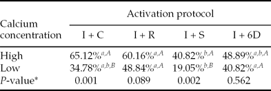
Significant differences were observed in the percentage of decondensed chromatin obtained after parthenogenetic activation of bovine oocytes using the four different protocols. For the oocytes activated in medium with high calcium concentration, the protocols with cycloheximide and roscovitine were superior to strontium and did not differ from the group treated with 6-DMAP (Table 1). In the oocytes activated at a low concentration of calcium, the protocols with 6-DMAP or roscovitine induced higher activation than strontium and did not differ from the group treated with cycloheximide (Table 1). When the oocytes were analysed after activation using the same artificial activation protocol with two different concentrations of calcium, significant differences were obtained only in the groups treated with ionomycin + cycloheximide (P = 0.001) and ionomycin + strontium (P = 0.002). In these conditions, high concentrations of calcium were beneficial to parthenogenetic development (Table 1).
Percentage of decondensed chromatin obtained 24 h after artificial activation of bovine oocytes using four protocols and two calcium concentrations

a,b Equal letters in the same line represent no significant difference at the level of significance α = 0.05 between the four protocols under the same concentration of calcium.
A,B Equal letters in the same row represent no significant difference at the level of significance α = 0.05 between the concentrations of calcium in the same protocol using the Z test for two proportions.
*Comparison at a significance level α = 0.05 of oocytes obtained with the same activation protocol with different calcium concentrations.
I, ionomycin; C, cycloheximide; R, roscovitine; S, strontium; 6D, 6-DMAP.
Artificial activation of equine oocytes
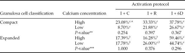
The analysis of equine compact cumulus oocytes obtained after artificial activation showed no significant differences between the three protocols tested when considering the same calcium concentration. Similar results were obtained comparing the different calcium concentrations in each protocol for compact and expanded oocytes. However, in media with high calcium concentrations, activation of expanded cumulus oocytes with 6-DMAP was higher than with cycloheximide and roscovitine, whereas for low calcium concentration, the protocol with 6-DMAP resulted in higher artificial activation than with cycloheximide, and no statistic differences with the group activated with roscovitine was observed (Table 2).
Percentage of decondensed chromatin obtained at 24 h of culture, after artificial activation of equine oocytes using three protocols and two calcium concentrations

*Equal letters represent no significant difference at the level of significance α = 0.05 among three protocols under the same calcium concentration.
**Comparison at a significance level α = 0.05 of compact oocytes obtained with the same activation protocol with different calcium concentrations.
***Comparison at significance level α = 0.05 of expanded oocytes obtained with the same activation protocol with different calcium concentrations.
I, ionomycin; C, cycloheximide; R, roscovitine; 6D, 6-DMAP.
Discussion
The results of the present study indicate that the dynamics of artificial activation differs between the studied species. For artificial activation of bovine oocytes, we observed that the associations of calcium ionophore (ionomycin) with drugs that inhibit protein synthesis such as cycloheximide, that inhibit protein phosphorylation like 6-DMAP, or that directly inhibit MPF like roscovitine, were effective in inducing oocyte activation, independently of the concentration of free calcium in the culture medium. We obtained activation rates of bovine oocytes from 35 to 65% (for low and high calcium concentration, respectively) using calcium ionophore in association with cycloheximide. Similar activation rates of cattle oocytes (around 55%) have been described for the same combination of drugs (Hinrichs et al., Reference Hinrichs, Schmidt and Selgrath1995). When ionomycin was associated with 6-DMAP or roscovitine, similar results were obtained (60.16% and 48.89% in the protocols with high calcium and 48.84% and 40.82% in the protocols with low calcium, respectively). These results are in agreement with those of a previous study, in which rates of artificial activation of bovine oocytes between 40 and 70% were reported (Presicce & Yang, Reference Presicce and Yang1994).
The ionomycin induces a single increase in intracellular calcium after 5 min (Nakada & Mizuno, Reference Nakada and Mizuno1998), which may not be sufficient for the maintenance of oocyte activation. During fertilization, an initial long peak of calcium is observed (521 nM), which is responsible for exocytosis of cortical granules and inducing resumption of meiosis (Kline & Kline, Reference Kline and Kline1992). This first peak is followed by a period of 1–3 h of minor peaks (368 nM) at intermittent intervals of 5–20 min (Nakada & Mizuno, Reference Nakada and Mizuno1998). To mimic calcium oscillations during fertilization, the best stimulus for artificial activation would be the one that induces a large number of calcium oscillations with high amplitude peaks, which is known to result in a significant improvement in the post deployment (Bos-Mikich et al., Reference Bos-Mikich, Whittinghan and Jones1997; Dolmetsch et al., Reference Dolmetsch, Lewis, Goodnow and Healy1997; Li et al., Reference Li, Lopis, Whitney, Zlokarnik and Tsien1998). Strontium has this feature, as it mobilizes calcium from cytoplasmic stores (Cuthbertson et al., Reference Cuthbertson, Whittingham and Cobbold1981) and has been shown to induce multiple peaks of calcium in mice (Bos-Mikich et al., Reference Bos-Mikich, Woods, Candy and Whittinghan1995), with low-frequency range but lasting longer when compared with those observed during fertilization of mouse eggs (Kline & Kline, Reference Kline and Kline1992; Jellerette et al., Reference Jellerette, He, Wu, Parys and Fissore2000). However, in the current investigation, the rates of artificial activation of bovine oocytes using ionomycin combined with strontium were the lowest ones (40.82 and 19.05% for low and high calcium, respectively). These activation rates are inferior (63.3%) to that obtained in a previous study in which the same association of drugs in a free calcium medium was used (Meo et al., Reference Meo, Yamazaki, Leal, Oliveira and Garcia2005), but similar to the 26.7% obtained when strontium was used alone (Meo et al., Reference Meo, Leal and Garcia2004).
Based on the results obtained with the use of cycloheximide, roscovitine, and 6-DMAP in bovine oocytes, these protocols were chosen for artificial activation of horse oocytes. We observed that the activation rates (pronucleus formation) of equine compact cumulus oocytes using ionomycin combined with 6-DMAP were 37.78% and 26.67% in high and low calcium concentrations respectively, corroborating the activation rate of 32% reported previously (Pimentel et al., Reference Pimentel, Bordignon and Smith2002). When oocytes classified as expanded were activated with this same combination of drugs, the rates of artificial activation were higher (59.46% and 44.74% for oocytes cultured in medium with high and low calcium concentrations, respectively), confirming the results of previous studies (45–60%; Carneiro et al., Reference Carneiro, Lorenzo, Pimentel, Pegorato, Bertolini, Ball, Anderson and Liu2001; Galli et al., Reference Galli, Colleoni, Duchi, Lagutina and Lazzri2007).
Hinrichs et al. (Reference Hinrichs, Schmidt and Selgrath1995) showed that the treatment with calcium ionophore combined with cycloheximide was more beneficial to oocytes classified as expanded in relation to the compact oocytes, because the culture of expanded oocytes for more than 30 h could lead to a ‘hyper maturation’, aging the oocytes. Those aged oocytes have a decreased ability to synthesize CSF and thus a simple stimulus should be sufficient to activate them (Hinrichs et al., Reference Hinrichs, Schmidt and Selgrath1995). Moreover, according to this study, the compact oocytes, even after 36 h of maturation, still have a reduced ability to respond to artificial activation. Similarly to the results described by Hinrichs et al. (Reference Hinrichs, Schmidt and Selgrath1995) and to those obtained in our study, Li et al. (Reference Li, Morris and Allen2000) reported rates of 56% for completion of meiosis and decondensation of chromatin resulting from the artificial activation of expanded horse oocytes. However in the present study, although the range of activation rates varied from 26.67 to 59.46%, no significant differences were observed between compact and expanded oocytes when ionomycin and 6-DMAP were used, independently of the calcium concentrations in the culture medium.
We also showed that, in the presence of high calcium concentrations, the combination of ionomycin and 6-DMAP resulted in higher activation rates (59.46%) of expanded horse oocytes compared with the other groups (16.28% and 17.39% for ionomycin in combination with cycloheximide and roscovitine, respectively). In medium with low calcium concentrations, this combination of drugs did not lead to statistically significant differences from the group treated with ionomycin and roscovitine (44.74% and 25.00%, respectively), but both groups presented higher activation rates than the group treated additionally with cycloheximide (17.78%). Apparently, the inhibition of protein phosphorylation promoted by 6-DMAP and the specific inhibition of MPF by the roscovitine were beneficial to the artificial activation of horse oocytes. The use of roscovitine for artificial activation has been reported in mice (Mitalipov et al., Reference Mitalipov, Nuisser and Wolf2001) and in Xenopus (Flament et al., Reference Flament, Bodart, Bertout, Browayes, Rousseau and Vilain2000) oocytes. However, in horses, this drug was used only for intracytoplasmic sperm injection together with sperm extract, showing no benefits to the rate of activation when compared with the use of sperm extract alone (Hinrichs et al., Reference Hinrichs, Choi, Love, Chung and Varner2006). Therefore, the results of the present study indicate, for the first time, that the use of roscovitine for the activation of equine oocytes is efficient, resulting in rates comparable with those obtained with the use of protocols already established, such as ionomycin in combination with 6-DMAP.
Although differences have been observed in other studies (Woods et al., Reference Woods, White, Vanderwall, Aston, Bunch and Campbell2002; Hinrichs et al., Reference Hinrichs, Choi, Walckenaer, Varner and Hartman2007), our results showed no significant differences between the rates of artificial activation of equine oocytes obtained when low and high calcium concentrations were used within the same protocol. However, when calcium concentrations were evaluated for artificial activation of bovine oocytes, the use of high concentrations was beneficial for oocytes activated with cycloheximide and strontium. These results are contrary to those reported previously, that demonstrated that culture of mouse oocytes activated with strontium in medium without calcium and magnesium was beneficial to chromatin decondensation. It was hypothesized by the authors that the inhibition of protein synthesis is more efficient when oocytes are cultured in media with low calcium and magnesium (Bos-Mikich et al., Reference Bos-Mikich, Woods, Candy and Whittinghan1995). This effect was not observed in our study, in either bovine or equine oocytes. It is possible that species-specific differences in the sensitivity of IP3 receptors and in the quality of oocytes in vitro contribute, at least in part, to the discrepancies observed between species (Bedford et al., Reference Bedford, Kurokawa, Hinrichs and Fissore2003).
Based on our results, it was possible to conclude that the parthenogenetic activation of bovine oocytes was equally efficient using the combination of ionomycin with cycloheximide, roscovitine, or 6-DMAP, with the best results being obtained in the presence of high concentrations of calcium. For equine oocytes, the use of high concentrations of calcium was not beneficial for parthenogenetic activation under the conditions tested in the present study. The combination of ionomycin with either 6-DMAP or roscovitine was effective in inducing parthenogenetic activation of equine oocytes, with the best results being obtained with oocytes classified as expanded.
We believe that our preliminary findings provide some clues for the development of a better AOA protocol to be used with these species.
Acknowledgements
FAPESP (Processes 04/00822-1 and 04/08782-9).