Staphylococcus aureus is a significant cause of invasive bacterial infections of skin and soft tissue, lung, bone, and bloodstream. S. aureus colonization of the skin and mucosa is the antecedent to invasive infection in the majority of cases.Reference von Eiff, Becker, Machka, Stammer and Peters 1 In the United States, approximately one-third of all people are colonized with S. aureus.Reference Kuehnert, Kruszon-Moran and Hill 2 Understanding factors related to S. aureus carriage and invasion, and the relationship between them, is important for developing effective approaches to reducing invasive infection. The worldwide development and spread of antimicrobial resistant strains, such as methicillin-resistant S. aureus (MRSA), further increases the importance of such knowledge. MRSA is a major focus for infection control programs due to the high incidence, morbidity, and mortality of MRSA infections.Reference Calfee, Salgado and Milstone 3
Colonization with S. aureus is now well-established as a risk factor for invasive infection. Multiple studies have established an association between S. aureus colonization and later invasive infection in various populations.Reference Nouwen, Fieren, Snijders, Verbrugh and van Belkum 4 – Reference Datta, Shah and Huang 12 With high rates of colonization, it is desirable to identify more specific characteristics of colonization that confer greater infection risk. One characteristic previously studied is the persistence of colonization, which has traditionally been classified into three groups: persistently colonized (PC), intermittently colonized (IC), and non-colonized (NC).Reference Nouwen, Fieren, Snijders, Verbrugh and van Belkum 4 – Reference Gupta, Martinello, Young, Strymish, Cho and Lawler 7 In these studies, persistent colonization markedly increased the risk of invasive infection. However, risk of invasive infection among IC persons varies widely; in some cases, the risk is similar to that of NC persons,Reference Nouwen, Fieren, Snijders, Verbrugh and van Belkum 4 , Reference Nouwen, Schouten and Schneebergen 5 and in others, the risk is similar to that of PC persons.Reference Sewell, Clarridge, Lacke, Weinman and Young 6 , Reference Gupta, Martinello, Young, Strymish, Cho and Lawler 7
MRSA produces disproportionately greater morbidity and mortality than methicillin-sensitive S. aureus (MSSA),Reference Engemann, Carmeli and Cosgrove 13 – Reference Safdar and Bradley 17 suggesting different virulence and pathogenicity characteristics that may influence conversion from colonization to infection. Therefore, research specific to MRSA may need to be considered separately from that involving MSSA. Only 1 of the prior studies evaluating persistence of colonization focused on MRSA.Reference Gupta, Martinello, Young, Strymish, Cho and Lawler 7
In the current study, we aimed to increase understanding of the relationship between persistence of colonization and the incidence of invasive MRSA infection. This study was conducted among patients who were actively screened for MRSA colonization during a 4-year period in a broad general hospital population.
METHODS
Study Design and Population
In this retrospective cohort study, the primary outcome was the occurrence of invasive MRSA infection. The study was conducted at the Department of Veterans Affairs Eastern Colorado Healthcare System (VA), a tertiary care medical center. The study protocol was approved by the Colorado Multiple Institutional Review Board. The study population consisted of all patients who received ≥5 MRSA nasal swab tests on different days between February 20, 2010, and July 26, 2013. There were no exclusion criteria; results for all nasal swabs and all hospitalizations during the study period were included. Decolonization therapy with mupirocin or rifampin was not routinely employed during the study period. Chlorhexidine baths were provided only to patients housed in an intensive care unit who had a central venous catheter.
Microbiologic Testing and Records
Beginning in 2007, all VA patients were screened for MRSA colonization with a nasal swab at admission, interward transfer, discharge, death, and weekly for patients who remained on a nursing unit ≥7 days. MRSA nasal swabs taken at admission were tested by real-time polymerase chain reaction (PCR) using the GeneXpert Infinity MRSA assay (Cepheid, Sunnyvale, CA) according to the manufacturer’s instructions to identify colonization within 24 hours of admission. MRSA nasal swab tests performed due to patient transfer, discharge, or death and those for weekly tests were cultured on selective MRSA agar plates (Remel, Lenexa, KS). Initially, only positive swab results were collected in an infection control database, but beginning in 2010, all positive and negative swab results were collected.
Cohort Definition
The study population was divided into three groups according to MRSA nasal swab results. The PC group included as patients in whom ≥80% of swabs were positive. The IC group included all patients with at least 1 but<80% positive swabs. The NC group included those for whom all swabs were negative. This division is similar to the classification schemes used in previous studies.Reference Nouwen, Fieren, Snijders, Verbrugh and van Belkum 4 – Reference Sewell, Clarridge, Lacke, Weinman and Young 6 , Reference Nouwen, Ott and Kluytmans-Vandenbergh 18 – Reference van Belkum, Verkaik and de Vogel 20 To further characterize the relationship between persistence of colonization and invasive infection, a secondary analysis was conducted using 6 groups: with 0%, 1%–19%, 20%–39%, 40%–59%, 60%–79%, and ≥80% positive swabs. To check for possible bias caused by differing quantity, frequency, or periods between nasal swabs, the median number of swabs was compared among groups, as were both median length of interval and longest interval between swabs for each patient.
Identifying the Primary Outcome
Electronic medical records of patients who received ≥5 MRSA nasal swab tests were screened to identify those who had an invasive MRSA infection during the study period. First, the records were searched for all MRSA positive cultures from any site other than nares. Records for these patients were then manually reviewed to determine whether each positive culture represented invasive infection. Reviewers were blinded to patient colonization status. Invasive infection was defined according to strict criteria developed by the Centers for Disease Control’s National Healthcare Safety Network in their surveillance definition of healthcare-associated infection.Reference Horan, Andrus and Dudeck 21 The first 20 records were reviewed by both physicians to establish reliability of classification and to identify factors that made classification difficult for some records. Any difficult-to-classify record encountered by either reviewer was flagged, and in these cases, classification was decided by group review of the chart (DIV, WDH, and MTB).
Covariates
Basic demographic information (age, gender, and race) and chronic disease states relevant to infection risk (eg, diabetes mellitus, end-stage renal disease, chronic heart failure, chronic obstructive pulmonary disease, immune deficiency, malignant neoplasm, and solid organ transplant) were also collected.
Statistical Analysis
The primary outcome, occurrence of invasive infection, was evaluated relative to days at risk using Cox proportional hazard and Kaplan-Meier time-to-event analyses. Days at risk were calculated from the date of the first nasal swab to the date of invasive infection, study end date, or death, whichever occurred first. Confirmatory analysis was conducting using an entry date of the fifth nasal swab rather than the first, excluding any patient with an infection prior to the fifth swab. The impact of covariates was tested in 2 steps. For univariate analysis, χ2 method was used to calculate 95% confidence intervals (CI) and P values. Multivariate analyses used Cox proportional hazard models. Both a full model with all variables and a parsimonious model with an entry criterion of 0.05 and exit criterion of 0.10 were reviewed. Hazard ratios were calculated for IC and PC with NC as the referent, and for PC vs IC. In the 6-group model, all hazard ratios were calculated with NC as the referent.
RESULTS
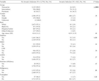
A total of 3,872 patients met inclusion criteria. Most patients were white (71.2%), nearly all were male (95.5%), and their mean age was 63.8 years (Table 1). Of the total cohort, 724 (18.7%) were either intermittently or persistently colonized. Analysis of quantity, frequency, and periods between nasal swabs showed no significant difference among groups in the median number of swabs, median time from first to second, second to third, third to fourth, or fourth to fifth swabs, nor the longest interval between any 2 swabs.
Population Characteristics and Proportions with Invasive Infection

NOTE. AA, African American; ESRD, end-stage renal disease; CHF, congestive heart failure; COPD, chronic obstructive pulmonary disease.
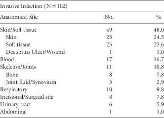
Among all included patients, 102 (2.6%) had invasive MRSA infection. Nearly half of all invasive infections were of skin or soft tissue (48.0%, Table 2). Blood, bone/joints, and respiratory infections made up 16.7%, 10.8%, and 9.8% of all invasive infections, respectively.
Anatomical Sites of Infection

Diabetes, congestive heart failure, and malignancy were the most common chronic diseases in the study population. In univariate analysis, the only variables significantly associated with invasive infection were colonization status (group) and diabetes (Table 1). In the parsimonious Cox regression model, colonization status was the first variable to enter the model, and with colonization status in the model, diabetes no longer met the inclusion criteria (hazard ratio [HR], 1.44; 95% confidence interval [CI], 0.98–2.14; P=.062).
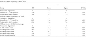
In this model, hazard ratios for developing invasive infection were 22.8 (95% CI, 13.2–39.3) for IC patients and 36.8 (95% CI, 18.4–73.6) for PC patients, with NC patients as the referent (P<.001 for both comparisons, Table 3). No statistically significant difference was detected between PC and IC in the risk for invasive infection (HR, 1.61; 95% CI, 0.94–2.78; P=.084). Results were unchanged in the full model. The risk for invasive infection among diabetics vs nondiabetics was significant (HR, 1.60; 95% CI, 1.08–2.35; P=.017) with diabetes alone and not significant when modeled along with the group (HR, 1.44; 95% CI, 0.98–2.14; P=.062). The Kaplan-Meier plot showed minimal occurrence of invasive infection among NC patients (<1%). According to Kaplan-Meier–based model estimates, 21% of PC patients and 13% of IC patients developed an invasive infection when followed for 2 years (Figure 1). This value was<1% for NC patients.
MRSA invasive infection-free survival stratified by colonization group.

Hazard Ratios for Invasive Infection, with Noncolonized Patients as the Referent Group

NOTE. HR, hazard ratio.
In the confirmatory analysis using the date of the fifth swab as the study entry date, 57 invasive infections were excluded, and analysis of the remaining cases confirmed the primary findings, with hazard ratios of 24.7 (95% CI, 14.3–42.6) for IC patients and 39.6 (95% CI, 19.8–79.2) for PC patients relative to NC patients (Table 3). When persistence was divided into 6 groups, the 1%–19% group had significantly more infection than the 0% (NC) group (HR, 13.8; 95% CI, 6.9–27.6; Table 3). A second notable increase occurred between the groups with 20%–39% and 40%–59% positive swabs; the direct comparison had statistical significance (P=.008).
DISCUSSION
Among the general patient population of this tertiary care medical center, the risk of MRSA infection in IC patients was much closer to that seen in PC patients than in NC patients, whereas the difference in invasive MRSA infection between PC and IC groups was not statistically significant. This relationship held with Cox modeling using all variables and with study entry beginning at the fifth swab rather than the first. When 6 levels of persistence were analyzed, 2 statistically significant increases occurred: (1) between the groups with 0% and 1–19% positive swabs and (2) between the groups with 20%–39% and 40%–59% positive swabs. These results suggest that any level of colonization is associated with markedly increased risk of infection but that a second threshold of persistence exists at which risk again rises significantly and then plateaus.
Diabetes, when tested independently, was significantly associated with increased risk of invasive infection. The correlation between diabetes and nasal colonization was also significant. However, the relationship between diabetes and infection was no longer significant when analyzed along with colonization status in the Cox regression model. While diabetes may be useful in assessing infection risk, nasal colonization was the more significant risk indicator and may be an important mediator between diabetes and invasive infection.
This study expands previous literature on S. aureus in several important ways. It represents one of the few studies examining colonization based on multiple swabs collected longitudinally rather than a single result, allowing a comparison between intermittent and persistent colonization. In our study, we evaluated MRSA rather than MSSA, and did so among a large general hospital population. Most previous studies that evaluated intermittent colonization focused on MSSA and were conducted among peritoneal dialysis patients.Reference Nouwen, Fieren, Snijders, Verbrugh and van Belkum 4 – Reference Sewell, Clarridge, Lacke, Weinman and Young 6 One prior study evaluated MRSA and intermittent colonization in a general hospital population; patients with either intermittent or persistent MRSA colonization were found to be at increased risk of positive clinical culture and of death.Reference Gupta, Martinello, Young, Strymish, Cho and Lawler 7 The results of the current study are consistent with these prior findings and add several other facets to understanding of MRSA colonization and infection. In this study, we used a primary outcome of confirmed invasive infection, with physician review of each patient record to carefully categorize each case based on National Healthcare Safety Network definitions. It also analyzed risk of infection according to proportion of positive swabs in the intermittently colonized group.
These results can inform considerations of risk-management strategies, including decolonization. Distinguishing IC from PC patients might not be of great importance in risk assessment. A single positive screening test places a patient in either the IC or PC category; both of these indicate substantially greater risk. Subsequent screening after an initial positive screen may allow more precise quantification of risk, but the clinical relevance of the small differences in risk may be minimal. However, because a single negative screening test does not rule out intermittent colonization and its concomitant risk, these data support policies of further periodic screening in patients with prior negative results.
We are aware of several limitations pertaining to this study. First, the VA study population is predominantly male, and its age distribution is older than the general population. Second, analysis of covariates does not include length of stay, antibiotic exposure, surgery or ICU admission. Third, the study methods only detected infections that were diagnosed at VA-ECHCS. Most of the VA patient population receives their care exclusively at the VA. Fourth, nasal and clinical isolates were not compared to assess strain type (clonality), and density of nasal colonization was not assessed. Previous studies have found a higher burden of organisms among PC patients,Reference Nouwen, Ott and Kluytmans-Vandenbergh 18 whereas studies of relationship between strain type and infection have had mixed results.Reference Nouwen, Schouten and Schneebergen 5 , Reference Feil, Cooper and Grundmann 22 , Reference Melles, Gorkink and Boelens 23 Fifth, the timing of nares screening tests was according to routine hospital practice rather than a uniform prospective study design. Finally, colonization sites other than nares were not screened.
In future studies, researchers should aim to provide information that could help characterize risk based on an initial patient assessment. Among patients with a negative initial MRSA nares screening, further investigation of factors that put individuals at risk for later colonization or adverse outcomes should be conducted, adding to the work of Gupta et al.Reference Gupta, Martinello, Young, Strymish, Cho and Lawler 7 Density of colonizationReference Datta, Shah and Huang 12 and strain typingReference Nouwen, Schouten and Schneebergen 5 , Reference Feil, Cooper and Grundmann 22 , Reference Melles, Gorkink and Boelens 23 are 2 other characteristics of colonization that may be useful in predicting risk; however, clinically practical methods of evaluating these must be developed. Among colonized patients, randomized trials of various management and decolonization plans should be conducted to determine clinical effectiveness in reducing risk.
ACKNOWLEDGMENTS
Financial support: This study was supported in part by VA Merit Review grant I01BX007080. DIV was supported by Grant No. D33HP25768 from the Health Resources and Services Administration (HRSA). WDH was supported by Grant No. D33HP25768 from HRSA and by Grant No. PTAPM-96-156-20-PTAPM (Physician Training Award in Cancer Prevention) from the American Cancer Society (ACS). The contents are solely the responsibility of the authors and do not necessarily represent official guidance issued by the Department of Veterans Affairs or its affiliates or the official views of HRSA or ACS.
Potential conflicts of interest: All authors declare no potential conflicts of interest relevant to this article.