INTRODUCTION
Actin dynamics play a critical role in the maintenance of eukaryotic cell surface structure, cell motility, cytokinesis, development, differentiation, signal transduction, encystation and excystation (Pollard and Borisy, Reference Pollard and Borisy2003; Bernstein and Bamburg, Reference Bernstein and Bamburg2010; Doi et al. Reference Doi, Shinzawa, Fukumoto, Okano and Kanuka2010; Makioka et al. Reference Makioka, Kumagai, Hiranuka, Kobayashi and Takeuchi2011). In parasites, actin dynamics can be also involved in invasion to or release from host cells (Sibley, Reference Sibley2004; Mehta and Sibley, Reference Mehta and Sibley2011). It is regulated by actin-binding proteins, including actin depolymerizing factors (ADFs), which are major regulatory molecules that usually bind to actin filaments (F-actin) to improve their depolymerization into actin monomers (G-actin) for turnover of actin filaments. ADF has been considered a function node in cellular biology (Bernstein and Bamburg, Reference Bernstein and Bamburg2010). For example, ADF from Toxoplasma gondii increases the re-circulation of F-actin, which is crucial for the development of parasites from tachyzoites to bradyzoites and for their maintenance of cyst structure (Allen et al. Reference Allen, Dobrowolski, Muller, Sibley and Mansour1997). Plasmodium berghei parasites lacking the ADF 2 gene developed normally to the ookinete stage and retained their motility, but they exhibited deficiencies in both the ookinete to oocyst and sporozoite to exo-erythrocytic form transformations, suggesting that ADFs play a pivotal role in the morphological regulation of the complex Plasmodium life cycle (Doi et al. Reference Doi, Shinzawa, Fukumoto, Okano and Kanuka2010). Over-expression of ADF/cofilin of Leishmania donovani impaired flagellum assembly and consequently cell motility by severely impairing the assembly of the paraflagellar rod, without significantly affecting vesicular trafficking or cell growth (Kumar et al. Reference Kumar, Srivastava, Mitra, Sahasrabuddhe and Gupta2012). When the ADF gene was knocked out, the flagella failed to wave, which resulted in impaired motility, sensation, invasion of host cells and cytokinesis (Tammana et al. Reference Tammana, Sahasrabuddhe, Mitra, Bajpai and Gupta2008, Reference Tammana, Sahasrabuddhe, Bajpai and Gupta2010). The ADF of Eimeria tenella regulates actin-based cell motility, which is critical for protozoa invading their hosts. Compared with the control, ADF-mRNA levels were down regulated by 63·86% in second-generation merozoites of E. tenella treated with diclazuril, which implied that the drug impaired parasite invasive ability by affecting its ADF (Xu et al. 2008; Zhou et al. Reference Zhou, Wang, Xue, Wang, Yang, Ban, Xin and Wang2010). Further experiments suggest that ADF/cofilin of Entamoeba invadens is involved in the regulation of actin dynamics, which is related to the encystation and excystation of the parasitic protozoa (Makioka et al. Reference Makioka, Kumagai, Hiranuka, Kobayashi and Takeuchi2011).
Cryptocaryon irritans is a ciliated parasite that causes cryptocaryonosis in marine fish, which severely affects mariculture in Southeast China. As described by Colorni and Burgess (Reference Colorni and Burgess1997), the life cycle of C. irritans is divided into 4 stages, i.e. trophont, protomont, tomont and theront stages. Trophonts parasitize in the epidermis of the skins and gills of various marine fishes and feed on interstitial fluid and cell debris through cytostomes, which result in pathological changes on hosts. When trophonts mature or are irritated by the death of their hosts they drop off their hosts into marine water and become mobile protomonts, which develop to tomonts post-encystation. Tomonts are the productive cysts with strong resistance to the environment and with high production rate, at which more than 200 infective theronts are hatched from a single tomont. Newly hatched theronts penetrate into skin, gills and eyes of fish hosts to develop into parasitic trophonts. Therefore, it would be crucial to prevent fishes from infective theront invasion, and to inhibit parasite development from trophonts to tomonts, the reproductive stage of the parasite, to reduce the ciliate population. As ADFs might play critical roles in both aspects, they were cloned from a cDNA library of C. irritans trophonts. Here, we report the molecular characterization and bioactivity of CiADF 2 (GenBank accession number: JQ906266), which would be useful for the development of effective measures to control outbreaks of cryptocaryonosis.
MATERIALS AND METHODS
Parasites and experimental animals
C. irritans trophonts were collected from the gills of infected Pseudosciaena crocea cultured in net cages during a cryptocaryonosis outbreak in the coastal area of Xiapu county, Fujian province, China, on 2 July 2009. The collection was carried out by soaking gills showing visible white spots in sterile seawater for 2 h. After discarding the gills, trophonts were collected in the sediment and allowed to develop into tomonts. Part of the tomonts was sampled for extraction of genomic DNA while part was incubated at 27 °C to encourage the development of theronts. On the one hand, the genomic DNA was used as template DNA to amplify the ribosomal DNA internal transcribed spacer 1 sequence (rDNA-ITS–1) for identification of the parasite strain used in this study. Results of the identification have been given by Sun et al. (Reference Sun, Zheng, Wu, Guo, Wang and Huang2011) and showed the strain to be the same as strains Chiayi and PYH4·12 based on the rDNA-ITS–1 sequences (Diggles and Adlard, Reference Diggles and Adlard1997; Sun et al. Reference Sun, Zhu, Xie, Wu, Li, Lin and Song2006). On the other hand, newly hatched theronts were used to infect Sebastiscus marmoratus maintained in an aquarium to continue the life cycle of the parasite. All animal experiments were conducted in accordance with the Guiding Principles for the Care and Use of Research Animals outlined by Fujian Normal University.
Collection of the parasites at different stages
Theronts were harvested by centrifugation at 1000 g for 5 min at 4 °C. Trophonts, the parasitic stage of the ciliate, were collected from S. marmoratus 3 days post-infection by scraping fish body surfaces with a glass slide in a dish of sterile seawater. Motile trophonts and protomonts were harvested individually and immediately using a pipette. Tomonts were collected 3 days post-infection by placing dishes on the bottom of the aquarium for 24 h, to allow mature trophonts to drop from the fish bodies into the dishes and to encyst. Parasites at each stage were washed in sterile seawater and stored immediately at −80 °C for further use.
Gene isolation from the cDNA library
The cDNA library was constructed as described previously (Huang et al. Reference Huang, Sun, Guo, Zheng, Xu, Yuan and Liu2012). To sequence expression sequence tags (ESTs), recombinant λTripIE × 2 was converted into recombinant plasmid λTripIE × 2 by transduction of the recombinant phage into E. coli BM25·8 strain expressing Cre recombinase. EST sequences were aligned with genes deposited in GenBank to detect clones carrying putative ADF genes. Two clones carrying a putative ADF gene were isolated and designated as CiADF 2 .
Examination of CiADF2 mRNA expression
For detecting the expression at different stages of life cycle, total RNA was extracted from the parasites at each stage by using a QIAGEN RNeasy Mini kit (QIAGEN) according to the manufacturer's instructions. Reverse transcription (RT)-PCR was performed using an RNA amplification kit according to the manufacturer's instructions (Takara Bio Inc.) with 500 ng of total RNA as a template for each 10 μl of reaction mixture. The reaction mixture was incubated at 30 °C for 10 min, 42 °C for 30 min, 95 °C for 5 min, and 5 °C for 5 min. The PCR conditions were 25 cycles of 95 °C for 30 s, 50 °C for 30 s, and 72 °C for 60 s, followed by 10 min of extension at 72 °C. The sequences of the gene-specific primers for amplification of CiADF 2 were 5′-ATGGTATCTACTGGAGTC-3′ and 5′-TCACATTAATAATTCCTTT-3′. An actin gene isolated from C. irritans (CiActin, GenBank Accession number: JN399999) was used as a control and amplified using the following primers, 5′-ATGGCCGAAGACTAACAAGCAG-3′ and 5′-TCAGAAGCATTTTCTGTGTACA-3′.
Site-directed mutagenesis of CiADF2 gene by PCR
One TAA codon was found in the open reading frame (ORF) of CiADF 2 . To allow gene expression in bacteria, the T in the TAA codon was mutated into C with a GeneTailor™ Site-Directed Mutagenesis System (Invitrogen) according to the manufacturer's instructions. Briefly, pTripIE × 2/CiADF 2 was methylated by DNA methylase and then amplified in a mutagenesis reaction with the overlapping primers 5′-TTATCATTGATATCTGTGGACAAAGAGAAGA-3′ and 5′-TCCACAGATATCAATGATAATGTTATTTCC-3′ (the underlined nucleotide was the target mutation). The mutagenesis reaction was carried out in a thermocycler. The cycling parameters were 20 cycles of 94 °C for 30 sec, 55 °C for 30 sec, and 68 °C for 1 min, followed by 10 min of extension at 68 °C. The mutagenesis mixture was then transformed into wild type E. coli, in which linear mutated DNA was circulated and the methylated plasmid DNA template was digested by inherent McrBC endonuclease.
Su cloning of the modified ORF from pTripIE × 2/CiADF2 into pGEX-4T-1
The modified ORF was amplified using a set of oligonucleotide primers, 5′- CGCGGATCCATGGTATCTACTGGAGTC -3′ and 5′-CGGAATTCTCACATTAATAATTCCTT T-3′ (the underlined sections in each primer indicate BamHI and EcoRI recognition sites, respectively), with pTripIE × 2/CiADF 2 as the template DNA. The PCR product was double-digested with BamHI and EcoRI (TaKaRa, Otsu, Japan), followed by ligation into the BamHI-EcoRI site of pGEX-4T-1 and then transformation into E. coli DH5α.
Expression, extraction and purification of recombinant CiADF2 protein
E. coli colony containing plasmid pGEX-4T/CiADF 2 was cultured in LB medium (1% Bacto Tryptone, 0·5% yeast extract, 1% NaCl, and 0·1% 5 M NaOH) with ampicillin sodium (50 μg/ml) at 37 °C. When the optical density at 600 nm reached between 0·3 and 0·5, E. coli was induced to express the recombinant CiADF2 fusion protein (G-rCiADF2) by the addition of 0·5 mM isopropyl-β-d-thiogalactopyranoside and incubation for another 4 h. G-rCiADF2 was extracted and purified using glutathione sepharose 4B (GE Healthcare Life Sciences, Uppsala, Sweden) according to the manufacturer's instructions. The recombinant CiADF2 protein (rCiADF2) was further purified by thrombin cleavage to remove glutathione-S-transferase (GST).
Production of antibodies against rCiADF2 and against lysate of trophonts
Fifteen specific pathogen-free Kunming mice (Shanghai Laboratory Animal Co. Ltd, China) were divided into 3 groups (5 per group) and immunized with rCiADF2 (100 μg/mouse), GST (100 μg/mouse) and lysate of trophonts, respectively. Mice were injected intraperitoneally with the corresponding protein emulsified with equal volume (200 μl/mouse) of Freund's complete adjuvant (Sigma-Aldrich, St Louis, USA). Two booster immunizations were performed using the same dose of antigen emulsified with an equal volume of Freund's incomplete adjuvant (Sigma-Aldrich). The intervals between the immunisations were both 14 days. Ten days after the last immunization, blood was obtained by cardiac puncture, sera against rCiADF2, sera against GST and sera against lysate of trophonts were then separated from the blood cells, respectively. They were stored at −30 °C for further use.
Expression of the native CiADF2 and antigenicity of rCiADF2 detected by Western blot analysis
To detect the expression of native CiADF2 and immunogenicity of rCiADF2, cell lysates of trophonts/protomonts, tomonts and theronts were sampled and denatured at 100 °C for 5 min in a sample buffer (62·5 mM Tris-HCl at pH 6·8, 2% SDS, 5% β-mercaptoethanol, 10% glycerol, and 0·02% bromophenol blue) and subjected to sodium dodecyl sulfate-polyacrylamide gel electrophoresis (SDS-PAGE) using 12% acrylamide gels as described previously (Huang et al. Reference Huang, Xuan, Yokoyama, Xu, Suzuki, Sugimoto, Nagasawa, Fujisaki and Igarashi2003). Subsequently, proteins were transferred to polyvinylidene fluoride membranes (Millipore, Billerica, USA) and analysed with mouse antibody against rCiADF2 or against GST as the primary antibody (1:100) and horseradish peroxidase-conjugated goat anti-mouse IgG antibody (ICN Biochemicals, Aurora, USA) as the secondary antibody (1: 4000). The reactions were visualized with 3,3′-diaminobenzidine (0·5 mg/ml) and 0·03% H2O2. On the other hand, to determine the reactigenicity of rCiADF2, purified rCiADF2 and lysate of trophonts (for positive control) were subjected to Western blot analysis with mouse antibody against the lysate of trophonts as the first antibody. The secondary antibody and the substrate were the same as those mentioned above.
Localization of native CiADF2 by immune fluorescent antibody testing (IFAT)
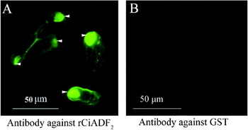
Newly hatched theronts were fixed in 0·15% formaldehyde for 30 min, and then spun down at 800 g for 5 min. After 3 washes with distilled water, the theronts were pipetted onto glass slides and dried on air. They were then incubated for 30 min with mouse antibody against rCiADF2 or against GST (1:100), respectively, followed by incubation with Alexa 488-conjugated goat anti-mouse IgG antibody (1:100 in PBS containing 3% fetal bovine serum) (Sigma) at 37 °C for 30 min. The results were observed under a fluorescence microscopy and photographed.
Detection on bioactivity of CiADF2 by pull-down assay using analytical ultracentrifugation
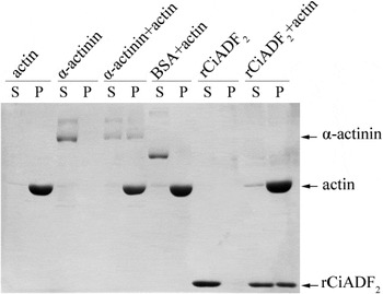
An actin-binding protein assay kit (Cytoskeleton, Denver, USA) was used to detect the F-actin binding and depolymerizing activity of rCiADF2 according to the manufacturer's instruction. Briefly, G-actin was first polymerized to F-actin, which was then mixed with rCiADF2, α-actinin (positive control) and bovine serum albumin (negative control), respectively. The 3 different protein mixtures and F-actin alone, rCiADF2 alone, α-actinin alone and BSA alone were all ultracentrifuged at 150 000 g for 1·5 h. After the separation of the supernatants from the pellets, each was sampled and subjected to SDS-PAGE analysis.
RESULTS
Characterization of gene sequence
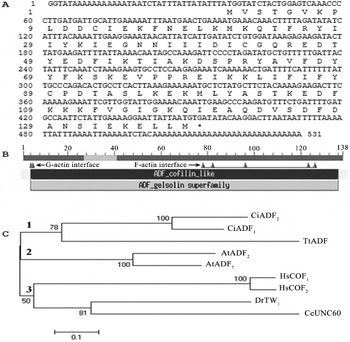
From the cDNA library of C. irritans trophonts, 14 clones carrying putative ADF genes were isolated and divided in 2 different ADF genes, CiADF 1 and CiADF 2 . Reported here is CiADF 2 with the GenBank accession number JQ906266. As shown in Fig. 1A, the full-length cDNA of CiADF 2 was 531 bp with an open reading frame (ORF) of 417 bp, which encoded a polypeptide of 138 aa with a predicted molecular weight of 16·2 kDa. There was 1 TAA codon in the ORF, which is a termination codon in the universal genetic code, but encodes glutamine in the ciliate genetic code. The ratio of AT to CG in CiADF 2 ORF was 71 to 29. The deduced polypeptide contained an ADF domain with amino acids typical for interactions with G-actin and F-actin. As shown in Fig. 1B, the 4th and 5th amino acids were a G-actin binding area, and the fragment from the 78th to 126th amino acids was an F-actin binding area. There were 23 acidic amino acids, 23 basic amino acids, 54 hydrophobic amino acids and 38 polar amino acids in the polypeptide, with a pI of 6·36. Ser3 and Tyr68 were found in CiADF2, which are typical for the ADF/cofilin protein family. There was 1 α-helix in a putative F-actin binding area, which is also a conserved trait of the family. The phylogenetic relationship of CiADF 2 with 9 other ADF/cofilins from different species is shown in Fig. 1C, which indicated that CiADF 2 is closely related to CiADF 1 (GenBank accession number: JQ906265) and an ADF from Tetrahymena thermophila with a similarity of 35%.
Characterization of the CiADF 2 gene and its deduced polypeptide. (A) The full-length cDNA sequence of CiADF 2 and its deduced amino acid sequence. (B) Schematic structure of CiADF2 showing the ADF/Cofilin-like domain and G-actin and F-actin interfaces. (C) Phylogenetic relationship of CiADF2 with ADFs from several different organisms. AtADF2 (AAB03697) and AtADF7 (AEE85080) are from Arabidopsis thaliana; DrTWI (NP_477034) from Drosophila twinstar; CeUNC60 (NP_503427) from Caenorhabditis elegans; HsCOF1 (NP_005498) and HsCOF2 (NP_068733) from Homo sapiens; TtADF (BAH84775) from Tetrahymena thermophila.

mRNA expression profile of CiADF2 detected by RT-PCR
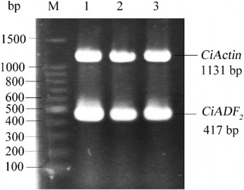
As shown in Fig. 2, in all stages of C. irritans 2 specific bands were found in the RT-PCR product. The lower band was around 417 bp, corresponding to the predicted size of CiADF 2 ORF, while that of the upper bands was approximately 1131 bp, consistent with that of CiActin ORF.
CiADF 2 mRNA expression was detected in parasites at different stages using reverse transcription-PCR. M, DNA ladder; lanes 1–3: RT-PCR products amplified from trophonts/protomonts, tomonts and theronts, respectively. Two sets of oligonucleotides were used to amplify CiADF 2 and CiActin (control), respectively.

Recombinant CiADF2 expressed in E. coli
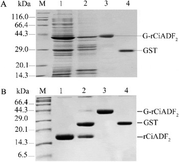
The modified CiADF 2 gene was expressed as the recombinant fusion protein with a GST tag, which was designated as G-rCiADF2. As shown in Fig. 3A, G-rCiADF2 was expressed in both soluble and insoluble fractions with a molecular mass of 42·2 kDa. Glutathione-Sepharose 4B combined with thrombin cleavage to remove the GST tag were used to purify rCiADF2 from the soluble fraction. As shown in Fig. 3B, the molecular mass of rCiADF2 was 16·2 kDa, which corresponded to the predicted result.
Expression and purification of the recombinant CiADF2 protein as detected by SDS-PAGE. (A) Expression and purification of the recombinant fusion protein, G-rCiADF2. M, proteins with standard molecular masses; Lanes 1 and 2: soluble and insoluble fractions of the bacterial lysate, respectively, Lane 3: purified G-rCiADF2; Lane 4: GST. (B) Removal of GST from G-rCiADF2 by thrombin cleavage. M, proteins with standard molecular masses; Lane 1: rCiADF2 after cleavage of GST, Lane 2: rCiADF2 and GST, Lane 3: G-rCiADF2 fusion protein, Lane 4: GST.

Expression of native CiADF2 and antigenicity of rCiADF2 detected by Western blot analysis
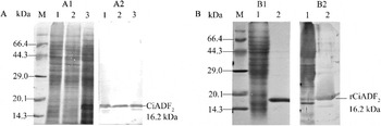
The purified rCiADF2 was used to immunize SPF mice. The resulting polyclonal antibodies against rCiADF2 were applied to detect native CiADF2 protein in C. irritans, with a polyclonal antibody against GST as a negative control. As seen in Fig. 4A, a specific positive reaction band presented in each well with lysates of trophonts/protomonts, tomonts and theronts, respectively, which suggested that rCiADF2 was immunogenic to mount an immune reaction in mice. The antibody against rCiADF2 could recognize native CiADF2, the molecular mass of which corresponded to the calculated molecular weight, 16·2 kDa, and the native CiADF2 was expressed at translation level in the entire life cycle of C. irritans. On the other hand, as shown in Fig. 4B, polyclonal antibodies against the cell lysate of C. irritans trophonts/protomonts reacted not only with native trophont proteins, but also with rCiADF2, suggesting that the reactigenicity of the recombinant protein.
Expression of native CiADF2 and antigenicity of rCiADF2 as detected by Western blot analysis. (A) Mouse antiserum against rCiADF2 recognized the native CiADF2, which was found in lysates of trophonts/protomonts, tomonts, and theronts. A1, proteins stained with amino black 10B. A2, the same proteins as those shown in A1 were analysed with mouse antiserum against rCiADF2 as the first detection reagent and horseradish peroxidase-conjugated goat anti-mouse IgG antibody as the secondary detection reagent (1: 4000). The reaction was visualized with 3,3′-diaminobenzidine and H2O2. M, proteins with standard molecular masses, Lanes 1–3: lysates of trophonts/protomonts, tomonts and theronts, respectively. (B) rCiADF2 reacted with mouse serum against lysate of Cryptocaryon irritans trophonts. B1, proteins stained with amino black 10B. B2, the same proteins as those shown in B1 were analysed with serum from mice immunized with native proteins from the lysate of trophonts as the first detection reagent and horseradish peroxidase-conjugated goat anti-mouse IgG antibody as the secondary detection reagent (1: 4000) to detect the reactigenicity of rCiADF2. M, proteins with standard molecular masses. Lane 1, native proteins from the lysate of C. irritans trophonts, Lane 2, rCiADF2.

Localization of native CiADF2 protein
The polyclonal antibody against rCiADF2 was used in an immunofluorescent antibody test (IFAT) to localize the native protein, with a polyclonal antibody against GST as a negative control. The results presented in Fig. 5 demonstrated that CiADF2 was scattered in the cytosol and primarily localized beneath the plasma membrane. However, it was especially enriched in a region about one-third to anterior of the organism, where the cytostome was localized.
Localization of native CiADF2 in theronts of Cryptocaryon irritans as detected by an immunofluorescent antibody test. Serum from mouse immunized with rCiADF2 (A) or serum from mouse immunized with GST (B) was used as the first antibody. Alexa 488-conjugated antibody against mouse IgG was used as the secondary antibody. The cytostomes of theronts in (A) were pointed out by the white arrows.

F-actin binding and depolymerizing activities of rCiADF2
The activities of actin binding and depolymerization of rCiADF2 were detected by using a pull-down assay combined with analytical ultracentrifugation. BSA and the known actin-binding protein α-actinin were used as the negative and positive controls, respectively. Each of them was incubated with or without F-actin for 30 min and followed by ultracentrifugation. As shown in Fig. 6, α-actinin, BSA, and rCiADF2 were all found in supernatants when they were not co-existing with F-actin, while F-actin alone was found in the sediment. However, after incubation with F-actin, approximately half of the α-actinin and rCiADF2, but none of the BSA, were presented in the sediments post-ultracentrifugation, which implicated F-actin binding activity of rCiADF2. Moreover, in the supernatant from the reaction tube of rCiADF2 plus F-actin, the actin amount was slightly increased when it was compared with those from the tube of F-actin alone and the tube of BSA plus F-actin, which suggested that rCiADF2 partially depolymerized F-actin into G-actin, which presented in the supernatant after ultracentrifugation.
F-actin binding assay of rCiADF2. Each of the following proteins, α-actinin (positive control), bovine serum albumin (BSA, negative control) and rCiADF2, was incubated with or without F-actin for 30 min, respectively. They were then centrifuged at 150 000 g for 1·5 h at 24 °C. Subsequently, the supernatants (S) and pellets (P) were collected for each reaction. They were sampled respectively, separated on an SDS-PAGE and stained with 0·1% Coomassie blue. The result showed that approximately half of α-actinin and rCiADF2, but none of the BSA, were co-precipitated with F-actin. Moreover, in the supernatant from the reaction of rCiADF2 plus F-actin, the amount of actin was a little more than those from the reactions of BSA plus F-actin, and F-actin alone. The result suggested that rCiADF2 could bind to and sever F-actin.

DISCUSSION
In this study, an ADF gene was cloned from a C. irritans trophont library. The sequence analysis revealed that CiADF 2 and its product had typical characteristics of ADF/cofilin, such as Ser3 and Tyr68, which are G-actin and F-actin binding sites respectively. Therefore, the activity of CiADF2 may be modulated in a way typical of the ADF family, i.e. by phosphorylation of Ser3 with LIM kinase or TES kinase, which would lead to most ADF/cofilin proteins losing their depolymerization activity, and dephosphorylation of Ser3 with SSH, which would restore the depolymerizing activity. Tyr68 is highly conserved in the family and its phosphorylation may enhance protein ubiquitination and degradation, so as to reduce the overall levels of ADF in the cells. CiADF 2 is closely related to an ADF from Tetrahymena thermophila (TtADF, Adf73p) with a similarity of 35%, although TtADF has no typical Ser3 and Tyr68, and no genes encoding LIM kinase and SSH were found in the genomic database of T. thermophila.
There are non-universal genetic codes in C. irritans genes. In CiADF 2 , one TAA codon was found to encode glutamine. However, it would be deciphered as a stop codon by bacterial cells so that the downstream sequence fragment would not be translated. In this study, the TAA codon was mutated successfully into CAA, the universal codon for glutamine. The results showed that the mutated gene was expressed well in bacterial cells. The purified recombinant protein mounted an immune reaction in mice, the antisera of which recognized the native CiADF2. On the other hand, rCiADF2 reacted to antisera against the cell lysate of C. irritans trophonts. These results suggested antigenicity of rCiADF2. Judging from the ADF structure, it is a cytosolic protein. However, parasitic trophonts of C. irritans have been reported to feed on interstitial fluid and cell debris through cytostomes, which would make uptake of host antibodies against recombinant CiADF2 into the parasitic cytosol possible. Therefore, anti-rCiADF2 might be a candidate for the development of vaccines or diagnostic reagents.
A bioactivity assay showed that rCiADF2 was not only capable of binding to F-actin, but also capable to depolymerize F-actin, which might help to elucidate the actin dynamics during the development of C. irritans and its invasion of fish hosts.
Cellular movements are powered by the assembly and disassembly of actin filaments. The assembly of actin filaments at the leading edge of motile cells pushes the plasma membrane forward. CiADF2 was located in the cytoplasm and was especially abundant at one end of the ciliate around the cytostome. It has been reported that microtubules exist around the ciliate cytostome. However, in the case of C. irritans, there might also be abundant actin in this area. The turnover of F-actin and G-actin under regulation of CiADF2 and some other actin-binding proteins might play an important role in the movement of the ciliate.
In conclusion, this study improves our understanding of the pathogen biology of C. irritans and the control of cryptocaryonosis.
ACKNOWLEDGEMENTS
This work was supported by grants from the National Natural Science Foundation of China (No. 31040084 and No. 31101032), the Natural Science Foundation of Fujian Province, China (No. 2008J004, No. 2010J01145), the Science Foundation for the Returned Overseas Chinese Scholars from Fujian province and Fujian Normal University.