Introduction
Definitive radiotherapy (RT) with concurrent cisplatin-based chemotherapy is the contemporary standard of careReference Pignon, le Maître, Maillard and Bourhis1 in the non-surgical curative-intent management of loco-regionally advanced head and neck squamous cell carcinoma (SCC). Conventional RT using two-dimensional techniques is commonly associated with significant acute and late toxicity such as mucositis, xerostomia, dysphagia and fibrosis with a consequent negative impact upon health-related quality of life (QoL).Reference Bentzen and Trotti2,Reference Ge, Liao, Yuan, Mao, Li and Yu3 Spurred by technological advances in treatment planning and delivery in the last decade, intensity-modulated radiation therapy has largely supplanted conventional RT in head and neck SCC with improved target volume coverage and sparing of surrounding organs at risk. The use of intensity-modulated RT provides significant reduction in RT-related late morbidityReference Gupta, Kannan, Ghosh-Laskar and Agarwal4 and potentially improved quality of survival.Reference Chen, Farwell, Luu, Vazquez, Lau and Purdy5 The impact of modern conformal techniques on some of the rare but highly morbid late severe complications of head and neck irradiation, such as necrosis of the bone, cartilage or cervical soft-tissues with attendant cosmetic and functional implications, is not well documented and described in contemporary indexed medical literature, possibly because of them being uncommon or even under-reported.
Osteoradionecrosis, defined as ‘the presence of more than 1 cm of exposed necrotic bone in a previously irradiated field, which fails to heal within 3 to 6 months’Reference Marx6 is the most well described definition, which originates from the orthovoltage era when there was a widely varying incidence of osteoradionecrosis ranging from 1 to 37 per cent.Reference Sciubba and Goldenberg7 Its incidence has dramatically reduced with the introduction of megavoltage RT in the 1960s and is typically now seen in around 5 per cent of patients with oral or oropharyngeal cancer.Reference Aarup-Kristensen, Hansen, Forner, Brink, Eriksen and Johansen8
Typical signs and symptoms of osteoradionecrosis include pain, swelling, trismus, orocutaneous fistula or exposed underlying bone.Reference Gevorgyan, Wong, Poon, Blanas, Enepekides and Higgins9 The initially proposed mechanism for osteoradionecrosis was the sequence of hypocellularity, hypovascularity and hypoxia induced by RT.Reference Marx6 Current understanding suggests osteoradionecrosis to be an RT-induced fibro-atrophic processReference Delanian and Lefaix10 that results from a combination of dying osteoblasts and excessive proliferation of myofibroblasts. The mandible is especially susceptible to osteoradionecrosis because of limited blood supply after RT.
Chondroradionecrosis is rarer with an estimated incidence of 1–5 per centReference Zbären, Caversaccio, Thoeny, Nuyens, Curschmann and Stauffer11,Reference Oppenheimer, Krespi and Einhorn12 and is seen largely in laryngo-pharyngeal cancers. It is a much more serious complication that may prove fatal as a result of airway collapse or aspiration if not identified early and managed aggressively. Chondroradionecrosis may present as hoarseness, respiratory distress, odynophagia, fistula, foetor oris, fixity to the skin, laryngeal oedema or airway obstruction.Reference Chandler13 Chondroradionecrosis occurs as a result of RT-induced inflammation that causes arteritis and thrombosis in vessels around the laryngeal framework leading to ischemia and fibrosis of the perichondrium progressing to tissue death. Susceptibility to chondroradionecrosis increases if the larynx is initially compromised by infection, trauma or direct tumour invasion.Reference Calcaterra, Stern and Ward14–Reference Keene, Harwood, Bryce and van Nostrand16 Cervical necrotising fasciitis following head and neck RT is an extremely rare entity which manifests as rapidly progressive cutaneous and superficial fascial necrosis of the cervical soft-tissueReference Mortimore and Thorp17 caused by infectious thrombosis of underlying small blood vessels.Reference Klabacha, Stankiewicz and Clift18 There are numerous predisposing factors associated with the development of necrosis of the bone, cartilage or cervical soft-tissues, such as site of the tumour, stage of disease, proximity to bone or cartilage, dental extraction, oral hygiene, RT technique, and dose fractionation. Use of concurrent chemotherapy, presence of co-morbidities, super-added infection, immuno-compromised status and habits (tobacco and alcohol) have also been identified as potentially aggravating systemic risk factors.Reference Lee, Koom, Lee, Kim, Yoo and Keum19–Reference Frankart, Frankart, Cervenka, Tang, Krishnan and Takiar26
Aims
This report described the clinico-demographic profile and treatment-related characteristics associated with RT necrosis (osteoradionecrosis, chondroradionecrosis or necrotising fasciitis) in a series of head and neck cancer patients treated on prospective clinical trials of definitive radio(chemo)therapy using high-precision conformal techniques and the management outcomes of such late morbidity.
Materials and methods
Medical records of patients with squamous cell cancers of the oropharynx, larynx and hypopharynx treated with definitive high-precision radio(chemo)therapy on prospective clinical trials between 2005 to 2019 in a single academic head and neck unit of a tertiary-care comprehensive cancer centre were reviewed retrospectively to identify patients with documented radiation necrosis. Waiver of consent was received in view of the retrospective nature of the study with less than minimal risk to participants. Patient characteristics that were studied included age, gender, habits, comorbidity, oral hygiene including dental extraction, anatomic site of the primary cancer, and American Joint Committee on Cancer tumour–node–metastases (TNM) staging or grouping. Treatment details included RT technique, dose fractionation and use of concurrent systemic chemotherapy.
Treatment details
Between 2005 and 2008, patients were treated with either three-dimensional conformal RT or static-field intensity-modulated RT using 6 MV photons on a linear accelerator (Siemens Primus Hi, Siemens Medical Systems, Erlangen, Germany). Since 2009, patients have been treated with rotational intensity-modulated RT using 6 MV photons on helical tomotherapy (Hi-ART II, Accuray, Sunnyvale, USA). RT dose-fractionation was variable based on the study protocol, ranging from normofractionated RT (70 Gy in 35 fractions over 7 weeks with 2 Gy per fraction and 5 fractions per week), normofractionated-accelerated RT (66 Gy in 33 fractions over 5.5 weeks with 2 Gy per fraction and 6 fractions per week) to slightly hypofractionated-accelerated RT (66 Gy in 30 fractions over 6 weeks with 2.2 Gy per fraction and 5 fractions per week or 70.2 Gy in 33 fractions over 6.5 weeks with 2.12 Gy per fraction and 5 fractions per week) to moderately hypofractionated-accelerated RT (55 Gy in 20 fractions over 4 weeks with 2.75 Gy per fraction and 5 fractions per week).
The planned objective was to restrict doses to the contralateral parotid gland (mean dose, less than or equal to 26 Gy) and spinal cord (maximum dose, less than 45 Gy) while ensuring that more than or equal to 95 per cent of the target volume was covered by at least 95 per cent of the prescribed dose. Patients with adequate renal function (defined as creatinine clearance more than 50 ml/minute) also received concurrent weekly low-dose cisplatin (30–35 mg/m2) with appropriate anti-emetic prophylaxis, adequate hydration and forced saline diuresis as per institutional protocol. All patients were referred for dental evaluation and appropriate prophylaxis including extraction if required prior to RT and subsequently underwent fluoride gel application periodically on follow up. Patients were scheduled for response assessment 18F-fluoro-deoxy-glucose positron emission tomography-computed tomography (FDG-PET-CT) at 8–12 weeks after completion of therapy. Patients with complete morphological and metabolic response at the primary site and neck on FDG-PET-CT were followed up clinically every 3 months for the first 2 years, every 6 months until 5 years and annually thereafter.
Diagnosis of RT necrosis was made on the basis of clinical evaluation, imaging findings and histopathology as required. Appropriate treatment was instituted in the form of antibiotics, debridement, salvage surgery, hyperbaric oxygen therapy and supportive care. A descriptive analysis of the events of interest (osteoradionecrosis, chondroradionecrosis or necrotising fasciitis) including time to development of necrosis, their management and outcomes are reported herewith. Baseline characteristics of the patient, disease and treatment of those developing RT necrosis were compared with patients without documented necrosis using the chi-square test and unpaired student t-test as appropriate. All statistical analysis was performed on GraphPad Prism® statistical software (version 6.01).
Results
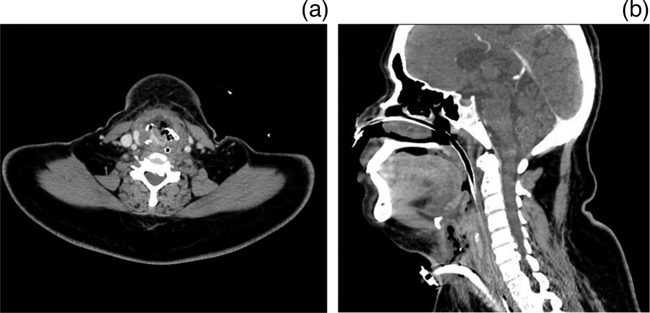
Between December 2005 and July 2019, a total of 290 patients with squamous cell cancers of the oropharynx (n = 139), larynx (n = 78) and hypopharynx (n = 73) were accrued on various prospective clinical trials and treated with high-precision definitive radio(chemo)therapy in a single academic head and neck unit of a tertiary cancer-care centre. Twelve patients had documented radiation necrosis in the form of osteoradionecrosis (n = 3), chondroradionecrosis (n = 7) and necrotising fasciitis (n = 2) and constitute the present study cohort. Imaging findings of representative cases of osteoradionecrosis and chondroradionecrosis from the study cohort are depicted in Figure 1 and 2, respectively. The overall rate of RT necrosis following definitive high-precision radio(chemo)therapy for head-neck cancer was 4.1 per cent with 95 per cent confidence interval ranging from 2.2 to 7.1 per cent.
Focal fluoro-deoxy-glucose uptake in the left lower alveolar region (a) with erosion on corresponding bone window of computed tomography scan (b) suggestive of osteoradionecrosis. Biopsy from the same area showed hyperplastic squamous epithelium with some necrotic bony spicules and dense inflammatory infiltrate confirming the diagnosis.

Axial (a) and sagittal (b) plane sections of contrast-enhanced computed tomography scan showing an ill-defined area with multiple foci of air involving the pre-epiglottic space with destruction of thyroid and cricoid cartilages and resultant chondroradionecrosis requiring dependence on tracheostomy for severe airway compromise.

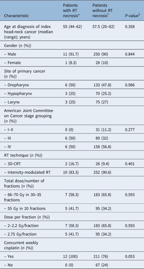
Baseline demographic, disease and treatment characteristics of patients with documented radiation necrosis (n = 12) are briefly summarised in Table 1. All 12 patients with RT necrosis had loco-regionally advanced (stage III–IV) head and neck SCC at diagnosis; 11 had habits in the form of smoking (n = 9), tobacco (n = 3) or alcohol use (n = 7), and only one had documented comorbidity in the form of diabetes and hypertension, both of which were controlled with medication. RT dose fractionation was variable, ranging from 55 Gy in 20 fractions over 4 weeks to 66–70.2 Gy in 33–35 fractions over 5.5–7 weeks. All 12 patients received concurrent weekly cisplatin (30–35 mg/m2) during RT. There was no significant difference in the baseline demographic (age, gender), disease (primary site, American Joint Committee on Cancer stage) and treatment characteristics (RT technique, total dose, fractionation) of patients developing RT necrosis versus those without necrosis (Table 1), except for the use of concurrent weekly cisplatin chemotherapy, which was somewhat higher in patients developing necrosis with borderline significance (p = 0.053).
Baseline patient, disease and treatment characteristics of the study cohort (developing radiation necrosis) compared with the patients without such necrosis

*n = 12; †n = 278; ‡reported p-values are based on chi-square test for all characteristics except for comparison of median age which is based on unpaired Student t-test. RT = radiotherapy; 3D-CRT = three-dimensional conformal radiotherapy
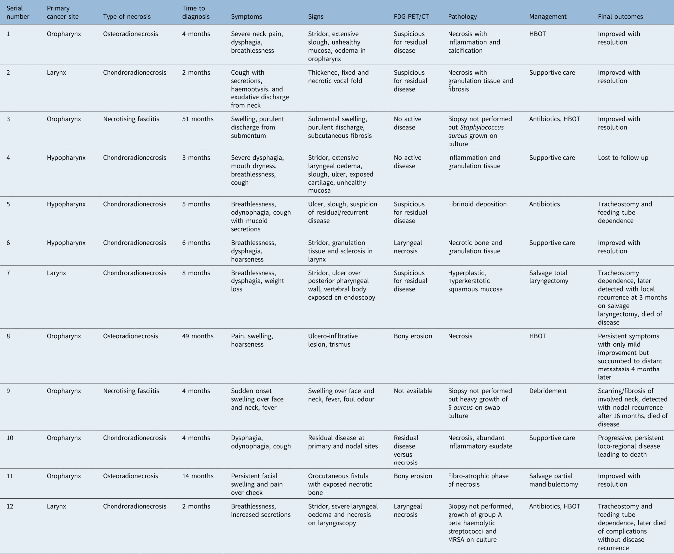
Radiation necrosis (osteoradionecrosis, chondroradionecrosis, necrotising fasciitis) was diagnosed based on signs and symptoms along with radiological or histopathological findings. Details of individual patients with RT necrosis are summarised in Table 2. The median time to development of symptoms of RT necrosis was 4.5 months (range, 2–51 months). Patients with osteoradionecrosis presented with painful swelling over the jaw, typically on the same side as the index primary tumour. Mandibular bone was exposed intra-orally in two of these cases, while intra-oral evaluation was suboptimal in one patient because of severe trismus. Patients with chondroradionecrosis presented with one or more of the following symptoms of breathlessness, dysphagia, throat pain, hoarseness and stridor. One patient expectorated small tissue bits of the laryngeal cartilage, which confirmed necrosis on histopathological evaluation. On microlaryngoscopy examination, six out of the seven patients with chondroradionecrosis had necrotic slough over the laryngeal mucosa, two had frank ulceration with exposed cartilage, three had severe laryngeal oedema and one had fixation of the vocal fold. Both patients with cervical necrotising fasciitis presented with rapidly progressive swelling in the neck and submentum, which are associated with desquamation, inflammation and sero-purulent discharge. One patient also had orocutaneous fistula communicating into the soft tissues in the neck.
Summary of presentation, work-up, management and outcomes of patients with necrosis after high-precision radiotherapy for head-neck cancer

FDG-PET/CT = fluoro-deoxy-glucose positron emission tomography/computed tomography; HBOT = hyperbaric oxygen therapy; MRSA = methicillin-resistant Staphylococcus aureus
Eleven of these 12 patients underwent whole-body 18F-fluoro-deoxy-glucose positron emission tomography-computed tomography with contrast-enhanced CT of the head and neck region at the time of presentation of these symptoms for more nuanced interpretation. Imaging was suspicious for residual or recurrent disease in four patients, demonstrated bony erosion in two patients and laryngeal necrosis in two patients, while it demonstrated absence of residual or recurrent disease in two patients. In one patient, fluoro-deoxy-glucose avidity in the lesion at the primary site could not be interpreted accurately (residual disease vs necrosis). Nine of the 10 patients with osteoradionecrosis or chondroradionecrosis underwent biopsy for histopathological correlation. Three of these specimens showed only inflamed granulation tissue with or without fibrinoid deposition whereas six patients had frank necrosis with or without associated inflammatory changes. None of the patients had any residual or recurrent tumour in the initial biopsy specimen. Both patients with necrotising fasciitis grew several micro-organisms on serial bacterial cultures obtained from cervical soft tissues. Initial management of necrosis included antibiotics or anti-inflammatory agents (n = 4), tissue debridement (n = 1) and tracheostomy (n = 7) as appropriate. Persistence of signs and symptoms necessitated further resective surgery in 2 patients. One patient underwent laryngectomy because of stridor and dysfunctional larynx, with the specimen showing recurrent tumour infiltrating underlying cartilage in addition to the necrosis. Another patient underwent segmental mandibulectomy for persistent pain and swelling over the cheek with a specimen confirming inflammatory infiltrate and erosion of the mandible. Four patients also underwent hyperbaric oxygen therapy for persistent signs or symptoms of radiation necrosis.
Outcomes following management of RT necrosis were variable and included significant symptomatic improvement and resolution in five patients, persistent symptoms with mild improvement in one patient, tube dependence (tracheostomy or feeding tube) in three patients and severe neck scarring or fibrosis in one patient. One patient with tracheostomy tube dependence succumbed to complications of laryngeal chondroradionecrosis without evidence of disease recurrence, four patients died of progressive disease in the form of loco-regional relapse (n = 3) or distant metastasis (n = 1), while one patient was lost to follow up after chondroradionecrosis precluding ascertainment of the final outcome.
Discussion
The impact of modern high-precision conformal techniques on the incidence and severity of late RT necrosis of bone, cartilage and cervical soft tissues following head and neck irradiation is not well described, possibly as a result of being uncommon and an under-reported phenomenon. The symptoms and signs of RT necrosis generally overlap with and mimic residual or recurrent tumour clinico-radiologically, making the diagnosis more difficult and challenging. The overall rate of RT necrosis in the present study was largely in keeping with single-institutional cohorts treated with contemporary high-precision conformal techniques.
Since the first description of bone necrosis following RT by Claudius Regaud nearly 100 years ago, the definition and staging or grading of osteoradionecrosis has evolved greatly,Reference Frankart, Frankart, Cervenka, Tang, Krishnan and Takiar26 but a common clinical presentation is a site of exposed bone in an irradiated field unrelated to tumour recurrence that develops a few months to several years after RT and fails to heal over a three to six month period.Reference Marx6,Reference Frankart, Frankart, Cervenka, Tang, Krishnan and Takiar26 The reported rates of osteoradionecrosis have varied considerably over time and across institutions with the most dramatic shifts reported after the introduction of megavoltage beams with further reduction in incidence with increasingly conformal techniques such as intensity-modulated RT in the last two decades. Overall, there has been a continued trend towards lower rates of osteoradionecrosis (around 5 per cent in the modern era) as treatment techniques have become more and more conformal with reduction of doses to normal oro-dental tissues.Reference Davis, Hanley and Cooper25,Reference Frankart, Frankart, Cervenka, Tang, Krishnan and Takiar26 Notable modifiable risk factors for osteoradionecrosis include continued tobacco and alcohol use during treatment, and appropriate counselling on these practices should be provided to all patients prior to RT.
Comprehensive evaluation of oral health prior to treatment is recommended with consideration of extraction of high-risk, non-restorable teeth prior to RT along with avoidance of extractions in the post-RT setting.Reference Frankart, Frankart, Cervenka, Tang, Krishnan and Takiar26 Goodrich and LenzReference Goodrich and Lenz27 reported the first case of laryngeal chondroradionecrosis over 70 years ago, and since that time only case reports or small case series have been published in the indexed medical literature.Reference Roh28–Reference Gessert, Britt, Maas, Wieland, Harari and Hartig30 The Chandler grading systemReference Chandler13,Reference Farneti, Fabbri and Prencipe31 is a clinical-based classification of post-RT laryngeal reactions with chondroradionecrosis almost always presenting as grade IV reaction. Sloughing of the arytenoid, fragmentation and collapse of the thyroid cartilage, and presence of gas bubbles around the cartilage are strongly suggestive CT-characteristics of chondroradionecrosis.Reference Farneti, Fabbri and Prencipe31,Reference Hermans, Pameijer, Mancuso, Parsons and Mendenhall32 Newer imaging modalities such as 18F-fluoro-deoxy-glucose positron emission tomography-computed tomography with contrast-enhanced CT might have improved diagnostic accuracy in distinguishing chondroradionecrosis from recurrent disease.Reference Greven, Williams, Keyes, McGuirt, Watson and Case33 Cervical soft-tissue necrosis including necrotising fasciitis is largely a clinical diagnosis with no specific imaging findings, but it can be supplemented by additional tests such as cultures with sensitivity tests and skin or soft-tissue biopsy.Reference Umbert and Winkelman34
Limitations
Despite the above descriptive analysis of consecutive patients with RT necrosis following high-precision definitive radio(chemo)therapy, several caveats remain. Although patients had been treated on prospective clinical trials, this was a retrospective analysis of RT necrosis and suffers from all the inherent biases and limitations of any retrospective study. Although baseline demographic, disease and treatment characteristics of patients developing late RT necrosis (n = 12) was compared with patients without necrosis (n = 278), the number of patients with necrosis was quite small, precluding robust statistical testing and comparison. Patients included in the study had been treated over a prolonged time-period with varying RT techniques and dose-fractionation which could be a potential confounding factor. Although meticulous attention was given to reducing hot-spots from bone and cartilage during treatment planning, RT plans were not reviewed subsequently to identify any such inadvertent hot-spots in patients developing RT necrosis to establish any dose-volume correlation.
• The impact of modern conformal techniques on the incidence and severity of late radiation necrosis of bone, cartilage and cervical soft tissues is not well documented
• Symptoms and signs of radio-necrosis generally mimic residual or recurrent tumours making the diagnosis challenging
• Use of megavoltage beams and introduction of intensity-modulated radiation therapy has led to a decline in the rates of radio-necrosis over the past two decades
• Multidisciplinary management is essential for the prevention, early diagnosis and successful treatment of such late radiation morbidity
Conclusion
Delayed necrosis of bone, cartilage or cervical soft tissues are uncommon and under-reported severe complications of definitive radio(chemo)therapy for head and neck cancers. Multidisciplinary management with inputs from radiation oncology, otolaryngology, oral surgery, imaging specialists, infectious disease experts and dental health professionals is the key in prevention, early diagnosis and successful treatment of such late radiation morbidity.
Acknowledgements
The authors would like to thank all patients and their caregivers for providing written informed consent for participation in institutional prospective studies of definitive high-precision radiotherapy and all members of the Head-Neck Disease Management Group of Tata Memorial Centre, Mumbai, India, for their logistic support towards successful conduct of the study.
Competing interests
None declared