Introduction
Attention-deficit hyperactivity disorder (ADHD) is a common neurodevelopmental disorder with high heritability (estimated as 70–80%), which is consistent across age and gender in childhood and adolescence (Larsson et al., Reference Larsson, Chang, D'Onofrio and Lichtenstein2014). A familial vulnerability has also been demonstrated in neuropsychological functions that both the ADHD probands and their unaffected siblings tend to have impaired performance in a wide range of neuropsychological functions such as sustained attention (Gau and Huang, Reference Gau and Huang2014; Pironti et al., Reference Pironti, Lai, Muller, Dodds, Suckling, Bullmore and Sahakian2014) and executive functions (Gau and Shang, Reference Gau and Shang2010), assessed by Cambridge Neuropsychological Test Automated Battery (CANTAB). Since unraveling the genetics of ADHD is still challenging, the endophenotype approach has been recognized as one of the promising attempts to provides insight into the mechanisms underlying the disorder (Glahn et al., Reference Glahn, Knowles, McKay, Sprooten, Raventos, Blangero, Gottesman and Almasy2014). Endophenotype, or intermediate phenotype, is relatively proximal to genetic underpinnings than the clinical symptoms (Glahn et al., Reference Glahn, Knowles, McKay, Sprooten, Raventos, Blangero, Gottesman and Almasy2014). An endophenotype consists of four criteria: it is (1) heritable; (2) associated with the disorder; (3) largely state independent (persistently exhibiting in the individual whether or not the illness is active); and (4) cosegregating with disorder within families (Gottesman and Gould, Reference Gottesman and Gould2003). Hence, asymptomatic carriers of the risk genes are vulnerable to manifest some endophenotypes; searching the neurobiological characteristics of unaffected relatives of ADHD probands as endophenotype candidates can help deciphering the pathogenetic mechanisms of this disorder (Glahn et al., Reference Glahn, Knowles, McKay, Sprooten, Raventos, Blangero, Gottesman and Almasy2014; Pironti et al., Reference Pironti, Lai, Muller, Dodds, Suckling, Bullmore and Sahakian2014).
In brain imaging studies, changes in brain activation patterns during neuropsychological tasks have been demonstrated to be similar in ADHD probands and their unaffected relatives. For example, decreased activation of the left superior, inferior frontal regions, temporal/parietal regions (van Rooij et al., Reference van Rooij, Hoekstra, Mennes, von Rhein, Thissen, Heslenfeld, Zwiers, Faraone, Oosterlaan, Franke, Rommelse, Buitelaar and Hartman2015b), anterior cingulate and cerebellar regions (Mulder et al., Reference Mulder, Baeyens, Davidson, Casey, van den Ban, van Engeland and Durston2008) during a response inhibition task were noted in ADHD probands as well as their unaffected siblings. As to the gray matter volumes, ADHD probands showed the volumetric reductions in the prefrontal cortex, cingulate cortex (Bralten et al., Reference Bralten, Greven, Franke, Mennes, Zwiers, Rommelse, Hartman, van der Meer, O'Dwyer, Oosterlaan, Hoekstra, Heslenfeld, Arias-Vasquez and Buitelaar2016), and total gray matter (Greven et al., Reference Greven, Bralten, Mennes, O'Dwyer, van Hulzen, Rommelse, Schweren, Hoekstra, Hartman, Heslenfeld, Oosterlaan, Faraone, Franke, Zwiers, Arias-Vasquez and Buitelaar2015) and their unaffected siblings had these volumes intermediate to ADHD probands and controls (Greven et al., Reference Greven, Bralten, Mennes, O'Dwyer, van Hulzen, Rommelse, Schweren, Hoekstra, Hartman, Heslenfeld, Oosterlaan, Faraone, Franke, Zwiers, Arias-Vasquez and Buitelaar2015; Bralten et al., Reference Bralten, Greven, Franke, Mennes, Zwiers, Rommelse, Hartman, van der Meer, O'Dwyer, Oosterlaan, Hoekstra, Heslenfeld, Arias-Vasquez and Buitelaar2016). Additionally, both impairments in sustained attention and volumetric abnormalities in the right inferior frontal gyrus were found to be neurocognitive endophenotypes in adults with ADHD (Pironti et al., Reference Pironti, Lai, Muller, Dodds, Suckling, Bullmore and Sahakian2014). While there is persuasive evidence supporting the abnormalities of the functional activations and gray matter volumes as the familial risk of ADHD, less is known about the altered microstructural property of white matter tracts connecting gray-matter functional networks.
The microstructural property of white matter tracts can be quantified by measuring the directional diffusion of water molecules along axonal pathways to present the structural brain connectivity. Diffusion tensor imaging (DTI) and diffusion spectrum imaging (DSI) are frequently used methods to measure the microstructural property of white matter. DTI reconstructs the water molecular diffusion in terms of a diffusion tensor based on the Gaussian model of the translational displacement (Basser et al., Reference Basser, Mattiello and LeBihan1994), while DSI reconstructs the water molecular diffusion in terms of an average propagator based on the Fourier relation between the diffusion signal and average propagator (Lin et al., Reference Lin, Wedeen, Chen, Yao and Tseng2003). In voxels with crossing fibers, the principal direction of the DTI points to a direction that is somewhere between the crossing fibers. DSI can address the limitation of DTI to show the directions of the maxima of the average propagator correspond to the underlying crossing fiber directions (Wedeen et al., Reference Wedeen, Wang, Schmahmann, Benner, Tseng, Dai, Pandya, Hagmann, D'Arceuil and de Crespigny2008).
Frequently used indices sensitive to tissue properties include generalized fractional anisotropy (GFA), fractional anisotropy (FA), mean diffusivity (MD), axial diffusivity (AD) and radial diffusivity (RD) (Alexander et al., Reference Alexander, Lee, Lazar and Field2007; Ozarslan et al., Reference Ozarslan, Koay, Shepherd, Komlosh, Irfanoglu, Pierpaoli and Basser2013; Hsu and Tseng, Reference Hsu and Tseng2018). GFA and FA refer to the degree of anisotropy of the diffusivity of water molecules. MD measures average diffusivity. More specifically, AD describes the diffusion along the axonal fibers, whereas RD reflects the average diffusion perpendicular to axonal fibers (Lawrence et al., Reference Lawrence, Levitt, Loo, Ly, Yee, O'Neill, Alger and Narr2013). These indices are sensitive microstructural property including axonal integrity, myelination, and homogeneity of axonal orientations (Jones et al., Reference Jones, Knosche and Turner2013).
The microstructural properties of the corpus callosum (CC), the superior longitudinal fasciculus (SLF), and the inferior fronto-occipital fasciculus (IFOF) have been frequently reported to be different between ADHD probands and typically developing controls (Lawrence et al., Reference Lawrence, Levitt, Loo, Ly, Yee, O'Neill, Alger and Narr2013; van Ewijk et al., Reference van Ewijk, Heslenfeld, Zwiers, Faraone, Luman, Hartman, Hoekstra, Franke, Buitelaar and Oosterlaan2014; Chiang et al., Reference Chiang, Chen, Lo, Tseng and Gau2015; Aoki et al., Reference Aoki, Cortese and Castellanos2018). Additionally, the microstructural property of the CC, SLF, and IFOF and the uncinate fasciculus (UF) is heritable in families enriched for ADHD within the eleven tracts evaluated by Sudre et al. (Reference Sudre, Choudhuri, Szekely, Bonner, Goduni, Sharp and Shaw2017). Recent studies reported that some diffusion indices of white matter tracts correlated with ADHD symptoms, but the tracts identified and the direction of correlations differ across these studies (Lawrence et al., Reference Lawrence, Levitt, Loo, Ly, Yee, O'Neill, Alger and Narr2013, van Ewijk et al., Reference van Ewijk, Heslenfeld, Zwiers, Faraone, Luman, Hartman, Hoekstra, Franke, Buitelaar and Oosterlaan2014, Chiang et al., Reference Chiang, Chen, Lo, Tseng and Gau2015). For example, while Chiang et al. (Reference Chiang, Chen, Lo, Tseng and Gau2015) found negative correlation between the alteration of the GFA value in the right SLF and inattentive symptoms, Lawrence et al. (Reference Lawrence, Levitt, Loo, Ly, Yee, O'Neill, Alger and Narr2013) reported the correlations between higher MD, AD, or RD values of the forceps minor and more inattentive symptoms.
To date, there are two studies using sibling design to explore shared alterations in white matter property between ADHD probands and unaffected siblings (Lawrence et al., Reference Lawrence, Levitt, Loo, Ly, Yee, O'Neill, Alger and Narr2013; van Ewijk et al., Reference van Ewijk, Heslenfeld, Zwiers, Faraone, Luman, Hartman, Hoekstra, Franke, Buitelaar and Oosterlaan2014). The first study focused on nine major white matter tracts with tract-based analysis and revealed elevated MD and AD in some tracts in both ADHD probands and their unaffected siblings (Lawrence et al., Reference Lawrence, Levitt, Loo, Ly, Yee, O'Neill, Alger and Narr2013). The same study also observed the severity of inattention symptoms were correlated with increased MD across groups (Lawrence et al., Reference Lawrence, Levitt, Loo, Ly, Yee, O'Neill, Alger and Narr2013). The second study using whole-brain voxel-wise approaches reported lower FA values among siblings as compared to ADHD probands, and similar MD values among siblings and controls (van Ewijk et al., Reference van Ewijk, Heslenfeld, Zwiers, Faraone, Luman, Hartman, Hoekstra, Franke, Buitelaar and Oosterlaan2014). However, the authors observed the severity of ADHD symptoms were positively associated with FA values and negatively associated with MD in ADHD probands in the voxels in which ADHD showed significantly lower FA and lower MD than controls, respectively (van Ewijk et al., Reference van Ewijk, Heslenfeld, Zwiers, Faraone, Luman, Hartman, Hoekstra, Franke, Buitelaar and Oosterlaan2014).
Owing to only two DTI studies with sibling design revealing mixed findings (Lawrence et al., Reference Lawrence, Levitt, Loo, Ly, Yee, O'Neill, Alger and Narr2013; van Ewijk et al., Reference van Ewijk, Heslenfeld, Zwiers, Faraone, Luman, Hartman, Hoekstra, Franke, Buitelaar and Oosterlaan2014), this exploratory study aimed to identify important loci of structural alterations related to clinical and cognitive phenotypes. Since the whole-brain voxel-wise approaches may be more sensitive to registration errors due to morphometric differences between groups (Vangberg et al., Reference Vangberg, Skranes, Dale, Martinussen, Brubakk and Haraldseth2006), we adopted tractography-based analyses to sample the microstructural property of the tracts more accurately and to cover tracts over the entire brain (Chen et al., Reference Chen, Lo, Hsu, Fan, Hwang, Liu, Chien, Hsieh, Liu, Hwu and Tseng2015). We compared the microstructural properties of white matter tracts among ADHD probands, their unaffected siblings and typically developing controls, and examined the associations between these properties and ADHD symptoms and neuropsychological functions. Based on the previous finding that the CC, SLF, IFOF, and UF may be white matter tracts endophenotype candidates reported being heritable within ADHD families (Sudre et al., Reference Sudre, Choudhuri, Szekely, Bonner, Goduni, Sharp and Shaw2017), we expected to find some shared alterations of the microstructural property of these fiber tracts in both ADHD probands and unaffected siblings. We also evaluated additional tracts that have not been characterized previously to identify microstructural alterations that may be unique to unaffected siblings as compensatory mechanisms.
Methods
Participants and procedures
The Research Ethics Committee of National Taiwan University Hospital approved this study prior to its implementation (approved number, 201204071RIC; ClinicalTrials.gov number, NCT01682915). We recruited 53 sibling pairs of medication-naïve youths with ADHD consecutively if they met the inclusion and exclusion criteria as stated below and their unaffected biological siblings. ADHD probands were clinically diagnosed with current ADHD based on the DSM-IV-TR diagnostic criteria by board-certified child psychiatrists at National Taiwan University Hospital, Taipei, Taiwan. Fifty-three controls were matched individually in age and sex with ADHD probands. Exclusion criteria for all the participants included psychosis, mood disorders, learning disability, substance use disorder, autism spectrum disorder, neurological disorders, major anxiety disorders, or a Full-scale IQ score less than 80.
The participants and their parents provided written informed consent after explanation of the procedures and purpose of the study. All participants' parents received psychiatric interviews about the diagnosis of the participants with the Chinese version of the Schedule for Affective Disorders and Schizophrenia for School-Age Children—Epidemiologic Version (K-SADS-E) (Gau et al., Reference Gau, Chong, Chen and Cheng2005). The unaffected siblings and controls were assessed with the K-SADS-E interview to confirm that they did not have ADHD. All the participants were assessed with the Wechsler Intelligence Scale for Children-third edition (WISC-III) and three tasks of the CANTAB (Chamberlain et al., Reference Chamberlain, Robbins, Winder-Rhodes, Muller, Sahakian, Blackwell and Barnett2011) for sustained attention (Rapid visual information processing, RVP), spatial short-term memory (Spatial Span, SSP) and nonverbal working memory (Spatial Working Memory, SWM).
Clinical measures
The Chinese version of the K-SADS-E
The preparation of the Chinese K-SADS-E included a two-stage translation and modification of several items with psycholinguistic equivalents relevant to the Taiwanese culture (Gau and Soong, Reference Gau and Soong1999). It was further modified to meet the DSM-IV (Gau et al., Reference Gau, Chong, Chen and Cheng2005) and DSM-5 (Chen et al., Reference Chen, Shen and Gau2017) diagnostic criteria. This semi-structured interview scale is a reliable and valid instrument to assess DSM-IV child and adolescent psychiatric disorders and has been widely used in a variety of studies regarding childhood mental disorders (e.g. (Gau and Huang, Reference Gau and Huang2014; Chiang et al., Reference Chiang, Chen, Lo, Tseng and Gau2015; Shang et al., Reference Shang, Lin, Tseng and Gau2018).
The Chinese version of the Swanson, Nolan, and Pelham, version IV scale (SNAP-IV) – parent form
The Chinese SNAP-IV is a 26-item rating scale with 9 items for inattention symptoms, 9 items for hyperactivity/impulsivity symptoms and 8 items for oppositional symptoms according to the DSM-IV symptom criteria of ADHD and oppositional defiant disorder (ODD). Items are rated as a 4-point Likert scale (0 for not at all, 1 for just a little, 2 for quite a lot, and 3 for very much). The norm and psychometric properties of the Chinese SNAP-IV–parent form have been established (Gau et al., Reference Gau, Shang, Liu, Lin, Swanson, Liu and Tu2008). The Chinese SNAP-IV has been widely used in assessing ADHD and ODD symptoms in clinical and research settings in Taiwan (e.g.(Chiang et al., Reference Chiang, Chen, Lo, Tseng and Gau2015; Han et al., Reference Han, Chen, Tsai and Gau2018).
Neuropsychological measurement
The CANTAB is a computerized test battery which has been validated and widely used worldwide (Chamberlain et al., Reference Chamberlain, Robbins, Winder-Rhodes, Muller, Sahakian, Blackwell and Barnett2011). It is designed to be administered by trained psychologists with standardized procedures. Three tasks involving sustained attention, spatial short-term memory and spatial working memory were administered to all the participants in this study (Gau and Huang, Reference Gau and Huang2014).
Rapid visual information processing (RVP)
The RVP, a 4-min visual continuous performance test, is designed to assess sustained attention. The participant had to detect three target sequences (2–4–6, 3–5–7, 4–6–8) and respond immediately, as the digits appeared one at a time at a rate of 100 digits per minute. The participant was instructed to detect as many target sequences as possible. In this study, the index of mean latency was used, which represented the mean time taken to respond in the correct responses. ADHD probands usually showed longer latency in the correct responses than the controls (Gau and Huang, Reference Gau and Huang2014).
Spatial Span (SSP)
The SSP is the visuospatial analog of the digit span test and is used to measure spatial short-term memory. There were 9 white boxes in fixed locations on the screen, and the color of the boxes was changed one after the other in a predetermined sequence. The participants were asked to remember the order of the boxes in which the color was changed, and reproduced the order by pointing to the boxes on the screen. The task began with a level of 2-box then gradually maximally up to a level of 9-box. The index used in the present study was span length, the longest sequence successfully recalled. The higher value of spatial length implied better performance in spatial short-term memory.
Spatial working memory (SWM)
The SWM a self-ordered search test to assesses nonverbal working memory (Luciana and Nelson, Reference Luciana and Nelson1998). Participants were asked to search through a number of colored boxes on the screen to find a blue token hidden inside. The participants had to memorize which boxes had been opened in each trial and the boxes in which the tokens had been found in the previous trial. The index used in this study was total errors: total times the participant visits a box that was sure not to have a blue token, i.e., either a token inside in the previous trial or empty in this trial. The lower value of spatial length implied better performance in spatial working memory.
MRI data acquisition
MRI data were acquired on a 3T MRI system (Trio, Siemens, Erlangen, Germany) with a 32-channel phased array head coil. A sagittal localizer was used to define a line between the anterior commissure and posterior commissure, which was then used to define transaxial planes orthogonal to the sagittal image. T1-weighted images covering the whole head were acquired using a 3D magnetization-prepared rapid gradient echo (MPRAGE) sequence, repetition time (TR) = 2530 ms; echo time (TE) = 3.4 ms; slice thickness = 1.0 mm; matrix size = 256 × 192 × 208; and field of view (FOV) = 256 × 192 × 208 mm3. Transaxial DSI data were acquired using a pulsed-gradient spin-echo EPI sequence with a twice-refocused balanced echo (Reese et al., Reference Reese, Heid, Weisskoff and Wedeen2003). An acquisition scheme which comprised 102 diffusion-weighted images (DWI) volumes corresponding to the grid points within a half sphere of the q-space (DSI 102) was applied with the maximum diffusion sensitivity value (bmax) set to 4000 s/mm2 (Kuo et al., Reference Kuo, Chen, Wedeen and Tseng2008). The acquisition scheme has been verified to be the optimal acquisition scheme of DSI at 3T (Kuo et al., Reference Kuo, Chen, Wedeen and Tseng2008), and has been used constantly in our previous studies (e.g.(Chiang et al., Reference Chiang, Chen, Lin, Tseng and Gau2017). Additionally, this imaging data can be used to reconstruct diffusion indices comprehensively, not only diffusion tensor indices (i.e. FA, AD, RD, and MD), but also a non-tensor index (i.e. GFA). Other acquisition indices were TR = 9600 ms; TE = 130 ms; matrix size = 80 × 80; FOV = 200 × 200 mm2; slice number = 56 and slice thickness = 2.5 mm without a gap. The scan time for DSI acquisition was approximately 16.5 min.
DSI image reconstruction
Before image reconstruction, the DSI datasets underwent image quality assurance entailing signal-to-noise ratio assessment, motion-induced signal dropout screening and within-subject spatial alignment between T1-weighted image and DSI images. Our DSI datasets underwent a quality assurance procedure by counting the number of diffusion-weighted images that had a significant signal dropout (Chen et al., Reference Chen, Lo, Hsu, Fan, Hwang, Liu, Chien, Hsieh, Liu, Hwu and Tseng2015). All of the acquired DSI datasets (3264 images per person) were scrutinized by calculating the signals in the central square (20 × 20 pixels) of each image. If there were more than 90 images of signal loss, which would lead to significant reductions in GFA values, the subject would be excluded for further analysis. In all participants, three out of 53 ADHD probands did not pass the criteria of quality assurance. These three ADHD probands, as well as their siblings and matched controls, were excluded in the final analyses.
The pipeline of DSI data reconstruction and automatic tract-specific analysis is illustrated in Fig. 1. The DSI raw data sets were reconstructed into the diffusion indices that represented the microstructural characteristics of white matter tracts. The diffusion indices including GFA, FA, AD, RD, and MD were estimated by using the regularized version of Mean Apparent Propagator MRI (MAP-MRI) framework (Ozarslan et al., Reference Ozarslan, Koay, Shepherd, Komlosh, Irfanoglu, Pierpaoli and Basser2013; Hsu and Tseng, Reference Hsu and Tseng2018). Specifically, the signal in diffusion-specific space (i.e. q-space) was fitted with an expansion series of Hermite basis functions. The term of zero-order in the expansion series contained the diffusion tensor that characterizes the Gaussian displacement distribution. The higher-order terms in the expansion series were the orthogonal corrections to the Gaussian approximation which enabled to estimate an orientational distribution function (ODF) that contained information about diffusion probabilities in different directions. The values of the AD, RD, and MD in each voxel were determined by calculating the first eigenvalue, mean of the second and third eigenvalues, and mean of the three eigenvalues of the diffusion tensor, respectively (Alexander et al., Reference Alexander, Lee, Lazar and Field2007). To quantify the degree of anisotropy, the FA calculated as the square root of the variance of the three eigenvalues of the diffusion tensor normalized by their norms (Alexander et al., Reference Alexander, Lee, Lazar and Field2007). Those diffusion indices represented various microstructural properties of white matter, such as the degree of myelination, fiber density and fiber damage (Cohen-Adad et al., Reference Cohen-Adad, El Mendili, Lehericy, Pradat, Blancho, Rossignol and Benali2011).
The pipeline of DSI data reconstruction and automatic tract-specific analysis.

Whole brain tract-specific analysis
To extract the tract-specific variables of white matter for statistical comparison, we used the tract-based automatic analysis (TBAA) to sample the diffusion indices from the pre-defined 76 major fiber tract bundles over the whole brain (Chen et al., Reference Chen, Lo, Hsu, Fan, Hwang, Liu, Chien, Hsieh, Liu, Hwu and Tseng2015). These 76 major fiber tract bundles were built in a DSI template, named NTU-DSI-122 (Hsu et al., Reference Hsu, Lo, Chen, Wedeen and Tseng2015), using deterministic streamline-based tractography with multiple regions of interest defined in the automated anatomical labeling atlas (Tzourio-Mazoyer et al., Reference Tzourio-Mazoyer, Landeau, Papathanassiou, Crivello, Etard, Delcroix, Mazoyer and Joliot2002). First, the sampling coordinates of the 76 tracts were transformed from NTU-DSI-122 to individual DSI datasets with the corresponding deformation maps. The deformation maps were achieved by the two levels of two-step registration. The registration garnered anatomical information provided by the T1-weight images (Ashburner and Friston, Reference Ashburner and Friston2011) and microstructural information provided by DSI datasets (Hsu et al., Reference Hsu, Hsu and Tseng2012). The two-level registration strategy involved the first level registration between the individual space and the subject-specific template (SST), and the second level registration between the SST and NTU-DSI-122 (Chen et al., Reference Chen, Lo, Hsu, Fan, Hwang, Liu, Chien, Hsieh, Liu, Hwu and Tseng2015). The registration strategy of SST provided less registration bias due to subject variabilities relative to the standard template. Second, the sampling coordinates were aligned with the proceeding direction of each fiber tract bundle, and the values of diffusion indices were sampled in native space along the sampling coordinates that were normalized and divided into 100 steps (Chen et al., Reference Chen, Lo, Hsu, Fan, Hwang, Liu, Chien, Hsieh, Liu, Hwu and Tseng2015). Finally, we obtained the output of tract-based analysis, called 3D connectogram (x-axis: 100 steps along sampling coordinates; y-axis: 76 white matter tract bundles; z-axis: subjects) (Chen et al., Reference Chen, Lo, Hsu, Fan, Hwang, Liu, Chien, Hsieh, Liu, Hwu and Tseng2015), for each diffusion index in the ADHD, sibling and control groups. In this paper, we took the average of diffusion indices over 100 steps for each tract to represent white matter property of each fiber tract.
Statistical analyses
Data analysis was conducted using SAS 9.2 version (SAS Institute, Cary, NC). The alpha value was preselected at the level of p < 0.05. The descriptive results were displayed as the mean and standard deviation for the continuous variables.
In order to address the lack of independence between the probands and their siblings within the same family in the comparisons of clinical measures, neuropsychological measurements and white matter microstructure indices among the three groups, we used a multi-level generalized linear model with mixed effects to analyze data with hierarchical structure through the inclusion of random effects in the model (Casals et al., Reference Casals, Girabent-Farres and Carrasco2014). The Proc Mixed Procedure was used with the Bonferroni correction method to adjust p values in post hoc analysis due to multiple comparisons for comparing white matter microstructure indices adjusted for age, gender, full-scale IQ due to their potential impact on white matter organization (Simmonds et al., Reference Simmonds, Hallquist, Asato and Luna2014). We also analyzed the Pearsons' correlations between the white matter properties and behavioral and neuropsychological manifestations and examined possible interactions among groups by Fisher r-to-z transformation test to test whether the Pearson correlations varied significantly across the groups. The correlations were first converted to z-scores with Fisher's r-to-z transformation and computed for the statistical significance of the group difference in the correlations. If there were no significant group differences in the correlations between microstructure properties and the clinical and neuropsychological measures, the indices for the whole sample were computed. If there was a significant group difference, the correlations were analyzed and presented for the three groups separately.
Results
Sample characteristics
We found no significant group differences in age, full-scale IQ and measurements of in-scanner head motion, but ADHD probands and controls had a higher percentage of male participants than unaffected siblings (Table 1). ADHD probands had the highest ADHD symptoms and worse performance in sustained attention (RVP), short-term spatial memory (SSP) and spatial working memory (SWM) among the three groups. Unaffected siblings showed intermediate performance in neuropsychological measurements (Table 1).
Demographics and characteristics of the participants

A, patients with ADHD; S, unaffected siblings; C, the controls.
a Pairwise comparison by post-hoc analysis with Bonferroni test.
b Controlling for age, gender, full-scale IQ.
Group comparisons in fiber tract microstructural property
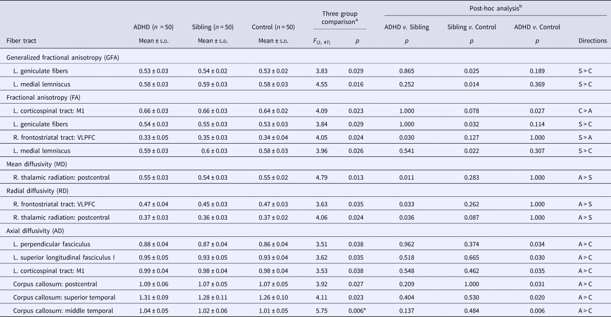
In the comparisons of microstructural property among the three groups, the calculated DSI indices from the white matter tracts with significant group differences in each index are presented in Table 2 and Fig. 2. ADHD probands had the highest AD values, while controls had the lowest AD values in the left perpendicular fasciculus, left SLF I, left corticospinal tract (connecting primary motor cortex, trunk component, M1) and CC connecting postcentral gyrus, superior temporal gyrus (STG), and middle temporal gyrus (MTG). Unaffected siblings showed intermediate AD values between ADHD probands and controls in these tracts as well as intermediate FA value of the left corticospinal tract. On the other hand, some alterations of DSI indices in unaffected siblings were opposite to what we found in the ADHD probands. Unaffected siblings showed increased FA and the decreased RD values of the right frontostriatal tract (connecting ventrolateral prefrontal cortex part, FS-VLPFC) and the decreased MD and RD values of the right thalamic radiation connecting postcentral gyrus (Table 2). Significant group difference remained in AD value of CC connecting MTG (q = 0.03) when using false discovery rate (FDR, q) to correct for multiple comparisons in the 5 indices of each white matter tract (online Supplementary Table S1).
Comparisons of the indices of white matter tract property among ADHD probands, their unaffected siblings and the controls

A, patients with ADHD; S, unaffected siblings; C, R, right; L, left; the controls; M1, the primary motor cortex; VLPFC, ventrolateral prefrontal cortex.
a Controlling for age, gender, full-scale IQ.
b Pairwise comparison by post-hoc analysis with Bonferroni test.
*Significant group difference remained (q = 0.03) when using false discovery rate (FDR, q) to correct for multiple comparisons in 5 indices of each white matter tract.
Reconstruction of the tracts and the connected regions. (a) Green: the superior longitudinal fasciculus; Purple: the corticospinal tract; Orange: perpendicular fiber. (b) Yellow: the corpus callosum connecting the postcentral gyrus; Purple: the corpus callosum connecting the superior temporal gyrus; Dark blue: the corpus callosum connecting the middle temporal gyrus. (c) Green: the right thalamic radiation connecting the postcentral gyrus; Light blue: the frontostriatal tract connecting the ventrolateral prefrontal cortex. L, left; R, right, M1, the primary motor cortex.

Correlations of fiber tract property with clinical and neuropsychological manifestations
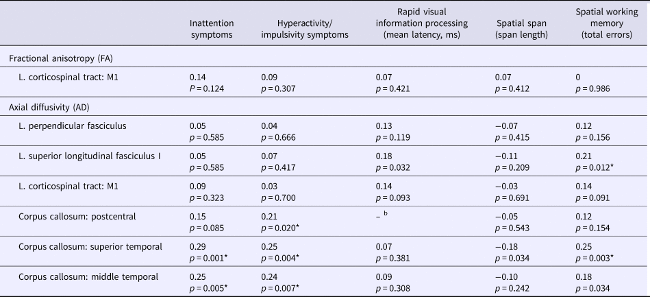
To elucidate the clinical and functional implications of tracts with endophenotype characteristics, we correlated the microstructural properties of their diffusion indices with ADHD symptoms and neuropsychological performance (Table 3). In the tracts without group interactions, the indices for all subjects were analyzed including all three groups. To correct for the number of clinical symptoms and neuropsychological measurements, a conservative adjustment of false discovery rate (FDR, q) was also provided to determine the significance of correlation analysis at q < 0.05. We found that higher AD values in the CC connecting the STG and MTG were significantly correlated with greater inattention (r = 0.29, q = 0.008 and r = 0.25, q = 0.017, respectively) as well as hyperactivity/impulsivity symptoms (r = 0.25, q = 0.024 and r = 0.24, q = 0.048, respectively). Higher AD values in the CC connecting the postcentral gyrus were only associated with higher hyperactivity/impulsivity (r = 0.21, q = 0.024). Higher AD values in the SLF I and the CC connecting the STG were associated with worse spatial working memory (r = 0.21, q = 0.018, and r = 0.25, q = 0.041, respectively).
Correlation analysis between white matter property and clinical and neuropsychological manifestations in the whole samplea

a Adjusted for age, gender and full-scale IQ.
b Here is a significant interaction among groups, so these three groups were computed separately (The results are shown in Fig. 2).
*The significance of correlation analysis remained when a false discovery rate (FDR, q) correction was set at q < 0.05.
There were significant group differences in the correlations between the diffusing indices of the CC connecting the postcentral gyrus of two hemispheres and the RVP performance across the three groups, so separate analyses for these three groups were conducted. Higher AD value in the tract of CC connecting the postcentral gyrus correlating with longer response latency in RVP assessing sustained attention was only observed in the ADHD proband group (r = 0.49, p < 0.001), but not in the unaffected sibling group (r = 0.18, p = 0.206) nor in the control group (r = 0.08, p = 0.556) (Fig. 3a). The group comparisons of these correlation coefficients using Fisher's r-to-z transformation test showed that the ADHD proband group was significantly different from the control group (z = 2.21, p = 0.027), but there was significant group difference neither between the ADHD proband group and the unaffected siblings group, nor between the unaffected sibling group and the control group.
Correlation analysis between white matter property and neuropsychological functions. *Significant difference in the correlations between the two groups.

In the right FS-VLPFC, where unaffected siblings showed highest FA and the lowest RD values among three groups, there were significant group differences in the correlations between the diffusing indices and performance of spatial span. Only in the sibling group, there were significant correlations between higher FA and lower RD values of the right FS-VLPFC and better performance of spatial span (r = 0.377, p = 0.007, Fig. 3b; r = −0.58, p < 0.001, Fig. 3c, respectively). When comparing FA values, we observed the correlations among unaffected siblings were significantly different from those among the controls (z = 2.31, p = 0.033, Fig. 3b). For the RD values of the right FS-VLPFC and spatial span, the correlation coefficients in sibling group were significantly different from those in the ADHD (z = 2.13, p = 0.033) and control (z = 2.62, p = 0.008) groups (Fig. 3c).
Discussion
This study used tract-based structural connectivity measures to identify neuroimaging endophenotypes of ADHD in a sample of ADHD probands, their unaffected siblings and control participants, and to explore their correlations with clinical and neuropsychological measures. The values of the AD in the perpendicular fasciculus, SLF I, corticospinal tract and CC are potentially important indices to be endophenotype candidates of ADHD. That is, higher AD values in ADHD probands than controls, and unaffected siblings lied intermediate between these two groups. Regarding the neural features associated with behavioral-cognitive phenotypes across three groups, the higher AD values tended to correspond to higher ADHD symptoms and worse neuropsychological performance. Significant associations with ADHD symptoms were observed in the CC connecting the postcentral gyrus, STG and MTG, and spatial working memory in the SLF I and the CC connecting the STG. In addition, the CC connecting the postcentral gyrus was associated with sustained attention only found in ADHD probands.
As an exploratory study including all major tracts rarely studied in the past, we present our analyses with and without correction for multiple comparisons in Tables 2 and 3, because we took these findings as hypothesis generating for future studies, rather than as confirmatory (Streiner and Norman, Reference Streiner and Norman2011). The correlations between the tract property and behavioral-cognitive phenotypes provided further support for the clinical significance for some of the tracts that we characterized, rather than randomly false positive findings.
The pathophysiological inference of the diffusion indices in white matter tracts of ADHD is still inconclusive duo to discrepant findings in the literature (Tamm et al., Reference Tamm, Barnea-Goraly and Reiss2012; van Ewijk et al., Reference van Ewijk, Heslenfeld, Zwiers, Faraone, Luman, Hartman, Hoekstra, Franke, Buitelaar and Oosterlaan2014). The diffusion anisotropy (e.g. GFA and FA) in white matter is likely to be influenced by a number of factors, including the degree of myelination, the density, diameter distribution, orientational coherence of axons, and potentially the diffusion barriers presented by glia, so it is not very specific to the type of changes (Alexander et al., Reference Alexander, Lee, Lazar and Field2007). As the AD represents the water diffusivity parallel to axonal fiber tracts, it provides more specific information on the property of axons, and dysmyelination alone has minimal impact on AD values (Song et al., Reference Song, Sun, Ramsbottom, Chang, Russell and Cross2002). Consistent with the previously reported higher AD values in ADHD probands, compared to the control group (Tamm et al., Reference Tamm, Barnea-Goraly and Reiss2012), our findings supported the AD index is important in evaluating the neuropathology of ADHD.
Abundant literature regarding the difference in white matter microstructural integrity between ADHD probands and controls in the SLF and the association between SLF tract property and working memory in children with ADHD support its relevance of ADHD neuropathology (Lawrence et al., Reference Lawrence, Levitt, Loo, Ly, Yee, O'Neill, Alger and Narr2013; van Ewijk et al., Reference van Ewijk, Heslenfeld, Zwiers, Faraone, Luman, Hartman, Hoekstra, Franke, Buitelaar and Oosterlaan2014; Chiang et al., Reference Chiang, Chen, Lo, Tseng and Gau2015). Our findings in unaffected siblings are also consistent with high heritability of the SLF in families enriched for ADHD (Sudre et al., Reference Sudre, Choudhuri, Szekely, Bonner, Goduni, Sharp and Shaw2017). However, the SLF was treated as a single large bundle in the majority of previous tractography studies (Lawrence et al., Reference Lawrence, Levitt, Loo, Ly, Yee, O'Neill, Alger and Narr2013; van Ewijk et al., Reference van Ewijk, Heslenfeld, Zwiers, Faraone, Luman, Hartman, Hoekstra, Franke, Buitelaar and Oosterlaan2014; Chiang et al., Reference Chiang, Chen, Lo, Tseng and Gau2015). Recent advances in DSI tractography enabled us to divide SLF into the SLF I, II and III (Chiang et al., Reference Chiang, Chen, Lin, Tseng and Gau2017). Specifically, a meta-analysis of functional MRI studies in healthy adolescents or adults concludes that the SLF I connects regions of the superior frontal lobe and superior parietal lobule, where its cortical projections involved in spatial working memory (Parlatini et al., Reference Parlatini, Radua, Dell'Acqua, Leslie, Simmons, Murphy, Catani and Thiebaut de Schotten2017). Although the association between the microstructure of the SLF and spatial working memory has been reported in previous studies in children with ADHD (Chiang et al., Reference Chiang, Chen, Shang, Tseng and Gau2016), the current study further demonstrated the specific role of the SLF I in ADHD that the microstructural property of the SLF I was correlated with spatial working memory across ADHD probands, unaffected siblings and typically developing controls.
The CC provides communication between bilateral hemispheres to integrate visuomotor and cognitive processes. Its alteration of white matter microstructural integrity in ADHD probands (van Ewijk et al., Reference van Ewijk, Heslenfeld, Zwiers, Faraone, Luman, Hartman, Hoekstra, Franke, Buitelaar and Oosterlaan2014; Aoki et al., Reference Aoki, Cortese and Castellanos2018) and its genetic heritability in ADHD families (Sudre et al., Reference Sudre, Choudhuri, Szekely, Bonner, Goduni, Sharp and Shaw2017) reported in previous studies are consistent with our findings that the microstructure of the CC was related to familial risks of ADHD. The primary somatosensory cortex, located in the postcentral gyrus, is responsible for performing multi-sensory integration processes. It is important because conflicting concurrent auditory and visual stimuli strongly impedes response inhibition performance (Chmielewski et al., Reference Chmielewski, Muckschel, Dippel and Beste2016). Consistent with this hypothesized mechanism, our finding that the microstructural property of the CC connecting bilateral postcentral gyrus was correlated with hyperactive/impulsive symptoms. A population-based study in children demonstrating correlations between thinner cortex in the right and left postcentral gyrus and more inattentive and hyperactive symptoms also provide collateral supports to our findings (Mous et al., Reference Mous, Muetzel, El Marroun, Polderman, van der Lugt, Jaddoe, Hofman, Verhulst, Tiemeier, Posthuma and White2014).
As to the STG, consisting of the primary auditory cortex and Wernicke's area, is important for auditory perception, language processing, and social perception. Children with ADHD showed delays in cortical maturation in bilateral STG (Shaw et al., Reference Shaw, Eckstrand, Sharp, Blumenthal, Lerch, Greenstein, Clasen, Evans, Giedd and Rapoport2007), in which significant activation deficits were also noted during the attention task (Orinstein and Stevens, Reference Orinstein and Stevens2014). On the other hand, the MTG is a functional nexus of the default-mode network (DMN) and executive networks (Davey et al., Reference Davey, Thompson, Hallam, Karapanagiotidis, Murphy, De Caso, Krieger-Redwood, Bernhardt, Smallwood and Jefferies2016). Failed suppression of the DMN during the task (Barber et al., Reference Barber, Jacobson, Wexler, Nebel, Caffo, Pekar and Mostofsky2015) and stronger connectivity between the executive networks and the DMN was associated with increased ADHD symptoms (van Rooij et al., Reference van Rooij, Hartman, Mennes, Oosterlaan, Franke, Rommelse, Heslenfeld, Faraone, Buitelaar and Hoekstra2015a). Moreover, an association has been reported between the lower cortical volume of the temporal lobe and worse working memory performance in families enriched for ADHD (Muster et al., Reference Muster, Choudhury, Sharp, Kasparek, Sudre and Shaw2019) and in a sample of adults with ADHD, unaffected siblings and controls (Duan et al., Reference Duan, Chen, Calhoun, Lin, Jiang, Franke, Buitelaar, Hoogman, Arias-Vasquez, Turner and Liu2018). Consistent with the previously reported association between microstructural property of the CC and inattention symptoms across children with ADHD, children with ASD and typically developing controls (Aoki et al., Reference Aoki, Yoncheva, Chen, Nath, Sharp, Lazar, Velasco, Milham and Di Martino2017), our findings further provide evidence to support that specific parts of the CC connecting the STG and MTG correlate with ADHD symptoms and spatial working memory.
The corticospinal tract plays important roles in controlling complex voluntary movements, and the M1 region activates in higher cognitive tasks, such as motor imagery and working memory (Tomasino and Gremese, Reference Tomasino and Gremese2016). The microstructural property of the left corticospinal tract has also been reported to be related to developmental improvement or persistence of hyperactive-impulsive symptoms in an adolescent cohort of ADHD (Francx et al., Reference Francx, Zwiers, Mennes, Oosterlaan, Heslenfeld, Hoekstra, Hartman, Franke, Faraone, O'Dwyer and Buitelaar2015). Our results did not provide evidence to support such correlations cross-sectionally. Our ongoing follow-up study will test the developmental perspective of such relationships and explore more clinical relevance of this tract in our future studies.
In the right FS-VLPFC and the right thalamic radiation connecting the postcentral gyrus, the direction of altered index values in unaffected siblings was opposite to that in the ADHD probands. Together with the observation that the higher FA and the lower RD values in the right FS-VLPFC were associated with better performance in spatial span, the right FS-VLPFC might be explained as protective factors against expressing the behavioral phenotype of ADHD or compensatory mechanisms for abnormalities in other brain systems in unaffected siblings carrying a familial risk for ADHD (Fassbender and Schweitzer, Reference Fassbender and Schweitzer2006; Chiang et al., Reference Chiang, Chen, Shang, Tseng and Gau2016).
Methodological limitations
There are some limitations to our study. First, we presented the original statistical results without correction for multiple comparisons to avoid over-correction (Veazie, Reference Veazie2006). Therefore the findings need further validation. Second, the sex distribution was not balanced in our three groups derived from the sibling group, although the effect of sex on white matter property is not well-established (Waddell and McCarthy, Reference Waddell and McCarthy2012). Lastly, the distinct patterns of white matter microstructure in unaffected siblings found in the current cross-sectional study warranted longitudinal follow-up studies to address the protective or compensatory hypotheses.
In conclusion, this study provides preliminary evidence that the AD values of the SLF I, CC and CST account for the underlying familial risks of ADHD and also the ADHD symptoms and/or neuropsychological functions. The finding that higher FA and the lower RD values in the right FS-VLPFC, the better performance in spatial short-term memory assessed by SSP in unaffected siblings suggests the potential role of FS-VLPFS in preventing the expression of the ADHD-related phenotype.
Author ORCIDs
Susan Shur-Fen Gau, 0000-0002-2718-8221
Supplementary material
The supplementary material for this article can be found at https://doi.org/10.1017/S0033291719001120
Financial support
All the authors report no financial relationships with commercial interests. This work was supported by the Ministry of Science and Technology (NSC101-2321-B-002-079; MOST103-2314-B-002-021-MY3), National Taiwan University Hospital (NTUH101-S1910), and National Health Research Institute (NHRI-EX107-10404PI), Taiwan.