Introduction
Recently, in vitro maturation (IVM) of canine oocytes has been performed with the aim to enhance the potential of nuclear maturation. The efficiency of IVM is still very low, however, in comparison with that of many other mammals (Bolamba et al., Reference Bolamba, Borden-Russ and Durrant1998, Reference Bolamba, Russ, Olson, Sandler and Durrant2002; Hewitt and England Reference Hewitt and England1999; Nickson et al., Reference Nickson, Boyd, Eckersall, Ferguson, Harvey and Renton2000; Saint-Dizier et al., Reference Saint-Dizier, Salomon, Pettit, Renard and Chastant-Maillard2001). This finding is thought to result from the unique canine reproductive biology. Unlike other mammals, canine oocytes are ovulated in the germinal vesicle stage and undergo maturation in the distal part of the oviduct, 2–3 days after ovulation (Pearson & Enders Reference Pearson and Enders1943; Holst & Phemister Reference Holst and Phemister1971; Phemister et al., Reference Phemister, Holst, Spano and Hopwood1973; Tsuisui et al., Reference Tsuisui, Shimada, Nishi, Kubo, Murao, Shimizu and Ogasa1989). Hence, several researchers have tried to increase the efficiency of IVM. However, the efficiency of in vitro nuclear maturation of canine oocytes is still very low compared with that of other mammals (Cinone et al., Reference Cinone, Ghneim, Caira, Dell'Aquila and Minoia1992; Hewitt & England, Reference Hewitt and England1998a–Reference Hewitt and Englandc, Reference Hewitt and England1999; Hewitt et al., Reference Hewitt, Watson and England1998; Otoi et al., Reference Otoi, Fujii, Tanaka, Ooka and Suzuki1999; Farstad Reference Farstad2000). IVM is a very important and basic tool for reproductive technology using canine oocytes. Therefore, researchers have tried to increase the efficiency of nuclear maturation of canine oocytes.
Although the efficiency of IVM is very low, a number of researchers have investigated in vitro fertilization (IVF) (Yamada et al., Reference Yamada, Shimazu, Awaji, Nakazawa, Naito and Toyoda1992; Theiss, Reference Theiss1997; Otoi et al., Reference Otoi, Muramaki, Fujii, Tanaka, Ooka, Suzuki and Une2000, Reference Otoi, Shin, Kraemer and Westhusin2004; England et al., Reference England, Verstegen and Hewitt2001; Rodrigues et al., Reference Rodrigues, dos Santos and Rodrigues2004; De los Reyes et al., Reference De los Reyes, Carrion and Barros2006). Several investigators have reported that embryos processed from IVF in canines were cleaved and developed, although the rate was low (Yamada et al., Reference Yamada, Shimazu, Awaji, Nakazawa, Naito and Toyoda1992; Otoi et al., Reference Otoi, Muramaki, Fujii, Tanaka, Ooka, Suzuki and Une2000, Reference Otoi, Shin, Kraemer and Westhusin2004; England et al., Reference England, Verstegen and Hewitt2001; Rodrigues et al., Reference Rodrigues, dos Santos and Rodrigues2004). Otoi et al., (Reference Otoi, Muramaki, Fujii, Tanaka, Ooka, Suzuki and Une2000)) produced a single blastocyst using a co-culture system with a bovine cumulus cell. Another study reported one pregnancy derived from IVF (England et al., Reference England, Verstegen and Hewitt2001). These experiments were performed under various conditions, for example, various estrus phases and various interval times of culturing (24–96 h). In these studies, the relationship of developmental efficiency following IVF with reproduction cycle of the bitches and other culture conditions remains unclear. The efficiency of IVF in canine oocytes investigated in these experiments was also very low, probably due to the low rate of IVM. So, the optimal protocol for IVF has not yet been established. Therefore, in order to determine the optimal protocol, we tried to develop a more effective method to increase the efficiency of IVF in canines. To increase the efficiency of fertilization in vitro, we used heparin as a factor for insemination stimulation in IVF in canines. Heparin binds to bovine sperm (Parrish et al., Reference Parrish, Susko-Parrish, Winer and First1988) and induces changes in the intracellular environment, including Ca2+ uptake, increases in intracellular free calcium and intracellular pH (Handrow et al., Reference Handrow, First and Parrish1989), increases in protein phosphorylation (Galantino-Homer et al., Reference Galantino-Homer, Viscconi and Kopf1997; Cary et al., Reference Cary, Madill, Farnsworth, Hayna, Duoos and Fahning2004; Li et al., Reference Li, Seidel and Squires2004) and induces acrosome reaction (Miller & Hunter Reference Miller and Hunter1986). Due to these heparin functions, we hypothesized that heparin could stimulate the efficiency of IVF in canine oocytes.
We investigated whether canine oocytes together with sperm in a fertilization medium containing heparin, which has been used as insemination factor in other mammals, progressed to fertilization, cleavage and subsequent development. The object of the present study was to investigate: (1) if the efficiency of IVM in canine oocytes increased with the addition of fetal bovine serum (FBS), to obtain the optimal conditions for IVF; and (2) if the efficiency of IVF and developmental ability of canine oocytes was increased by the addition of a capacitation factor, heparin, along with insemination time.
Materials and Methods
Effect of fetal bovine serum on in vitro maturation
Collection of oocytes
Reproductive tracts obtained after sacrificing bitches, aged more than 6 months, at a private abattoir, were kept in physiological saline solution (PSS) composed of 0.9% NaCl, 0.06 mg/ml potassium penicillin G and 0.1 mg/ml streptomycin sulphate maintained at 38.5°C and were transported to the laboratory within 3 h. Ovaries removed from the ovarian tracts were washed three times in fresh PSS and maintained at 38.5°C. Cumulus–oocyte complexes (COCs) were aspirated from the ovaries with a 10 ml syringe containing a washing medium, consisting of TCM199 (Invitrogen) supplemented with 25 mM HEPES, 0.1% polyvinyl alcohol (PVA), 0.1 mM glutamine, 2.5 mM sodium pyruvate, 0.06 mg/ml streptomycin sulphate and 0.06 mg/ml potassium penicillin G adjusted to 38.5°C. The COCs were washed twice in the washing medium and examined under a stereoscopic microscope (SZ51, Olympus). COCs were allocated into three groups according to the standard reported by Hewitt et al. (Reference Hewitt, Watson and England1998). Grade 1 COCs were darkly pigmented and surrounded by one or more layers of cumulus cells. Grade 2 COCs were lightly pigmented with incomplete layers of cumulus cells. Grade 3 COCs were disrupted or broken oocytes. Among these COCs, only grade 1 oocytes were washed three times in the washing medium prior to use in experiments. All chemical reagents were obtained from the Sigma–Aldrich Corporation, unless otherwise indicated.
In vitro maturation
Grade 1 COCs (n = 123) were cultured randomly in the following two groups: Group 1 cells (n = 64) were cultured in IVM medium I consisting of TCM199 (Invitrogen) supplemented with 0.1% PVA, 0.1 mM glutamine, 2.5 mM sodium pyruvate, 0.06 mg/ml streptomycin sulphate and 0.06 mg/ml potassium penicillin G; Group 2 cells (n = 59) were cultured in IVM medium II consisting of IVM medium supplemented with 5% fetal bovine serum (FBS). COCs were cultured in 10 µl droplets of each medium (up to 20 oocytes per drop) under mineral oil at 38.5°C and 5% CO2 with maximum humidity for 48 h.
Assessment of nuclear stage of maturation
After 48 h of IVM, all oocytes in both groups were denuded of cumulus cells with TCM199 supplemented with 0.1% hyaluronidase for 5 min. These oocytes were washed with a phosphate-buffered solution (PBS) consisting of 8 mg/ml sodium chloride, 0.2 mg/ml potassium chloride, 1.44 mg/ml Na2PO4 and 0.24 mg/ml KH2PO4. After washing, denuded oocytes were preserved in a fixation solution consisting of 2% formalin, 0.1% polyvinyl pyrrolidone (PVP) and 0.25% glutaraldehyde for 20 min. After fixing, oocytes were stained with 1.9 µM Hoechst 33342 medium in glycerol and observed using a fluorescence microscope (BX50, Olympus) fitted with a digital camera (Coolpix4500, Nikon). Nuclear morphology in the oocytes was classified as germinal vesicle (GV), germinal vesicle breakdown (GVBD), metaphase I (MI), metaphase II (MII) and degenerated.
Effect of heparin on in vitro fertilization
Collection of oocytes
In this experiment, 1676 COCs were collected from sacrificed bitches aged more than 6 months. Canine oocytes used in this experiment were collected using the same method as used in the previous experiment. Of these oocytes, 940 Grade 1 COCs were used for IVM/IVF. In this experiment, the collected COCs were matured in TCM199 without FBS (IVM medium I). COCs were cultured in 100 µl droplets of each medium (up to 20 oocytes per drop) under mineral oil at 38.5°C and 5% CO2 with maximum humidity for 48 h. These COCs were distributed into the following four categories: Group A cells (n = 235) were not treated with heparin during IVF; Group B cells (n = 220) were treated with 10 µg/ml heparin during IVF; Group C cells (C = 217) were treated with 20 µg/ml heparin during IVF; Group D cells (n = 268) were treated with µg/ml heparin during IVF.
Sperm preparation
Reproductive tracts containing testes and epididymides were removed from adult male dogs sacrificed at a private abattoir. The individual reproductive tracts were stored in sterile steel containers with tight sealing lids and maintained at 38.5°C. Epididymal spermatozoa were collected with the modified method of epididymodeferentectomy used by Cary et al. (Reference Cary, Madill, Farnsworth, Hayna, Duoos and Fahning2004). The cauda epididymis and vas deferens were isolated from each testicle, washed with PBS solution and sterilized with 70% ethanol. After catheterization of the lumen of the vas deferens with an 18 gauge syringe, retrograde flushing of the vas deferens and cauda epididymis with 5 ml of the washing medium maintained at 38.5°C was performed. The flushed suspensions were collected in warmed sterile 50 ml conical tubes (Nunc) at 38.5°C. The collected sperm of each dog was centrifuged at 120 g for 5 min. The supernatant was discarded and the sperm pellet was resuspended with 10 ml of the washing medium. Suspended sperms were incubated to swim-up for 30 min at 38.5°C and 5% CO2 with maximum humidity. The supernatant (swim-up layer) was carefully collected and centrifuged at 120 g for 5 min. The supernatant was discarded and the pellet was resuspended with 500 µl of the washing medium. Sperm collected from dogs were evaluated for motility and viability using a microscope. The percentage of total motile sperm (number of sperm with progressive motility divided by the total number of sperm observed) was assessed in 5 µl drops of sperm suspension in a Makler counting chamber (Self-medical Instruments), using an inverted biological microscope (CKX31, Olympus). Only sperm with a motility of more than 90% were used for IVF. After assessment for motility, the sperm concentration was adjusted to 1 × 10⁷ sperm/ml with fertilization medium I (Fert I). Fert I consisted of 114 mM sodium chloride, 3.2 mM potassium chloride, 25 mM NaHCO3, 0.3 mM NaH2PO4, 10 mM lactate sodium salt, 2 mM CaCl2, 0.5 mM MgCl3, 0.02 mM sodium pyruvate, 5 mM glucose, 0.3 mM bovine serum albumin (BSA), 2 mM potassium penicillin G, 0.001 mg/ml phenol red and the modified Fert-TALP used by Parrish et al. (Reference Parrish, Susko-Parrish, Winer and First1988).
In vitro fertilization
The oocytes for IVM were processed for 48 h, randomly allocated to four groups and cultured under different culture conditions. Groups A–D were fertilized in fertilization medium I (Fert I, n = 235), fertilization medium II (Fert II, n = 220), fertilization medium III (Fert III, n = 217) and fertilization medium IV (Fert IV, n = 268) respectively. Fert II consisted of Fert I supplemented with 10 µg/ml heparin. Fert III or Fert IV consisted of Fert I supplemented with 20 or 30 µg/ml heparin. Each group was inseminated in each medium for 12, 18 or 24 h, respectively. Firstly, in vitro matured oocytes were added to each 50 µl droplet of fertilization medium. These oocytes were added at the rate of 20 oocytes per droplet and prepared sperm (50 µl) were added to each droplet to maintain 5 × 10⁶/ml as final concentration. Oocytes and sperm were co-incubated at 38.5°C and 5% CO2 with maximum humidity for 12, 18 or 24 h.
In vitro culture
For in vitro culture (IVC), an IVC medium consisting of IVM medium supplemented with 5% FBS was used. IVC was maintained at 38.5°C and 5% CO2 with maximum humidity. At 48 h of IVC, cumulus cells were removed from COCs. The IVC medium was changed to a fresh medium and cultured for additional 96 h.
As well as embryo cleavage, 2-, 4-, 8-, 16-cell, morula and blastocyst were assessed. Cell morphology was observed using a fluorescence microscope (BX50, Olympus) fitted with a digital camera (Coolpix4500, Nikon).
Statistical analysis
All experiments on the effects of FBS, insemination and heparin treatment, three replicates of each, were evaluated by chi-squared analysis.
Results
Effect of fetal bovine serum on in vitro maturation
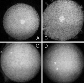
COCs cultured in either IVM medium I or II were fixed and stained. As shown in Table 1, the percentage of oocytes that underwent maturation to the MII stage in the two culture media was very low. The rate of oocytes developing to the MII stage was higher in IVM medium I (4.7%) compared with in IVM medium II (1.7%). Additionally, the rates of meiotic resumption in oocytes were higher in IVM medium I (62.5%) than in IVM medium II (33.9%). Especially, the rate of unidentified or degenerated oocytes was significantly higher in IVM medium II (42.4%) compared with IVM medium I (14.1%). Oocytes observed after IVM are shown in Fig. 1.
Effect of FBS for meiotic rates of canine oocytes

GV, germinal vesicle; GVBD, germinal vesicle breakdown; MI, metaphase I; MII, metaphase II.
a,bValues with different superscripts in the same column differ significantly (p < 0.05).
Nuclear morphology in canine oocytes. Germinal vesicle (A), germinal vesicle breakdown (B), metaphase I (C), metaphase II (D).

Effect of heparin on in vitro fertilization
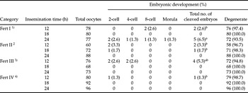
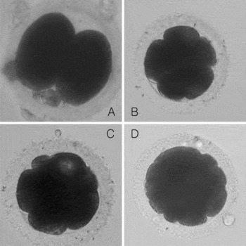
The rate of embryonic development after IVF/IVC in all groups is summarized in Table 2. The embryonic development rate after IVF/IVC was detected at 144 h. The cleavage rates were very low under all culture conditions. The overall cleavage rate of oocytes cultured in Fert I was the highest of all groups. However, each group was not significantly different. Of note, embryonic development of canine oocytes cultured in Fert I for 24 h insemination (6.5%, p < 0.05) had the highest rate of all of the culture conditions used. Canine embryos cultured in Fert I for 24 h were observed as various morphologies for each developmental stage of cleavage.
Effect of heparin on embryo development of canine oocytes

1Fert I, modified Fert-TALP from Parrish et al. (Reference Parrish, Susko-Parrish, Winer and First1988) without heparin.
2Fert III, modified Fert-TALP from Parrish et al. (Reference Parrish, Susko-Parrish, Winer and First1988) with 10 µg/ml heparin.
3Fert III, modified Fert-TALP from Parrish et al. (Reference Parrish, Susko-Parrish, Winer and First1988) with 20 µg/ml heparin.
4Fert III, modified Fert-TALP from Parrish et al. (Reference Parrish, Susko-Parrish, Winer and First1988) with 30 µg/ml heparin.
a, bValues with different superscripts in the same column differ significantly (p < 0.05).
Only one embryo in the morula stage was detected when oocytes were cultured for 24 h, as shown in Fert I (Fig. 2). Canine embryos cultured in Fert III for 12 h insemination time progressed to the early embryonic development stage (2-cell, 4-cell), although the rate of embryonic development is relatively higher (5.3%). Additionally, in our study, the rates of embryonic cleavage in canine oocytes were decreased in the heparin-treated groups in comparison with the heparin-free group. As summarized in Table 2, the rates of embryonic cleavage in the heparin-free group fertilized for 24 h (6.5%) and the 20 µg/ml heparin treatment group fertilized for 12 h (5.3%) were high when compared with any other culture conditions.
Canine embryos resulting from in vitro fertilization of oocytes matured in vitro. 2-cell (A), 4-cell (B), 8-cell (C), early morula (D).

Discussion
Previous studies investigated the efficiency of IVM of canine oocytes under various conditions, such as a culture time of 48–72 h, serum supplementation and hormone addition (Cui et al., Reference Cui, Jin, Shen, Lee, Lee, Yin, Kong and Kim2006; De los Reyes et al., Reference De los Reyes, de Lange, Miranda, Palominos and Barros2005; Kim et al., Reference Kim, Fibrianto, Oh, Jang, Kim, Lee, Kang, Lee and Hwang2004, Reference Kim, Lee, Hyun, Nam, Lee, Jeong, Kim, Kim, Kang, Lee and Hwang2005; Rodrigues et al., Reference Rodrigues, dos Santos and Rodrigues2006; Vannucchi et al., Reference Vannucchi, de Oliveira, Marques, Assumcao and Visintin2006) found that the rate of IVM of canine oocytes was still very low.
In the present experiment, IVM medium II consisting of TCM199 supplemented with 5% FBS did not significantly influence the nuclear maturation of canine oocytes. In a previous study, exposure of COCs to culture medium supplemented with FBS (Bolamba et al., Reference Bolamba, Russ, Olson, Sandler and Durrant2002) or FCS (Fujii et al., Reference Fujii, Otoi, Murakami, Tanaka, Une and Suzuki2000; Saint-Dizier et al., Reference Saint-Dizier, Reynaud and Chastant-Maillard2004) exerted a stimulating effect on oocyte maturation, with an increase in the number of oocytes reaching MII stage. The present study found that IVM medium supplemented with FBS decreased the rates of IVM compared with IVM medium without FBS. This result is similar to previous reports that showed that FBS negatively affected both nuclear and cytoplasmic maturation (Funahashi & Day Reference Funahashi and Day1993; Wood et al., Reference Wood, Montali and Wildt1997; Ali & Sirard Reference Ali and Sirard2002). We propose, therefore, that FBS does not have to be added for IVM of canine oocytes. However, the role of FBS for canine IVM remains unclear because of differences in research design by many investigators. It is hypothesized that the low MII efficiency and degeneration percentages in canine oocytes are caused by inadequate culture conditions or culture medium, along with other variables. Until now, there have been few investigations using canine embryos generated by IVF/IVC after IVM. This fact may be due to the low rate of IVM of canine oocytes (Yamada et al., Reference Yamada, Shimazu, Awaji, Nakazawa, Naito and Toyoda1992; Otoi et al., Reference Otoi, Muramaki, Fujii, Tanaka, Ooka, Suzuki and Une2000, Reference Otoi, Shin, Kraemer and Westhusin2004; England et al., Reference England, Verstegen and Hewitt2001; De los Reyes et al., Reference De los Reyes, Carrion and Barros2006). England et al. (Reference England, Verstegen and Hewitt2001) obtained a single pregnancy after transplanting two embryos (2-cell stage) acquired from IVF following the IVM of oocytes for 24–72 h. In their experiment, a very small number of embryos was obtained. Until now, the only blastocyst after IVF following IVM was achieved by Otoi et al. (Reference Otoi, Muramaki, Fujii, Tanaka, Ooka, Suzuki and Une2000), who accomplished IVF after canine oocytes for IVM were cultured on a feeder layer of bovine cumulus cells. This experiment showed first that canine oocytes could develop to the blastocyst stage following IVM, IVF and IVC.
Recently, several researchers have used heparin as a stimulator to increase the efficiency of IVF in mammals, such as for bovine, porcine and goat (Gliedt et al., Reference Gliedt, Rosenkrans, Rorie and Rakes1996; Kano et al., Reference Kano, Miyano and Kato1998; Katska-Ksiazkiewicz et al., Reference Katska-Ksiazkiewicz, Rynska, Gajda and Smorag2004; Lie et al., 2004; Kim et al., Reference Kim, Lee, Hyun, Nam, Lee, Jeong, Kim, Kim, Kang, Lee and Hwang2005). Kano et al. (Reference Kano, Miyano and Kato1998) verified that heparin does not increase development efficiency after IVF in porcine. Katska-Ksiazkiewicz Reference Katska-Ksiazkiewicz, Rynska, Gajda and Smoraget al. (2004) did not prove an effect of heparin for capacitation activity on fresh caprine sperm. However, Kim Reference Kim, Lee, Hyun, Nam, Lee, Jeong, Kim, Kim, Kang, Lee and Hwanget al. (2005) insisted that heparin supports preimplantation of porcine embryos. Gliedt et al. (Reference Gliedt, Rosenkrans, Rorie and Rakes1996) observed that heparin increased the cleavage rate, but only numerically improved subsequent development to the morula or blastocyst stage. Li et al. (Reference Li, Seidel and Squires2004) contended that heparin not only plays an important role in sperm capacitation and the acrosome reaction, but also improves pronuclear formation and early embryonic cleavage in bovines. As previously mentioned, although many researchers have investigated the effect of heparin in IVF in various mammals, how heparin acts in the IVF of canines is not fully understood. Therefore, this study investigated whether heparin plays a role as a stimulator of embryonic development in canines. Embryonic development in the heparin-free condition for 24 h reached the morula stage, indicating that embryonic fertilization, cleavage and development in canines can be accomplished in heparin-free culture conditions. In addition, differences between groups along with various concentrations were not revealed. From our result, to our knowledge, heparin does not need to be present in IVF of canine oocytes. This result is similar to the assertion that heparin did not stimulate fertilization (Tanghe et al., Reference Tanghe, Van Soom, Duchateau, Nauwynck and de Kruif2004), which is in agreement with observations made by Miller & Hunter (Reference Miller and Hunter1986).
The present study shows that viable canine oocytes obtained from bitches in various phases of the estrus cycles were cultured in TCM199 medium without FBS for IVM. Although the rates of nuclear maturation in canine were very low, we tried to achieve embryonic development. One embryo in the morula stage was obtained following IVM, IVF and IVC. An embryo in the morula stage was obtained in heparin-free culture condition. Therefore, it is concluded that embryonic development from IVF after IVM in canine oocytes can be induced from IVF medium without heparin.
Our experiments, for the first time, used abandoned oocytes obtained after a routine sacrifice of various bitches. Therefore, we propose that our results are applicable to the IVM and IVF of wild dog. Our results may be an especially important tool to restore endangered and distinct canidae, such as the red wolf and the bush dog.
Additionally, we, for the first time, investigated the function of heparin in the IVF of canine oocytes, although the efficiency of embryonic development by heparin is similar to that of heparin-free condition. Embryonic development in canine oocytes derived from IVF is likely to be arrested at the 8- to 16-cell stage. Although canine oocytes were cultured in various culture conditions (Yamada et al., Reference Yamada, Shimazu, Awaji, Nakazawa, Naito and Toyoda1992; Theiss Reference Theiss1997; Otoi et al., Reference Otoi, Muramaki, Fujii, Tanaka, Ooka, Suzuki and Une2000, Reference Otoi, Shin, Kraemer and Westhusin2004; England et al., Reference England, Verstegen and Hewitt2001; Rodrigues et al., Reference Rodrigues, dos Santos and Rodrigues2004; De los Reyes et al., Reference De los Reyes, Carrion and Barros2006) for insemination in vitro, these oocytes failed to develop into morulae and blastocysts. Therefore, it is hypothesized that the embryonic development in canine oocytes has a developmental block that we call the 8-cell block. A similar developmental block exists in other mammals, such as mice (Whittingham & Biggers, Reference Whittingham and Biggers1967), bovines (Thibault, Reference Thibault1966), pigs (Davis & Day Reference Davis and Day1978) and sheep (Gandolfi & Moor Reference Gandolfi and Moor1987). Embryonic development is arrested at earlier stages after IVF in those animals. Such developmental blocks can give rise to resistance to the components of the culture medium and culture condition. It is thought that the 8-cell limit can be overcome by alternating the culture condition and the components of medium. It is still unknown whether the 8-cell block exists in the embryonic development in canine oocytes. This question may be resolved by further study in various culture conditions.