Anthracycline-related cardiotoxicity remains a major concern for survivors of childhood acute myeloid leukemia (AML). Reference Temming, Qureshi and Hardt1 Predictors of cardiotoxicity include age at the time of diagnosis, cumulative anthracycline dosage, and cardiovascular risk factors. Reference McGowan, Chung and Maulik2 Three distinct types of cardiotoxicities are recognised acute, occurring after a single dose with a low cumulative dose, with onset within 2 weeks; Reference Cardinale, Colombo and Lamantia3 early-onset chronic, developing within 1 year, most frequent form, presenting as dilated and hypokinetic cardiomyopathy; and late-onset chronic, developing years, after the end of chemotherapy. Reference Cardinale, Colombo and Lamantia3
Mechanism of cardiotoxicity leading to necrosis and apoptosis of cardiac myocytes followed by myocardial fibrosis is multifactorial. Reference McGowan, Chung and Maulik2 Doxorubicin-induced cardiotoxicity involves several processes; formation of iron-dependent oxygen-free radicals and lipid peroxidation in the membrane of myocardial mitochondria, leading to suppression of DNA, RNA, and proteins; with altered adenylyl cyclase activity and disrupted calcium homoeostasis. Reference Zhang, Liu and Bawa-Khalfe4–Reference Lipshultz, Lipsitz and Sallan6
Early detection and treatment of cardiotoxicity in patients receiving anthracycline, even when asymptomatic, is critical for cardiac function recovery and for the reduction of associated adverse cardiac events. Reference Cardinale, Colombo and Lamantia3
Case presentation
A 2.5-year-old Omani boy presented with a history of fever, progressive pallor, and lethargy for 1 week with haemoglobin 3.8 g/dL and white blood cell count 48.2 10^9/L. Bone marrow aspiration revealed AML with complex cytogenetics (47, XY, t (3; 12) (q24; p12), r (7), + 21 (cp11)/46, XY (14)) and the patient was planned for chemotherapy (cytarabine, daunorubicin, and etoposide).
Baseline chest X-ray revealed mild cardiomegaly. Echocardiography revealed a structurally normal heart, good size ventricles, normal systolic function, left ventricular ejection fraction (LVEF) 55–60%, and mild left ventricular dilatation with trivial MR.
The patient was febrile during admission and was initiated on antibiotics. His BPs which were in the 95th–99th centile for age (124/59–114/51 mmHg while awake and 118/75–101/59 mmHg sleeping, respectively) went unnoticed without intervention. Septic work up was unremarkable. Chemotherapy with cytarabine was initiated on day 3. Fever continued in the second week of admission. Repeat LVEF was 55%.
By third week, the patient became afebrile and was initiated on etoposide and daunorubicin. Within a few hours of receiving the second dose of daunorubicin, the patient developed tachycardia, tachypnoea with desaturation followed by a brief seizure, which terminated in bradycardia and hypotension. After initial resuscitation, the patient was intubated, ventilated, and started on ionotropic support (milrinone and dobutamine) for the next 48 hours. LVEF dropped to 35%. chest X-ray revealed bilateral basal infiltrates. Biochemical analysis revealed CK-MB 33.3 (0–25) U/L and troponin I 0.06 (<0.04) ug/L. In view of depressed cardiac function, signs of congestive cardiac failure and evident hypertension, diuretics, and ACE inhibitors (frusemide, spironolactone, and captopril) were started. The patient was successfully extubated after 5 days. Etoposide (two doses) and the last dose of daunorubicin were skipped. A week later, echocardiography revealed global dyskinesia (LVEF 35%); digoxin was started.
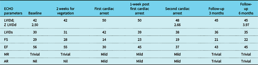
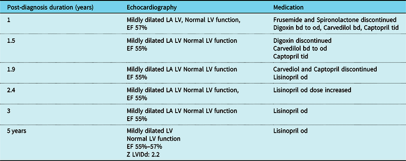
Post-induction patient was in remission. He received three more courses of anthracycline-free chemotherapy. The patient had another cardiac arrest during central line insertion and was promptly resuscitated. The patient was continually monitored (Table 1) and his medication was tailored, based on echocardiographic findings (Tables 2–3). He is currently 7.5 years old and continues to be in remission 5 years post-diagnosis.
Serial echocardiographic findings with a time frame

AR=aortic regurgitation; EF=ejection fraction; FS=fractional shortening; MR=mitral regurgitation; LVIDd=left ventricular internal dimension-diastole; LVIDs=left ventricular internal dimension-systole.
Follow-up cardiac monitoring with medication

EF=ejection fraction; LA=left atrium; LV=left ventricle.
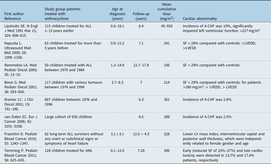
Description of study groups evaluating antracycline-related cardiotoxicity in children

A-CHF=anthracycline-induced congestive heart failure; SF=shortening fraction; LVEDD-left ventricular end-diastolic diameter; LVESD=left ventricular end-systolic diameter.
Discussion
Acute anthracycline-cardiotoxicity affects less than 1% of patients and is manifested by paroxysmal arrhythmia, pericardial exudate, left ventricular dysfunction, and episodically sudden cardiac death and can be fatal. Reference Sadurska7
Our patient developed acute cardiotoxicity post-second dose of daunorubicin (cumulative dose of 120 mg/m2). Although cardiomyopathy correlates strongly with a cumulative dose of anthracyclines; lower doses (<300 mg/m2) have also been implicated. Reference Fraschini, Galbiati and Capra8 There is probably no completely safe dose, and even the lowest dose can induce severe cardiotoxicity, suggesting an important role of individual susceptibility to anthracyclines. Reference Sadurska7,Reference Kremer, van der Pal and Offringa9
Genetic variation may contribute to individual susceptibility. Reference Chow, Leger and Bhatt10,Reference Chow, Antal, Constine and Gardner11 The RARG S427L variant (rs2229774) is highly associated with paediatric patients. If validated, genetic predictors of ACT could be incorporated as part of clinical risk predictors and screening algorithms. Reference Chow, Leger and Bhatt10 Further independent studies are needed to confirm these findings in a cohort of patients with AML.
In our patient, notable risk factors included very young age, uncontrolled hypertension, and sepsis contributing to the acute cardiac decompensation, aggravating the cardiotoxicity.
Very young age (<4 years) at diagnosis and treatment with anthracycline-based therapy has been correlated with increased risk of cardiac disease. Reference Kremer, van der Pal and Offringa9,Reference Blanco, Sun and Landier Chen12 Expert panels recommend close monitoring for young patients who receive anthracyclines. Reference Rathe, Carlsen and Oxhøj13,Reference Fulbright14
It is known that cardiotoxicity may be induced or increased by risk factors for cardiovascular diseases with a higher risk for hypertension when compared to other factors. Reference Sadurska7,Reference Vyskočil, Petráková and Jelínek15 The combination of exposure to anthracyclines and hypertension genetic susceptibility loci puts this subgroup of individuals at greater risk of developing cardiotoxicity. Reference Hildebrandt, Monica and Wu16
Patients with AML have a higher sepsis incidence. Reference Malik, Cardenas-Turanzas and Gaeta17 Sepsis-induced myocardial dysfunction (SIMD) is considered to be a major risk associated with sepsis. Reference Vallabhajosyula, Jentzer and Geske18 SIMD could further contribute to the severity of cardiac dysfunction after anthracycline exposure.
Markers of necrosis as T-troponin, I-troponin, and CKMB are elevated very early after the first administration of anthracyclines, Reference Sadurska7 and were noted in our patient. In nearly 50% of children receiving moderate dose anthracyclines, subclinical troponin elevations related to myocardial injury has been seen. Reference Lipshultz, Miller and Scully19
Besides treatment of the underlying sepsis, prompt identification and treatment of cardiac failure and hypertension with discontinuation of further anthracycline exposure contributed to complete recovery in our patient. Treatments included diuresis, afterload reduction, beta-adrenoceptor antagonists, and improving myocardial contractility.
The Scottish Intercollegiate Guidelines Network (SIGN) has developed a long-term follow-up of survivors of childhood cancer guidelines. They recommend echocardiograms at regular intervals during treatment with anthracyclines and every 3 years thereafter in patients who have received a modest dose. Reference Fulbright14
Conclusion and recommendations
We conclude that in the presence of known risk factors for cardiac dysfunction like sepsis or hypertension, there probably remains a risk of cardiotoxicity even with a low dose of anthracycline, especially in very young patients. For early identification of cardiac dysfunction, we recommend monitoring by echocardiography prior to each dose of anthracycline; baseline cardiac markers with repeat levels after each course of anthracycline.
Acknowledgments
All authors approved the final manuscript as submitted and agree to be accountable for all aspects of the work. Authors’ contributions: S.T. was directly involved in the clinical care of the patient, detected the association of anthracycline-induced cardiotoxicity, and drafted the initial manuscript. R.M. performed a literature review, reviewed, and revised the final manuscript. N.J. performed the cardiac assessment of the patient, including ECG and ECHO monitoring, and critically reviewed the final manuscript. All authors approved the final manuscript as submitted.
Financial support
This research received no specific grant from any funding agency, commercial, or not-for-profit sectors.
Conflict of interest
None.