Introduction
Mastitis is an inflammation of udder tissues due to physical damage, chemical irritation, or infection (Ruegg et al., Reference Ruegg, Erskine and Morin2014). An overview of bovine mastitis, illustrating impact, diagnostic methods, treatment strategies, and future aspects, is shown in Fig. 1. Mastitis is one of the most common and prevalent diseases affecting the dairy sector (Hussain et al., Reference Hussain, Javed and Khan2012). Along with other losses, the costs of preventive measures should also be estimated (van Soest et al., Reference van Soest, Santman-Berends, Lam and Hogeveen2016). Due to various contributing factors such as milk reduction, milk price, treatment, and replacement costs, the financial losses vary among different countries (Halasa et al., Reference Halasa, Huijps, Østerås and Hogeveen2007). Dairy farmers usually underestimate these costs due to evaluation difficulties (Huijps et al., Reference Huijps, Lam and Hogeveen2008). Reduced milk production due to mastitis leads to notable financial losses. In a recently reported study, the financial losses were compared for various causative agents, and it was concluded that losses were not the same for all pathogens. It was found that Escherichia coli and Staphylococcus aureus caused more damage in terms of reduced milk production as compared to other pathogens. Minor pathogens, such as Corynebacterium bovis and non-aureus staphylococci species also cause such effects and should not be ignored. In conclusion, reduced milk production in case of mastitis is due to the type of pathogen, stage of lactation at the onset of disease, and severity of infection (Heikkilä et al., Reference Heikkilä, Liski, Pyörälä and Taponen2018).
Comprehensive overview of factors related to bovine mastitis.

Control of mastitis is a challenge worldwide, posing a risk to the dairy industry and making animal health management difficult (Gomes et al., Reference Gomes, Saavedra and Henriques2016). Global concern is evident regarding this issue as it is not only fatal for animals but poses a serious economic threat for dairy farmers (Sørensen et al., Reference Sørensen, Mark, Sørensen and Østergaard2010). Mastitis has the highest clinical and economic significance in dairy animals. Its causes are of multi-factorial nature, and the involvement of a large number of pathogens makes it very difficult to control (Ruegg et al., Reference Ruegg, Erskine and Morin2014).
Causes
Mastitis is an inflammation of udder tissue which affects milk production and brings pathological changes such as swelling, pain, edema, inflammation, and fibrosis of the udder (Shaheen et al., Reference Shaheen, Tantary and Nabi2016). The teat orifice is made up of smooth-muscled sphincter, and its role is to keep the teat canal closed and to stop milk from flowing. This function also prevents pathogen invasion of the teat canal. Keratin is produced by the teat lining which has bacteriostatic action and serves as a barrier against bacteria. Physical injury to the teat makes it more vulnerable to bacterial invasion, colonization, and infection because of damage to keratin or mucous membranes lining the teat sinus.
The degree of the inflammatory response depends on the invading pathogen, and host factors such as stage of lactation, age, immune status of the cow, genetics, and nutritional status (Harmon, Reference Harmon1994). The milk production system includes the involvement of both local and systemic hormones which play an important role along with other milk-producing factors (Bhutto et al., Reference Bhutto, Murray and Woldehiwet2012). It causes phenomenal changes in physical, chemical, and bacteriological properties of milk. Decrease in lactose, fat, protein, and milk urea nitrogen levels in mastitis milk is observed in various mastitis infections mainly due to coagulase-negative staphylococcus (CNS) Staphylococcus aureus, coagulase-negative staphylococci (CNS), and other bacterial pathogens (Kayano et al., Reference Kayano, Itoh, Kusaba, Hayashiguchi, Kida, Tanaka, Kawamoto and Gröhn2018). Immunological reactions triggered by pathogen invasions result in intramammary (IM) infection and inflammation (Abera et al., Reference Abera, Demie, Aragaw, Regassa and Regassa2010).
Financial losses
Dairy farmers believe that bovine mastitis is one of the major reasons for their personal economic fluctuation that ultimately risks the national economy as well (Pol and Ruegg, Reference Pol and Ruegg2007a). Major concerns by the global dairy industry toward mastitis in dairy animals are due to its widespread scale and constantly increasing costs to counter these diseases (Seegers and Fourichon, Reference Seegers and Fourichon2003). It is regarded as the most important disease of the dairy sector worldwide (Hogeveen et al., Reference Hogeveen, Pyorala, Waller, Hogan, Lam, Oliver, Schukken, Barkema and Hillerton2011). Control and management schemes are focused on the prevention and cure of mastitis (Gilbert et al., Reference Gilbert, Cunha, Jensen, Glass, Foucras, Robert-Granié, Rupp and Rainard2013).
Mastitis is generally considered as the most serious threat to the dairy industry, causing economical losses as well as a negative environmental effect (Hospido and Sonesson, Reference Hospido and Sonesson2005). It is also reported as the most common cause of death of dairy animals (Kossaibati and Esslemont, Reference Kossaibati and Esslemont1997). The financial losses associated with mastitis cannot be accurately measured. However, financial losses are attributed to treatment cost, labor, and veterinary services including diagnostics, early cow replacement, reduced milk production, and discarded milk (Halasa et al., Reference Halasa, Huijps, Østerås and Hogeveen2007). Another indirect cost burden, due to risk and transmission of disease to other dairy animals, is often ignored (Down et al., Reference Down, Green and Hudson2013). Studies to evaluate the financial losses due to bovine mastitis are mainly conducted in developed countries. According to these studies, 78% of the economic losses are due to reduced milk production, 8% include treatment costs, and 14% are due to culling (Halasa et al., Reference Halasa, Huijps, Østerås and Hogeveen2007).
Types
Bovine mastitis is an intricate disease and can be broadly classified into two types, clinical mastitis and subclinical mastitis. Clinical mastitis can be further categorized as peracute, acute, and subacute mastitis depending on the severity of symptoms. Various factors which contribute to the level of infection include the disease-causing agent, age of the animal, its immunological health, and lactation stage (Hurley and Theil, Reference Hurley and Theil2011) Clinical mastitis is a severe condition in which local and systemic stages including redness and inflammation of affected areas, pain, loss of appetite, increased body temperature, reduction in milk yield, and changes in milk composition are evident (De Vliegher et al., Reference De Vliegher, Fox, Piepers, McDougall and Barkema2012). In severe cases, abnormal teat secretions such as blood clots are observed (Lehtolainen et al., Reference Lehtolainen, Røntved and Pyörälä2004).
Subclinical mastitis results in the normal appearance of the mammary gland as well as milk. Somatic cell count is one of the primary indications of subclinical mastitis. The other indicators of subclinical mastitis include increased bacterial population in milk, decreased milk production, and change in composition and quality of milk (Bian et al., Reference Bian, Lv and Li2014). Detection of subclinical mastitis is crucial for executing mastitis control and management strategies (Hoque et al., Reference Hoque, Das, Talukder, Alam and Rahman2015). Laboratory diagnosis is required for the isolation and identification of the pathogen involved (Madouasse et al., Reference Madouasse, Huxley, Browne, Bradley and Green2010).
Etiology and epidemiology
The etiology of complex diseases such as mastitis is not completely known and new pathogens are continuously being detected and reported (Shaheen et al., Reference Shaheen, Tantary and Nabi2016). It can be caused by pathogenic microorganisms and chemical irritants that can trigger an immune response or any kind of physiological injury. However, pathogenic microorganisms are the major cause of mastitis (Bramely et al., Reference Bramely, Cullor, Erskine, Fox, Harmon, Hogan, Nickerson, Oliver, Smith and Sordillo1996). Almost 200 microorganisms that cause bovine mastitis have been identified, including bacteria, yeast, fungi, and viruses (Wellenberg et al., Reference Wellenberg, Van Der Poel and Van Oirschot2002).
Bacterial mastitis is most common and significant from physiological and economic perspectives (Blowey and Edmondson, Reference Blowey, Edmondson, Blowey and Edmondson2010). More than 150 bacterial species are identified as mastitis pathogens (Kuang et al., Reference Kuang, Tani, Synnott, Ohshima, Higuchi, Nagahata and Tanji2009). Both Gram-positive and Gram-negative bacteria can cause mastitis. Gram-positive bacteria include various species of staphylococci and streptococci and the most common Gram-negative bacteria are E. coli and Klebsiella pneumoniae (Contreras and Rodríguez, Reference Contreras and Rodríguez2011).
There are three major categories of bacteria that can infect bovine mammary gland including contagious, environmental, and opportunistic pathogens (Hawari and Hassawi, Reference Hawari and Hassawi2008). Contagious pathogens live on the udder and are transmitted from infected to uninfected teats during the milking process. They mainly include Streptococcus agalactiae, S. aureus, and Mycoplasma bovis. Opportunistic pathogens have very strong adhesive properties that help them invade the inner lining of the gland. They can cause periodic episodes of clinical mastitis (Fox and Gay, Reference Fox and Gay1993).
Environmental pathogens usually reside in the housing and bedding and tend to enter the teat canal during the milking process. E. coli are the major cause of environmental mastitis (Günther et al., Reference Günther, Esch, Poschadel, Petzl, Zerbe, Mitterhuemer, Blum and Seyfert2011). S. agalactiae, S. dysgalactiae, and S. uberis are the common streptococcal species causing mastitis (Dmitriev et al., Reference Dmitriev, Bhide and Mikula2006). Other environmental coliforms include Klebsiella spp., Citrobacter spp., Enterobacter spp., including E. faecalis and E. faecium., and other Gram-negative bacteria such as Serratia, Pseudomonas, and Proteus (Radostitis et al., Reference Radostitis, Gay, Blood and Hinchcliff2000).
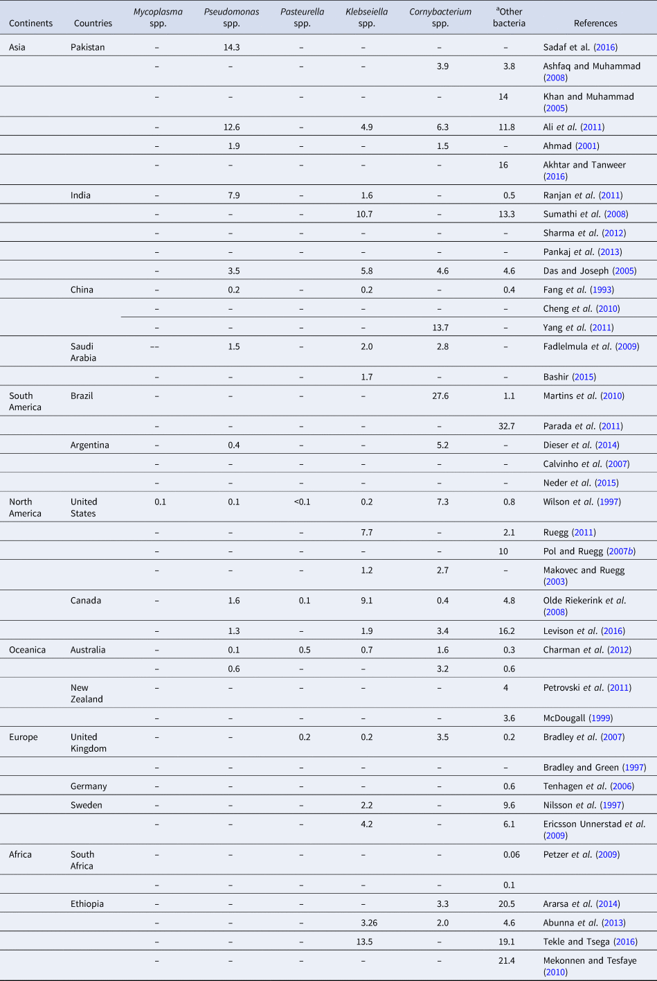
Coagulase-negative staphylococci (CNS; S. epidermidis, S. simulans, S. saprophyticus, and S. chromogenes) (dos Santos Nascimento et al., Reference dos Santos Nascimento, Fagundes, de Paiva Brito, Dos Santos and de Freire Bastos2005) are opportunistic pathogens that colonize the lining of the teat or udder skin. Staphylococcal mastitis is the most frequent cause of economic losses in terms of low milk production in South Asia, and is the leading cause of mastitis in Europe (Wilson et al., Reference Wilson, Gonzalez and Das1997; Shaheen et al., Reference Shaheen, Tantary and Nabi2016). The most prevalent pathogens of bovine mastitis throughout the world are S. aureus and coagulase-negative staphylococci species (Graber et al., Reference Graber, Casey, Naskova, Steiner and Schaeren2007). This complex etiology due to a large number of possible pathogens makes it difficult to control. Epidemiological data of common and less common bacterial pathogens involved in mastitis worldwide are given in Tables 1 and 2. The data show the percentage of prevalence of various bacterial species worldwide. According to the data provided in Table 1, S. aureus is the most common cause of bovine mastitis worldwide, followed by CNS and E. coli. However, there are limited variations observed in different studies based on the outbreak in specific cases. According to the given data, the prevalence of these commonly found pathogens varies among the countries of the same continent and among geographically closely situated countries. As very limited epidemiological data are reported worldwide, no clear conclusion can be drawn from these studies.
Percentage of prevalence of common mastitis pathogens worldwide

Percentage of prevalence of less common mastitis pathogens worldwide

a Other bacteria: Bacilli, Micrococcus, and Pyogenes.
For the less prevalent bovine mastitis pathogens, very few studies are reported. Mycoplasma spp. have not been reported as a cause of bovine mastitis in any of the Asian and African countries. The reason behind this could be difficult cultivation and isolation techniques for Mycoplasma, which if present are not detected due to the non-availability of resources or otherwise it is assumed that these species do not cause bovine mastitis in these areas. Few studies are reported regarding the detection of Mycoplasma in various European countries (Tschopp et al., Reference Tschopp, Bonnemain, Nicolet and Burnens2001; Aebi et al., Reference Aebi, van den Borne, Raemy, Steiner, Pilo and Bodmer2015).
Pseudomonas aeruginosa is associated with sporadic clinical mastitis, resulting in acute cases, and the source of this is usually contaminated water (Kirk and Bartlett, Reference Kirk and Bartlett1984). Bovine mastitis is also caused by Nocardia spp., which have an environmental origin and are usually caused by soil contamination. Occasionally, bovine mastitis is caused by Bacillus cereus, Streptococcus pyogenes, and S. pneumonia (Quinn et al., Reference Quinn, Carter, Markey and Carter1999).
Non-bacterial mastitis is mainly due to the yeast belonging to the genus Candida or to the algae of the genus Prototheca (mostly P. zopfii). Bovine mastitis due to fungal infections is continuously increasing (Spanamberg et al., Reference Spanamberg, Wunder, Brayer Pereira, Argenta, Sanches, Valente and Ferreiro2008; Tarfarosh and Purohit, Reference Tarfarosh and Purohit2008). Infections with Aspergillus fumigatus and Candida spp. are of an acute nature and can cause death; the severity of these infections depends on the load of pathogenic organisms (Pengov, Reference Pengov2002).
Pathophysiology or immunology
Pathophysiological studies on mastitis are mainly focused to develop a better understanding of pathogen and host response, so that it will further lead to better preventive actions for control of disease (Schukken et al., Reference Schukken, Günther, Fitzpatrick, Fontaine, Goetze, Holst, Leigh, Petzl, Schuberth, Sipka and Smith2011). The innate and acquired immune systems of mammary glands work together and provide maximum protection against pathogenic microorganisms causing mastitis (Sordillo et al., Reference Sordillo, Shafer-Weaver and DeRosa1997; Rivas et al., Reference Rivas, Tadevosyan, Quimby, Coksaygan and Lein2002).
Inflammation in a mammary gland is the response of the host immune system toward invading microorganisms and it varies depending on the type of infectious agent (Oviedo-Boyso et al., Reference Oviedo-Boyso, Valdez-Alarcón, Cajero-Juárez, Ochoa-Zarzosa, López-Meza, Bravo-Patino and Baizabal-Aguirre2007). This response varies among Gram-negative and Gram-positive bacteria. Infections due to E. coli, a Gram-negative bacterium, generate a very quick and aggressive immune response by host mammary cells, while S. uberis, a Gram-positive bacterium, is responsible for slow or moderate immune response (Rambeaud et al., Reference Rambeaud, Almeida, Pighetti and Oliver2003; Bannerman, Reference Bannerman2009; Genini et al., Reference Genini, Badaoui, Sclep, Bishop, Waddington, van der Laan, Klopp, Cabau, Seyfert, Petzl and Jensen2011a, Reference Genini, Badaoui, Sclep, Bishop, Waddington, van Der Laan, Klopp, Cabau, Seyfert, Petzl, Jensen, Glass, de Greeff, Smith, Smits, Olsaker, Boman, Pisoni, Moroni, Castiglioni, Cremonesi, Del Corvo, Foulon, Foucras, Rupp and Giuffra2011b). S. aureus-related mastitis results in a very slow or at times non-observable innate immune response (Petzl et al., Reference Petzl, Zerbe, Gunther, Yang, Seyfert and Schuberth2008; Bannerman, Reference Bannerman2009). Further investigation revealed that the probable cause behind this discriminative behavior of the intramammary immune response is due to the Toll-like receptor (TLR) signaling induced by Gram-negative bacteria. Gram-positive bacteria do not induce TLR signaling response, and that is why they result in a slow or moderate response of the host immune system (Petzl et al., Reference Petzl, Zerbe, Günther, Seyfert, Hussen and Schuberth2018).
Studies on signaling pathways of pattern recognition receptors such as TLR, NOD-like (NLR), and RIG-1-like (RLR) receptors could be helpful for better understanding of host–pathogen interplay (Bhattarai et al., Reference Bhattarai, Worku, Dad, Rehman, Gong and Zhang2018). It has been proposed that effective strategies for improvement in mammary gland defense can help us control mastitis, which will further lead to less use of antibiotics at farm level (Sordillo, Reference Sordillo2018). In another reported study, some of the genes such as exsbP1, iihK, iirR slp, and exsbP2 were identified in S. uberis which are upregulated at the early stage of host–pathogen interaction. These genes showed an association with adhesion and internalization of bacteria to host cells. The immunogenic effect of these proteins was confirmed which can further lead to the use of these recombinant proteins as vaccine antigen for S. uberis–caused mastitis (Dego et al., Reference Dego, Almeida, Saxton, Abdi, Ensermu and Oliver2018).
Detection methods
Early disease detection results in better control and management strategies (Ashraf and Imran, Reference Ashraf, Imran, Yaqub, Tayyab, Shehzad, Mingala and Chang2018). Primary diagnosis is based on the physiological signs such as swelling and inflammation of mammary gland or changes in the milk quality and quantity. Severe cases can lead to systemic illness and signs such as fever, weakness, dehydration, and inappetence (Royster and Wagner, Reference Royster and Wagner2015). But clear signs are usually evident at the chronic or clinical state of mastitis. The most commonly employed methods for the diagnosis of mastitis in dairy animals is the measurement of somatic cell count. It is an indicator of disease status, severity, and stage (Harmon, Reference Harmon1994). Somatic cell counting can be done by direct enumeration under a microscope using methylene blue staining; however, this direct counting method has many limitations. The other way is the indirect estimation of somatic cell number using the California mastitis test (Schalm, Reference Schalm1957) and Surf field mastitis test (Muhammad et al., Reference Muhammad, Athar, Shakoor, Khan, Rehman and Ahmad1995). These tests are based on the principle that by adding the detergent, a high somatic cell count can be detected by the release of nucleic acid and other constituents which result in the formation of a gel. But these simple methods are subject to false-positive and negative results (Smolenski et al., Reference Smolenski, Haines, Kwan, Bond, Farr, Davis, Stelwagen and Wheeler2007). Further, these tests do not produce a numerical result, but only indicate low or high counts (Viguier et al., Reference Viguier, Arora, Gilmartin, Welbeck, O'Kennedy and O'Kennedy2009).
Other indirect methods include the Coulter Milk Cell Counter which counts cells or particles as they pass through an electric field, the Fossomatic counting method based on fluorescent cells, and SomaScope which involves flow cytometry. However, automated milking systems cannot use these detection methods (Miller et al., Reference Miller, Paape and Acton1986; Moon et al., Reference Moon, Koo, Joo, Jeon, Hur, Chung, Jo and Park2007).
None of the above-mentioned assays identify the causative agent and the severity of the infection. For better control and treatment strategies, identification of the causative pathogen is critically important. Culture method is still the major criterion for the detection of microorganisms, but it is very time-consuming labor-intensive, and expensive (Viguier et al., Reference Viguier, Arora, Gilmartin, Welbeck, O'Kennedy and O'Kennedy2009); moreover, it can only detect viable bacteria. Due to this reason, false-negative results may be obtained. However, molecular detection methods have the potential to detect the pathogenic organisms from those culture-negative milk samples (Taponen et al., Reference Taponen, Salmikivi, Simojoki, Koskinen and Pyörälä2009).
For better control of this devastating disease of dairy animals, a precise and economical diagnostic tool is needed (Volling and Krömker, Reference Volling and Krömker2008). Over the last few decades, molecular diagnostic methods have been developed with the ability to identify the organisms with great specificity, even if the organisms are very closely related (Muellner et al., Reference Muellner, Schaefer and Lahaye2011; Gurjar et al., Reference Gurjar, Gioia, Schukken, Welcome, Zadoks and Moroni2012). These molecular diagnostic tests have many advantages over the traditional bacteriology techniques in terms of low cost and more accurate detection (Gurjar et al., Reference Gurjar, Gioia, Schukken, Welcome, Zadoks and Moroni2012). With the advancement in molecular techniques, quick and accurate diagnosis of veterinary diseases has become possible (Biek et al., Reference Biek, O'Hare, Wright, Mallon, McCormick, Orton, McDowell, Trewby, Skuce and Kao2012).
Conventional diagnostic methods cannot identify every microorganism due to their existence as a complex community (Schlaberg et al., Reference Schlaberg, Simmon and Fisher2012). As the microbes do not exist in pure culture, the growth of an individual strain may be suppressed by the presence of other microorganisms (Cressier and Bissonnette, Reference Cressier and Bissonnette2011). So, it is not possible to comprehensively catalog all the microorganisms from milk, which is a complex biological fluid (Phuektes et al., Reference Phuektes, Mansell and Browning2001; Taponen et al., Reference Taponen, Salmikivi, Simojoki, Koskinen and Pyörälä2009). However, identification of the pathogen is necessary for the implementation of suitable treatment and management strategies (Oliver et al., Reference Oliver, Gonzalez, Hogan, Jayarao and Owens2004).
Among molecular techniques, the polymerase chain reaction (PCR) holds promise for efficient and accurate microbial identification (Meiri-Bendek et al., Reference Meiri-Bendek, Lipkin, Friedmann, Leitner, Saran, Friedman and Kashi2002). The PCR has become a regular diagnostic tool for various human and veterinary diseases (Schmitt and Henderson, Reference Schmitt and Henderson2005). The various features contribute to the choice of detection techniques, including specificity, sensitivity, time, and expense (Clarridge and Alerts, Reference Clarridge and Alerts2004). In general, the earlier the diagnosis is made, the less time will be needed to cure the disease. It is essential to identify the pathogen not only for antibiotic treatment but also to monitor the severity of infection at farm level (Milner et al., Reference Milner, Page and Hillerton1997). Use of chemical disinfectants, antiseptic, or herbal teat dips is recommended for the control of mastitis (Sharma and Maiti, Reference Sharma and Maiti2005). Antibiotic infusion is among the most frequent and well-established practices for the treatment of bovine mastitis. The excessive use of antibiotics without the prior identification of pathogens has resulted in the development of multidrug resistance in various bacteria (De Vliegher et al., Reference De Vliegher, Fox, Piepers, McDougall and Barkema2012; Wichmann et al., Reference Wichmann, Udikovic-kolic and Andrew2014). Due to the involvement of a wide variety of pathogenic microorganisms, vaccination remains ineffective for bovine mastitis.
For the diagnosis of mastitis, many PCR-based tests have been developed (Lee et al., Reference Lee, Quesnell, Fox, Yoon, Park, Davis, Falk, Deobald and Bohach1998; Baird et al., Reference Baird, Carman, Dinsmore, Walker and Collins1999). A very large number of pathogens are involved in mastitis, and so it is very difficult, time-consuming, and expensive to perform individual tests for each one of them. Accordingly, research has focused on the development of multiplex PCR that can detect many pathogens in a single reaction (Phuektes et al., Reference Phuektes, Browning, Anderson and Mansell2003; Bottero et al., Reference Bottero, Dalmasso, Soglia, Rosati, Decastelli and Civera2004). Various multiplex PCR-based assays have been developed targeting more than five mastitis pathogens in a single test. The newly developed diagnostic assays showed high specificity and sensitivity (Shome et al., Reference Shome, Das Mitra, Bhuvana, Krithiga, Velu, Shome, Isloor, Barbuddhe and Rahman2011; Ashraf et al., Reference Ashraf, Imran, Yaqub, Tayyab, Shehzad and Thomson2017). Progress in diagnostic methods will improve the well-being of animals through rapid diagnosis and treatment. The 16S ribosomal RNA gene sequencing together with PCR is a better diagnostic tool as compared to the traditionally used methods and has many advantages (Petti et al., Reference Petti, Polage and Schreckenberger2005). Reliable identification of the etiological agent is crucial in mastitis diagnostics. Real-time PCR is a fast, automated tool for detecting the most common udder pathogens directly from milk (Hiitiö et al., Reference Hiitiö, Riva, Autio, Pohjanvirta, Holopainen, Pyörälä and Pelkonen2015). Loop-mediated isothermal amplification (LAMP) is another simple and rapid diagnostic test. Many studies based on LAMP were conducted in the last few years for the detection of bovine mastitis pathogens such as M. bovis (Ashraf et al., Reference Ashraf, Imran, Yaqub, Tayyab, Shehzad, Mingala and Chang2018), S. agalactiae (Bosward et al., Reference Bosward, House, Deveridge, Mathews and Sheehy2016), and S. aureus (Tie et al., Reference Tie, Chunguang, Xiaoyuan, Xinghua and Xiuhui2012; Sheet et al., Reference Sheet, Grabowski, Klein and Abdulmawjood2016).
Matrix-assisted laser desorption/ionization time-of-flight mass spectrometry is a robust, rapid, and reliable method for the identification of common microorganisms of bovine mastitis. Expansion of the database with more species further improves the results (Nonnemann et al., Reference Nonnemann, Lyhs, Svennesen, Kristensen, Klaas and Pedersen2019). Identification of acute phase proteins as biomarkers in mastitis caused by different pathogens is also studied and promising results were obtained in a pilot study (Thomas et al., Reference Thomas, Geraghty, Simões, Mshelbwala, Haining and Eckersall2018). Biosensor-based diagnostic methods based on plasmin protease as a biomarker coupled with a gold nanoparticle for the detection of mastitis pathogens are reported. According to the study, this biosensor has the ability to efficiently and rapidly detect mastitis pathogens from milk and milk products (Chinnappan et al., Reference Chinnappan, Al Attas, Kaman, Bikker and Zourob2017).
Treatment
Disease management involves the diagnosis and treatment of mastitis, and control of the spread of infection (Ali et al., Reference Ali, Rahman, Qureshi, Hussain, Khan, Uddin, Iqbal and Han2014). Mastitis cases during lactation are usually treated with antibiotics; however, antibiotic dry cow therapy is also very effective in mastitis control (Halasa et al., Reference Halasa, Nielen, Whist and Osterås2009). It is generally believed that 80% of antibiotics used in dairy animals are administered for control and treatment of mastitis worldwide (Saini et al., Reference Saini, McClure, Léger, Dufour, Sheldon, Scholl and Barkema2012; Ganda et al., Reference Ganda, Bisinotto, Lima, Kronauer, Decter, Oikonomou, Schukken and Bicalho2016a). Different pathogens respond differently to antibiotic treatment, e.g. coliform infections have a high cure rate even when an antibiotic is not administered. Detection of the pathogens before starting antibiotic treatment is crucial. A drug sensitivity test should be performed before choosing an appropriate antibiotic (Koskinen et al., Reference Koskinen, Wellenberg, Sampimon, Holopainen, Rothkamp, Salmikivi, Haeringen, Lam and Pyörälä2010). The treatment efficacy generally depends on the pathogen involved and the antibiotic selected (Owens et al., Reference Owens, Ray, Watts and Yancey1997; Sol et al., Reference Sol, Sampimon, Barkema and Schukken2000; Gillespie et al., Reference Gillespie, Moorehead, Lunn, Dowlen, Johnson, Lamar, Lewis, Ivey, Hallberg, Chester and Oliver2002).
The decision and choice of antibiotic treatment is a debatable issue due to the risks associated with it. The trend toward the use of non-antibiotic treatment is increasing due to the negative impact of the development of antibiotic resistance and adverse effects on the soil and water environment (Wichmann et al., Reference Wichmann, Udikovic-kolic and Andrew2014). Detection of the pathogen is important before antibiotic treatment is initiated, and culture results show that 10–40% of the cases have no detectable bacterial colonies, so in those cases, antibiotics are likely of no use because mastitis has a very complex etiology and different pathogens respond differently to certain drugs. In some very severe cases, segregation and culling are the better, and may in fact be the only choice. In short, all these factors should be considered before treating mastitis with antibiotics (Bartlett et al., Reference Bartlett, Miller, Lance and Heider1992).
In the last couple of decades, many studies have focused on an alternative treatment plan for mastitis, but still lack decisive peer-reviewed scientific evidence. For example, polymer-based external and internal sealant is a prophylactic approach and offers promising results as an alternative to antibiotic dry cow therapy (Timms, Reference Timms2004). Research by Woolford et al. (Reference Woolford, Williamson, Day and Copeman1998) and Huxley et al. (Reference Huxley, Green, Green and Bradley2002) has reported the effectiveness of this product for mastitis prevention in uninfected teats at drying off. An alternative approach such as the use of bacteriocin has many advantages over conventional antibiotic treatment (Twomey et al., Reference Twomey, Wheelock, Flynn, Meaney, Hill and Ross2000).
Recent research has focused on finding alternative approaches for the treatment of mastitis, the ones with less drawbacks and side effects. One such simple study was conducted which involved the administration of a subcutaneous injection containing different concentrations of zinc, manganese, selenium, and copper (Machado et al., Reference Machado, Bicalho, Pereira, Caixeta, Knauer, Oikonomou, Gilbert and Bicalho2013). These injections were found to have a positive impact on udder health and reduced the cases of subclinical mastitis compared to the control group. No effect was observed on milk production and composition. However, further studies are required to confirm these findings (Machado et al., Reference Machado, Bicalho, Pereira, Caixeta, Knauer, Oikonomou, Gilbert and Bicalho2013). Another supporting study reported that there was no effect on the quantity and quality of milk produced when these trace minerals supplement was administered (Ganda et al., Reference Ganda, Bisinotto, Vasquez, Teixeira, Machado, Foditsch, Bicalho, Lima, Stephens, Gomes and Dias2016b).
To minimize the use of antibiotics to dairy animals suffering from mastitis, certain studies have been conducted to find the best suitable alternative. In one such study, antimicrobial activity of 13 lactic acid bacteria isolated from the honey crop of honey bees was investigated in vitro against mastitis-causing pathogens and promising results were obtained (Piccart et al., Reference Piccart, Vásquez, Piepers, De Vliegher and Olofsson2016). Similarly, platelets can actively regenerate glandular tissue; in a recent study, platelet concentrate was administered along with antibiotic and alone. Significantly better results were obtained in mastitis cases treated with platelet concentrate and antibiotic (Lange-Consiglio et al., Reference Lange-Consiglio, Spelta, Garlappi, Garlappi, Luini and Cremonesi2014). A study was conducted to analyze the effect of silver ions on the mastitic pathogens E. coli and S. aureus through proteomic analysis. Silver ions have bactericidal activity against these organisms and may be an alternate treatment of mastitis (Kang et al., Reference Kang, Cho, Kim and Cho2016). Silver ions have a high affinity for proteins and are very reactive (Coward et al., Reference Coward, Carr and Rosenkranz1973). Due to this reason, they most likely bring structural changes in the bacterial cell wall and show antibacterial activity. Silver ions usually attract negatively charged bacterial cells due to the electrostatic attraction (Ovington, Reference Ovington2001) and their attachment on the protein part of cell membrane makes the cell more permeable which further leads to cell death (Liau et al., Reference Liau, Read, Pugh, Furr and Russell1997). Copper is also known for its inhibitory action against various pathogens. Various concentrations of copper have been tested against pathogens (E. coli, CNS, S. aureus, and S. uberis) isolated from bovine mastitis. The results were satisfactory, but further studies on pathogen response to copper in vivo are needed before practical application (Reyes-Jara et al., Reference Reyes-Jara, Cordero, Aguirre, Troncoso and Figueroa2016).
Bacteriophage cocktails have also been used to prevent coliform mastitis in dairy farms. Four bacteriophages were isolated from the samples of primary effluent of wastewater from a local sewage treatment plant. Phage plaques of different morphological features were characterized and used in this study. In vitro studies in raw milk and mammary gland tissue culture showed that the growth of E. coli isolates from mastitis may be inhibited by these bacteriophages. Inhibition results of this novel cocktail were comparable to some of the commonly used antibiotics. This method has the potential to be introduced as a treatment method for E. coli-induced mastitis. However, in vivo trials must be carried out to estimate the dose and test for any deleterious effects involved (Porter et al., Reference Porter, Anderson, Carter, Donjacour and Paros2016). Citrus-derived oil (CDO) has been tested. CDO efficiently killed S. aureus cells and its invasion in bovine mammary cells treated with CDO was also inhibited. As this study was performed in vitro and confined to S. aureus, more comprehensive research is required (Federman et al., Reference Federman, Joo, Almario, Salaheen and Biswas2016). Cinnamon cassia oil has been tested against a few of the mastitic pathogens. It has bactericidal activity and inhibits the growth of major bacterial species most common in bovine mastitis. It is safe and resistance is not developed even after prolonged use. With these advantages, it has the potential to replace some of the antibiotics particularly in organic dairies (Zhu et al., Reference Zhu, Du, Fox and Zhu2016).
Cell-free supernatants (CFS) of Lactococcus lactis strains have been tested against various mastitis pathogens. L. lactis strains LL11 and SL153 inhibit the growth of major pathogens. These strains produced nisin A, a class I bacteriocin. The immune response of a bovine mammary epithelial cell line BME-UV1 against bacteriocin containing CFS was tested. The CFS stimulates lysosomal activity and so may be a candidate to treat and control bovine mastitis (Malvisi et al., Reference Malvisi, Stuknyte, Magro, Minozzi, Giardini, Noncty and Piccinini2016). In a recent study, bovine single chain variable region fragment (scFv) cDNAs were detected in lymphocytes suffering from S. aureus-induced mastitis. scFvs with a high affinity for S. aureus antigen were screened and tested in culture medium where they inhibited S. aureus growth. These scFvs were also effective against S. aureus-induced mastitis in a mouse model. These results suggest that scFvs may be used as a preventive agent for S. aureus-induced mastitis but, further study of risk factors is required (Wang et al., Reference Wang, Zhang and Zhu2016).
Insertion of certain genes coding for lysozyme and lysostaphin that can cure different types of mastitis was tested in a reported study. This is an alternative approach to induce the production of an antibacterial agent by mammary glands and kill the pathogen (Fan et al., Reference Fan, Plaut, Bramley, Barlow and Kerr2002). To further progress in gene therapy technique for the treatment of bovine mastitis, a new mammary-specific vector containing human lysozyme cDNA and kanamycin resistance gene was constructed. Clinical studies showed noticeable therapeutic effects on clinical and subclinical bovine mastitis cases (Sun et al., Reference Sun, Xue, Qian, Fang, Qiu, Zhang and Yin2006). Antimicrobial peptides which exist naturally in living organisms can be used as a tool of gene therapy (Zasloff, Reference Zasloff2002). Lingual antimicrobial peptide is a member of the β-defensin family in bovines. It is a part of the innate immune system and it can kill a variety of microorganisms (Isobe et al., Reference Isobe, Hosoda and Yoshimura2009).
Milk microbiota are complex microbial populations and changing patterns of milk peptides can serve as potential biomarkers for characterization studies (Mansor et al., Reference Mansor, Mullen, Albalat, Zerefos, Mischak, Barrett, Biggs and Eckersall2013). Meta-transcriptomics, meta-proteomics, and meta-metabolomics are contributing largely in building new concepts. Milk microbiota has a role to play in physiology and health of the udder and animal as well as the quality and quantity of milk (Addis et al., Reference Addis, Tanca, Uzzau, Oikonomou, Bicalho and Moroni2016).
In recently reported studies, Chitosan, a polysaccharide (Felipe et al., Reference Felipe, Breser, Bohl, da Silva, Morgante, Correa and Porporatto2019), and phospholipases such as sPLA2 and PLA2G1B (Seroussi et al., Reference Seroussi, Blum, Krifucks, Lavon and Leitner2018) have shown promising results for the treatment of mastitis infections caused by various bacterial pathogens. Chitosan has shown the potential to affect the biofilm formation and thus can be used in the control and treatment of bovine mastitis caused by Staphylococcus species (Felipe et al., Reference Felipe, Breser, Bohl, da Silva, Morgante, Correa and Porporatto2019). Another study also showed positive results in in vitro analysis. It involves the use of the nitric oxide-releasing solution as a liquid formulation, whereby release of nitric oxide has shown antibacterial effect. This could be a reliable alternative to antibiotics for the treatment of bovine mastitis (Regev et al., Reference Regev, Martins, Sheridan, Leemhuis, Thompson and Miller2018). Photodynamic therapy (PDT) has also been tested for its efficacy in the treatment of bovine mastitis caused by S. dysgalactiae and coagulase-negative Staphylococcus species and has shown improvement in PDT-treated group. This method can be used in routine at the farm level (Moreira et al., Reference Moreira, de Souza, de Lima, Salgado, Fernandes, Andreani, Villaverde and Zângaro2018).
More than 30 different studies based on the use of various homeopathic medicine for the prevention and treatment of bovine mastitis have been reported. These studies showed that homeopathic alone or in combination with antibiotics might serve as a good alternative for the control of bovine mastitis (Zeise and Fritz, Reference Zeise and Fritz2019).
Effects of mastitis on human health
Clinical and severe subclinical mastitis not only cause damage to udder tissues and milk production and cause financial damage by affecting animal health but also pose a serious threat to human health by affecting adversely the nutritional quality of milk (Gröhn et al., Reference Gröhn, Wilson, González, Hertl, Schulte, Bennett and Schukken2004; Schukken et al., Reference Schukken, Hertl, Bar, Bennett, González, Rauch, Santisteban, Schulte, Tauer and Welcome2009; Gurjar et al., Reference Gurjar, Gioia, Schukken, Welcome, Zadoks and Moroni2012). Mastitis alters physical, chemical, bacteriological, and organoleptic properties of milk. High somatic cell counts due to mastitis are associated with a reduction in lactose and non-fat solids in milk; however, the level of change is dependent on the causative agent (dos Reis et al., Reference dos Reis, Barreiro, Mestieri, de Felício Porcionato and dos Santos2013). Similarly, proteins and calcium levels are lowered in mastitic milk, and damaged epithelial cells in milk promote the release of certain enzymes, having potential to serve as a biomarker for the diagnosis of mastitis (Oliszewski et al., Reference Oliszewski, Nunez de Kairuz, Gonzalez De Elias and Oliver2002).
Antibiotic residues in milk can cause allergic reactions when consumed, but this can be avoided by discarding milk during antibiotic treatment. Resistant strains of bacteria are continuously developed due to antibiotic use and their transfer to humans cannot be ignored (Virdis et al., Reference Virdis, Scarano, Cossu, Spanu, Spanu and De Santis2010). Identification of antimicrobial-resistant clones at an early stage can help to control its transmission from livestock to humans (Spoor et al., Reference Spoor, McAdam, Weinert, Rambaut, Hasman, Aarestrup, Kearns, Larsen, Skov and Fitzgerald2013). Mastitic milk obtained from the antibiotic-treated dairy animal can be the source of antimicrobial resistance in humans. To ensure safety and quality of milk, the excessive use of antibiotics must be controlled (Shamila-Syuhada et al., Reference Shamila-Syuhada, Rusul, Wan-Nadiah and Chuah2016).
Control measures and future aspects
The establishment of dairy herd improvement (DHI) system at the national level is important for the record of phenotypic data, which will allow scientists to further explore the genetic components responsible for them. By the enlargement of DHI system, genetic selection and genome prediction will be highly improved (Martin et al., Reference Martin, Barkema, Brito, Narayana and Miglior2018). Genetic selection for mastitis resistance can lead to better prevention of this disease. In order to improve the efficiency of this work, a study was performed to identify the genomic markers such as single nucleotide polymorphism (SNP) set to be used for genomic feature best linear unbiased prediction model. Using such a model, we can improve the genomic prediction for mastitis resistance traits (Fang et al., Reference Fang, Sahana, Ma, Su, Yu, Zhang, Lund and Sørensen2017).
Identifying specific high-density SNPs associated with mastitis traits together with genomic breeding values, it would be expected that resistance to mastitis could be increased over consecutive generations of breeding (Deb et al., Reference Deb, Singh and Kumar2012; Mustafa et al., Reference Mustafa, Khan, Heather, Kuthu, EuiSoo, Ajmal, Ain and Sonstegard2018). Further progress in genomic selection technology will allow genetic improvement in terms of mastitis-resistant animals (Vukasinovic et al., Reference Vukasinovic, Bacciu, Przybyla, Boddhireddy and DeNise2017).
Use of selenium and vitamin E supplements were also reported to reduce the incidence of mastitis (Vasil et al., Reference Vasil, Zigo, Elečko, Zigová and Farkašová2017). Vitamin D minimizes cell viability and bacterial invasion in the case of S. aureus-induced mastitis (Yue et al., Reference Yue, Hymøller, Jensen, Lauridsen and Purup2017). However, these studies are not enough, and more research is needed for the practical implication to control bovine mastitis.
The control strategies must be adopted to avoid the use of antibiotics and greater losses in terms of culling. Control measures mainly rely on detailed screening and inspection of dairy farms, evaluation of welfare plans, and the use of prognostic diagnostic tests. These monitoring actions can identify animals at risk, and the spread of disease can also be controlled. The use of immuno-modulators may improve immuno-competence and disease resistance in animals (Trevisi et al., Reference Trevisi, Zecconi, Cogrossi, Razzuoli, Grossi and Amadori2014).
In recent studies, killed bacteria have been used as a vaccine to induce immunity against infection. For example, when an ultraviolet-killed E. coli bacterin was injected for IM immunization at dry off, partial prevention against E. coli-induced mastitis was observed. When compared with the J-5 bacterin vaccine evaluated by Gurjar et al. (Reference Gurjar, Klaessig, Salmon, Yancey and Schukken2013), it gave better protection and increased milk production. Further progress in immunization strategies and related challenges in the field can lead to improved results (Pomeroy et al., Reference Pomeroy, Gurjar, Sipka, Klaessig, Salmon, Quesnell and Schukken2016).
Another major target for vaccine development is S. aureus, and for this purpose, virulence factors have been studied. The relationship between virulence factors of pathogen and host has been explored to study the immune response (Scali et al., Reference Scali, Camussone, Calvinho, Cipolla and Zecconi2015). Expression of antigens in non-conventional hosts such as plants is of interest. Development of new vaccines can help minimize the antibiotic use and control S. aureus mastitis (Festa et al., Reference Festa, Brun, Piccinini, Castagliuolo, Basso and Zecconi2013). S. uberis is another pathogen causing mastitis worldwide. Vaccine antigens from the protein of S. uberis were evaluated as a potential target and tested in murine models (Collado et al., Reference Collado, Prenafeta, González-González, Pérez-Pons and Sitjà2016).
Comparative study of conventional and organic dairy farms can be very useful to identify the factors involved in the control of mastitis. This could lead to the development of better management strategies and promote organic farming or otherwise implementation of contributing factors in conventional dairy farms (Levison et al., Reference Levison, Miller-Cushon, Tucker, Bergeron, Leslie, Barkema and DeVries2016). Major mastitic pathogens have an ability to form a biofilm which makes them more resistant and difficult to control by causing recurrent infections. In-depth knowledge of biofilm formation and control mechanisms could lead to better control (Gomes et al., Reference Gomes, Saavedra and Henriques2016).
There is an old belief (and it is still true) that ‘prevention is better than cure’. Accordingly, various management and control strategies have been adopted for the prevention of mastitis. In the last few decades, studies have been directed to farm management. These include genetic selection for better milk production and disease prevention, health care facilities with better hygiene, and early diagnosis and understanding of host response which may lead to improved treatment of mastitis (Shook, Reference Shook2006; Arif et al., Reference Arif, Rehman, Bhat, Mir, Bhat, Mir, Bilal and Hussain2015).
Biosensors can be used as monitoring tools for animal health management. Such tools are used to monitor human health status and disease diagnosis. With some modifications, these can be used for animals. Such advanced techniques, including microfluidics, sound analyzers, sweat and saliva sensing, and nano-biosensors, are gaining attention due to various advantages. Together with these techniques, a comprehensive online system can contribute to better farm management (Neethirajan, Reference Neethirajan2017). Understanding of host response to various mastitis pathogens by a meta-analysis of available literature discloses various vital themes including genes responsible for biological functions and pathways, which can be very useful for better control (Genini et al., Reference Genini, Badaoui, Sclep, Bishop, Waddington, van der Laan, Klopp, Cabau, Seyfert, Petzl and Jensen2011a, Reference Genini, Badaoui, Sclep, Bishop, Waddington, van Der Laan, Klopp, Cabau, Seyfert, Petzl, Jensen, Glass, de Greeff, Smith, Smits, Olsaker, Boman, Pisoni, Moroni, Castiglioni, Cremonesi, Del Corvo, Foulon, Foucras, Rupp and Giuffra2011b).