Introduction
Radiation-induced malignancies are a rare phenomenon.Reference Cahan1 CahanReference Cahan1 and MurrayReference Murray, Werner, Greeff and Taylor2 proposed the following criteria for radiation-induced malignancies: (1) the radiation must have been given previously, and the sarcoma that subsequently developed must have arisen in the area included within the 5% isodose line; (2) no evidence that the sarcoma was likely present before the onset of radiotherapy (RT); (3) all sarcomas must be proven histologically and must clearly be of a different pathology than that of the primary condition.
Post-radiation sarcoma accounts for 0·5−5·5% of all sarcomas.Reference Sheppard and Libshitz3 Osteosarcoma (OS), malignant fibrous histiocytoma and fibrosarcoma are most common subtypes.Reference Murray, Werner, Greeff and Taylor2,Reference Sheppard and Libshitz3 Adjuvant RT after surgery plays a significant role in the treatment of breast cancer.Reference Veronesi4 Sarcomas of the breast, chest wall, sternum, axilla or supraclavicular region have been reported as a rare complication of RT for breast cancer.Reference Seinen5-Reference Erel, Vlachou, Athanasiadou, Hassan, Chandrasekar and Peart7 OS of the sternal bone is a rarely reported entity.Reference Downey, Hums and Martini8 OS of the sternum secondary to therapeutic ionising radiation is an even rarer diagnosis, and no such cases have been reported in India as per our literature search. Here we report such a case of post-radiation sarcoma after breast cancer treatment—OS at the sternum and both the second ribs in a young lady.
Case history
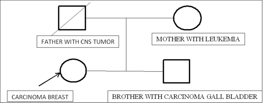
A 33-year-old G3A2 (gravida 3, abortion 2) premenopausal female presented with triple-positive carcinoma of the right breast (cT2N+M0) in 2010, with a significant family history as shown in Figure 1.
Chart depicting significant family history of malignancies.

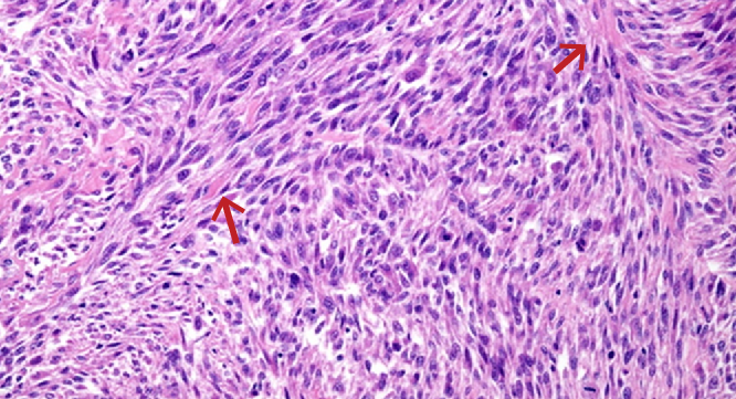
Histopathological image showing features suggestive of OS, pointers at the filigree-like osteoid formation.

She underwent modified radical mastectomy (MRM) in 2010. Histopathology report revealed an invasive ductal carcinoma with a Nottingham Prognostic Index grade III. The largest tumour dimension was 1·5 cm, and 6 out of 51 dissected lymph nodes were positive for tumour invasion. She received adjuvant chemotherapy with six cycles of Fluorouracil, Adriamycin and Cyclophosphamide followed by locoregional RT to the right chest wall and supraclavicular fossa. The treatment was planned by 2D simulation and delivered using tangential fields on a Tele-Cobalt machine. A total dose of 50 Gy in 25 fractions was delivered over 34 days. In view of oestrogen receptor positivity of the tumour, she also received Tamoxifen next 5 years. She conceived her first child in October 2017 with assisted reproductive techniques. In March 2018, during the third trimester of pregnancy, she noticed a tender bony swelling over her chest (8 years after completion of breast RT). Clinically, its size was 4 × 5 cm, and it was situated over right second rib and sternal junction. Ultrasound-guided fine-needle aspiration cytology suggested a low- to intermediate-grade giant cell lesion, and a core biopsy was positive for malignancy, favouring sarcoma. Immunohistochemical staining was negative for GCDFP (gross cystic disease fluid protein), CD34 (cluster of differentiation), smooth muscle actin, Cytokeratin7 and epithelial membrane antigen, and Ki-67 was 30%. MRI chest and abdomen was suggestive of a well-defined heterogeneous mass involving the manubrium sterni and sternal ends of bilateral second ribs measuring about 7·3 × 6·5 × 7·2 cm with positive axillary lymph nodes on both sides. The tumour was extending into the perivascular space posteriorly, abutting the aorta and other great vessels and was also invading the overlying subcutaneous tissues, causing a contour bulge. As the patient insisted, in view of her ongoing pregnancy, no intervention was done until the delivery of her child. She underwent elective lower-segment caesarean section at 32 weeks of gestation. Brain and whole-body 18-FDG PET-CT was done after the delivery, which displayed an abnormally higher metabolic activity in the heterogeneously avid expansile lytic lesion with SUVmax (maximum standardised uptake value) of 11.5, which increased to 18·8 in delayed images. At this stage, she had severe pain in the sternal area, which was managed with analgesics and opioids. As per the decision of the multidisciplinary tumour board, she was started on neoadjuvant chemotherapy initially with one cycle of the AIM (Adriamycin, Ifosfamide and MeSNa [sodium methanethiolate]) protocol. A review of core biopsy blocks and slides at a reference laboratory showed tumour composed of sheets of atypical oval to spindle cell proliferation with many foci of partly calcified filigree-like osteoid formation (Figure 2) —impression of OS; IHC (immunohistochemistry)—vimentin and SATB2 (special AT-rich binding protein) positive in the majority of tumour cells; Desmin positive in occasional cells; negative for CK (cytokeratin), EMA (epithelial membrane antigen), SMA (smooth muscle actin), GATA-3 (GATA-binding protein-3), ER (oestrogen receptor), PR (progesterone receptor), and Ki-67 was 40%.
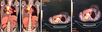
In view of change in histological diagnosis, the multidisciplinary tumour board decided in favour of change in chemotherapy—Ifosfamide, Carboplatin, Etoposide protocol. She received a total of six cycles of inj. Ifosfamide 1800 mg/m2, Carboplatin AUC5, Etoposide 100 mg/m2 along with inj. MeSNa 360 mg/m2 over the next 24 weeks. Post-chemotherapy response assessment using contrast-enhanced CT and PET/CT was suggestive of a partial response (as per RECIST criteria with reduction in size and metabolic activity of the sternal lesion to SUVmax of 3.5). Excision and reconstruction was planned. Video bronchoscopy dated 14 December 2018 was suggestive of extrinsic compression right bronchus 1, 2, 3. However, the patient was unwilling for surgery and hence was offered RT as an alternative for local control. She was treated by re-irradiation of the chest wall by volume-modulated image-guided arc therapy to a total dose of 60 Gy in 30 fractions to the PTV. Dose constraints to the organs at risk (OARs) were as follows: heart V25 (volume receiving 25 Gy) <10%, V20 (volume receiving 20 Gy) <20%. However, re-assessment with PET/CT post-RT (Figure 3) was suggestive of a progressive disease with FDG avid patchy areas of consolidation in the right lung, right-sided pleural effusion and two pleural-based metastatic deposits in the left lung as new findings.
Multiple pleural-based soft-tissue mass lesions in the lungs, largest measuring 7·8 × 7·7 × 10·8 cm (SUVmax 11.35) in the superior segment of right lower lobe, infiltrating the mediastinum and indenting the trachea with architectural distortion of lung parenchyma on the right side.

The surgical oncologist opined inoperability and referred her for palliative chemotherapy. There was a mild FDG avid lytic lesion with soft-tissue component in the manubrium and upper part of sternum (SUVmax 2.44) causing its near-complete destruction, and a lymph nodal mass involving left subpectoral lymph nodes with peripheral FDG avidity measuring approx. 3·4 × 4·6 × 2·6 cm (SUVmax 2.39). She received two cycles of i.v. inj. Doxorubicin 30 mg and i.v. inj. Cisplatin 50 mg till date, but succumbed to her advanced disease.
Discussion
This patient developed sarcoma within a previously irradiated field. The latent period was 7·5 years. Because >5% of the prescribed dose had been delivered to the axillary area and there was no evidence of tumour on a previous PET scan, this case also fulfilled the revised criteria for radiation-induced malignancies suggested by Murray et al.Reference Murray, Werner, Greeff and Taylor2 She underwent radical excision of the breast in the form of MRM. Although the survival rates after breast cancer treatment are typically high, the risk of secondary malignancies, particularly sarcomas, increases. Therapeutic radiation for a childhood malignancy has been associated with malignant thyroid neoplasms.Reference Acharya9 But a population-based retrospective cohort study regarding the risk of thyroid carcinoma after RT for breast cancer showed no significant increase in the risk of thyroid carcinoma in either the RT cohort or the non-RT cohort compared with the general population.Reference Huang, Walker, Groome, Shelley and Mackillop10 According to a large retrospective study of 16,705 patients previously treated for breast cancer, adjuvant RT significantly increased the rate of sarcomas and lung cancers compared with a non-RT group (p = 0·020 and 0·022, respectively).Reference Kirova, Gambotti, De Rycke, Vilcoq, Asselain and Fourquet11 Compared with the general population, the standardised incidence ratio is 10·2% and 1·3% for patients with breast cancer who did or did not receive RT, respectively. Another study using surveillance, epidemiology and end-result data reported a cumulative incidence of sarcoma at 15 years of 0·32% for cases receiving RT, which is more frequent than cases not receiving RT (0·23%, p = 0·001).Reference Seinen5 The mean latency periods of the above two studies were 8·7 and 7·5 years, respectively, which is similar to our case. Angiosarcoma is the most prevalent histology among radiation-induced sarcomas.Reference Seinen5,Reference Kirova, Vilcoq, Asselain, Sastre-Garau and Fourquet6 Higher radiation doses would increase the risk of soft tissue and bone sarcomas after breast cancer.Reference Rubino12 Compared with patients who received <14 Gy, the odds ratios were 1·6 and 30·6 for patients who received 14–44 and >44 Gy, respectively, at the site of the sarcoma. Intensity-modulated RT has been found to improve target coverage and reduce normal tissue complication for breast cancer.Reference Krueger, Fraass, McShan, Marsh and Pierce13 According to this finding, our patient who received 50 Gy at the site of sarcoma was at a high risk of developing post-radiation sarcoma. There were no distinguishable imaging features of post-radiation sarcomas.Reference Sheppard and Libshitz3 However, the presence of bony destruction with a soft tissue mass, tumour matrix mineralisation at a previously irradiated area and an appropriate latency period could be important clues for a diagnosis of post-radiation sarcoma. In addition to imaging features, a low incidence after a long latency period makes it difficult to diagnose the disease accurately. Thus, a high index of clinical suspicion is warranted. The prognosis of post-radiation sarcomas is generally poor, with 5-year survival rates of 27–36%.Reference Seinen5-Reference Erel, Vlachou, Athanasiadou, Hassan, Chandrasekar and Peart7 The standard treatment is surgical resection, but this is often made challenging by tumour location.Reference Kirova, Vilcoq, Asselain, Sastre-Garau and Fourquet6,Reference Erel, Vlachou, Athanasiadou, Hassan, Chandrasekar and Peart7,Reference Tabone14 Only a complete surgical resection can guarantee long-term survival.Reference Tabone14 Chemotherapy or RT has limited roles in treatment; therefore, early detection is important to enable curative resection. In the present case, the patient was unwilling for surgery or chemotherapy since she was pregnant. She received chemotherapy only after delivering the child. The knowledge regarding post-radiation sarcomas is limited to a few case reports. Despite well-organised nationwide breast cancer databases from Korea,Reference Jung15 no large-scale study has been conducted regarding post-radiation sarcomas. The younger age at diagnosis, increased use of RT as a primary treatment for breast cancerReference Veronesi4,Reference Jung15 and longer survival after treatment could lead to more cases of post-radiation sarcoma. In total, 1,831 cases of radiation-induced sarcomas of the breast have been published in the English literature.Reference Sheth, Cranmer, Smith, Grasso-LeBeau and Lang16
Another key factor is the high possibility of familial/hereditary cancer inheritance syndrome like Li-Fraumeni in our patient. Though she was never tested for p53 mutations, her young age at first diagnosis (26 years), extremely strong positive family history and spectra of cancers affecting her first-degree blood relatives (brain tumours, leukaemia) strongly hint at the possibility of such a cancer syndrome. Though the guidelines for cancer treatment in Li-Fraumeni or other hereditary syndromes are not different from non-syndromic patients, preclinical data has suggested the possibility of increased radiosensitivity in patients with such congenital disorders. Similarly, clinical studies have suggested that Li-Fraumeni families might be at a higher risk of secondary radio-induced malignancies. According to a French study, if a germline mutation is detected, then it should be taken into account for decision-making concerning local treatment: (1) Adjuvant RT for localised breast cancer should be extensively discussed and prohibited whenever the risk–benefit ratio is doubtful. (2) Both mastectomy of the cancer-bearing breast and contralateral prophylactic mastectomy (with immediate reconstruction, as frequently as possible) should be advised and discussed with the patient, as is the case for BRCA1/2 mutation carriers, with the additional advantage of potentially avoiding RT if conservative treatment could be avoided.Reference Heymann17 Also, there is a role of tumour suppressor protein P53 in the complex response to RT by modulating the radiosensitivity in tissues as well as sensitising cells to chemotherapy. Hence, the P53 pathway remains an attractive target for exploitation in the war on cancer.Reference Fei and El-Deiry18 Retrospectively, the question certainly arises, given her young age and family history, whether this patient was a right candidate for RT even once as compared to the fact that she received radiation twice.
Future studies should investigate the incidence or distribution of sarcomas after RT for breast cancer based on the analyses of a large database.
Conclusion
We report here a case of post-radiation sarcoma after breast cancer treatment after a latency period of 7·5 years. Post-radiation sarcomas are a rare complication after such long latency periods. Curative surgical resection is the standard treatment despite a poor prognosis. Enhanced clinician awareness and early detection are essential to improve clinical outcomes.
Acknowledgements
Department of Nuclear Medicine and Molecular Imaging, Command Hospital Central Command, Lucknow, Uttar Pradesh, India.
Conflicts of Interest
The authors declare that they have no competing interests.