Introduction
Acute rheumatic fever is characterised by an aberrant immune response and develops following Group A haemolytic streptococcus infection. The joints and the heart are commonly affected, manifesting as carditis with variable severity. In some cases, the carditis in the acute state of acute rheumatic fever does not completely resolve and may progress to rheumatic heart disease. Although there is a declining trend in the number of new cases in developed countries, the burden of the disease remains high in developing and underdeveloped countries.Reference Arvind and Ramakrishnan1 Thus, the global prevalence of rheumatic heart disease ranges widely from 3.4 to 444 per 100000.Reference Watkins, Johnson and Colquhoun2 Recently, the Jones criteria used for diagnosing acute rheumatic fever underwent a review from an epidemiological perspective and were revised to include the findings of Doppler echocardiography.Reference Gewitz, Baltimore and Tani3 Conversely, no consensus has been reached for the diagnosis of rheumatic heart disease. Although cardiac involvement in acute rheumatic fever typically manifests as pancarditis, it often presents as endocarditis, primarily affects the left-sided cardiac valves.Reference Reményi, Wilson and Steer4 The carditis in acute rheumatic fever, which affects 30–70% of the cases and classified based on the degree of cardiac involvement, determines the prognosis. The risk of rheumatic heart disease is high in cases with severe carditis and valve impairment during the active state of the disease. Therefore, the World Heart Federation has elaborated on echocardiographic evaluation to assess valve regurgitation. heumatic heart disease-associated valvular stenosis develops due to fibrotic thickening of valves in recurring episodes and advancing age.Reference Arvind and Ramakrishnan1,Reference Reményi, Wilson and Steer4
The etiopathogenesis of acute rheumatic fever is grounded in a delayed immune response to the Group A haemolytic streptococcus infectionReference Bright, Mayosi and Martin5; hence genetic factors contribute to the susceptibility to acute rheumatic fever and rheumatic heart disease. Research has shown associations between acute rheumatic fever/rheumatic heart disease and HLA class II alleles (DR, DQ), specific B cell alloantigen (D8/17), and polymorphisms in genes like IL-4 and IL-10,Reference Yousry, Sedky and Sobieh6 TNF-α (1), and Ficolin-1.Reference Catarino, Andrade and Boldt7 Epigenetic changes, such as DNA methylation, have also been suggested in rheumatic heart disease pathogenesis (8). Disease progression may be predicted by an increase in the inflammatory cytokines (TNF-α) and a decrease in anti-inflammatory cytokines (IL-4),Reference Yousry, Sedky and Sobieh6 with symptoms prominently due to the inflammation of the tissues.Reference Bright, Mayosi and Martin5
Micro-RNA (miRNA) molecules, single-stranded, small, and non-coding have been known to exert regulatory effects by suppressing their target genes. Research on miRNAs, particularly their role as diagnostic and prognostic biomarkers, has gained interest. MiRNAs with plausible roles on the genes expressed in the immune system and inflammatory processes have been reported for cardiovascularReference Asulin, Volinsky and Grosman-Rimon9–Reference Siddeek, Mauduit and Yzydorczyk12 and rheumatologic diseases.Reference Wang, Zhang and Zhu13 MiR-155-5p has recently been reported to play a critical role in the progression of rheumatic heart disease.Reference Chen, Wen and Lu14 Using next-generation sequencing, miR-145-5p, miR-199a-5p, and miR-5701 were identified with elevated expressions in rheumatic aortic valves.Reference Asulin, Volinsky and Grosman-Rimon9 Another study suggested the involvement of the IL-1 pathway augmentation by identifying lower expressions of miR-205-3p and miR-3909 in rheumatic valve tissues.Reference Lu, Sun and Duan15 Children with rheumatic carditis showed significant downregulations of miR-16-5p, miR-223-3p, and miR-92a-3p.Reference Gumus, Giray and Bobusoglu16 Downregulation of miR-101 leading to higher TLR2 expression levels was found to be associated with rheumatic heart disease.Reference Dong, Sun and Shan17 miR-1183 and miR-1299 were identified with distinct roles in rheumatic heart disease pathology, and their use as potential biomarkers was suggested.Reference Li, Lian and Zhao18
The literature on miRNA profiles in the etiopathogenesis of acute rheumatic fever and rheumatic heart disease in the paediatric age group remains scarce. Here, we aimed to investigate the expressions of miR-101, miR-1183, and miR-1299 in children with acute rheumatic fever and rheumatic heart disease and determine their potential as diagnostic biomarkers.
Materials and methods
This cross-sectional study was conducted in the Department of Pediatric Cardiology, Xxx University Medical School between January 2008 and December 2017. The study group included patients with acute rheumatic fever treated in the inpatient clinics. Jones criteria were used for diagnosis. The hospital records indicated 173 patients, and prior power analysis revealed that 72 cases would be sufficient to achieve 80% power with a 95% confidence interval.
Study groups
The medical charts were reviewed for comorbid conditions such as CHD, autoimmune/autoinflammatory conditions like systemic lupus arteriosus, familial Mediterranean fever, or chronic liver/kidney diseases. The presence of comorbid conditions, family history of premature myocardial infarction/death, pulmonary hypertension, and recurring acute rheumatic fever episodes were accepted as exclusion criteria. The eligible patients were contacted by phone and were informed about the study. Seventy patients agreed to participate in the study. The control group consisted of age and gender-matched 35 children without chronic diseases or autoimmune disorders, and in whom physical examination, ECG, and echocardiographic studies indicated no signs of cardiovascular pathology.
Clinical evaluation
The medical charts were reviewed, and prior physical examination notes, results of laboratory tests, ECG, and echocardiography were recorded. The patients with carditis during the active state were categorised considering the severity of carditis, as previously described.19
All participants were evaluated by echocardiography using GE Vingmed Vivid Pro 7 (GE Vingmed Ultrasound, Horten, Norway) with a 3 MHz probe. Standard 2D, pulsed-Doppler, colour Doppler, and M-Mode were used at precordial position and assessed as previously reported.Reference Reményi, Wilson and Steer4
Molecular genetic studies for miRNA
Peripheral venous blood samples were obtained from participants used for RNA isolation using Quick-RNA Miniprep Plus (#R1057, Zymo, USA) according to the manufacturer’s protocols. Total RNA was converted into cDNA using ID3EAL miRNA qPCR Assay Kit (#1103101, MIRXES, Singapore) using the protocol in the product pamphlet. 1:10 diluted cDNA samples were amplified using Qiagen Rotorgene 6000. The normalisation of data was performed using U6 snRNA.
Statistical analysis
All statistical analyses were performed using SPSS 25.0 software. Kolmogorov–Smirnov and Shapiro–Wilk tests were used for determination of normal distribution. Continuous variables were defined by the mean ± standard deviation; median (min–max values) and categorical variables were defined by number and percent. For independent groups comparisons, we used independent samples t test and one-way ANOVA (post hoc: Tukey) when parametric test assumptions were provided, Mann–Whitney U test and Kruskal–Wallis variance analysis (post hoc: Mann–Whitney U test with Bonferroni correction) were used when parametric test assumptions were not provided. Difference between categorical variables were analysed with chi-square analysis. Also, Roc analysis was performed for determining the diagnostic performance of variables. Statistical significance was determined as p < 0.05.
Results
The study comprised 69 patients and 27 children in the control group. During the active state of acute rheumatic fever and the study, the mean age of children was 10.6 ± 2.5 (range: 5–16) and 13.4 ± 2.8 years (range: 6–20), respectively. The mean age of children in the control group was 11.8 ± 3.3 years (range: 6–18). The ratio of boys to girls in the patient and control group was 37:32 and 17:10, respectively, with no significant difference between the groups in terms of age and gender (p > 0.05). The family history of acute rheumatic fever was positive in 13% (n : 9) of the patients. There was no consanguinity between the parents of children in both groups.
Clinical findings
Major clinical findings included isolated arthritis in 21.7% (n : 15), carditis in 78.2% (n : 54), Sydenham chorea in 7.2% (n : 5), and erythema marginatum in 1.4% (n : 1) of the patients. The mean age and gender distribution between patients with isolated arthritis and those with carditis showed no significant difference (p > 0.05). The patients with carditis (n : 54) were subdivided into three groups based on the echocardiographic findings. The mean age of patients with mild, moderate, and severe carditis was 10.3 ± 2.8, 11.5 ± 2.3, and 10.4 ± 2.5 years, respectively, and no statistical difference was present among groups (p > 0.05). The male to female ratio of patients with mild, moderate, and severe carditis was 9:8, 11:10, and 8:8, respectively, and no statistical difference was present among groups (p > 0.05). In most of the carditis patients (n : 36, 66.7%), both the mitral and aortic valves were affected, while isolated mitral and aortic valve involvement was present in 17 (31.5%) and 1 (1.8%) patients, respectively.
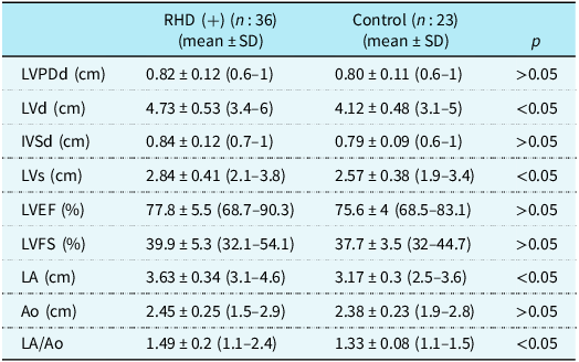
The mean duration of follow-up for acute rheumatic fever patients was 3.7 ± 2.4 years (range: 10 months–10 years). Out of 54 patients with carditis during the active state, 18 (33.3%) showed resolution of cardiac involvement, while 36 (66.7%) developed rheumatic heart disease. The medical records revealed that 16 patients out of the 36 who developed rheumatic heart disease belonged to the severe carditis group, while the previous carditis severity was moderate and mild in 14 and 6 of the rheumatic heart disease patients, respectively. In the second part of the study, the control group consisted of 23 children matched for gender, age, and BMI to the rheumatic heart disease patients. Repeat echocardiography findings showed that mitral valve involvement was present in most of the rheumatic heart disease patients (n : 21, 58.3%), while the involvement of both mitral and aortic valves, and isolated aortic valve was present in 11 (30.4%) and 4 (11.1%) rheumatic heart disease patients, respectively. The dilatation of left-sided cardiac chambers was found during the echocardiography of 11 patients (30.6%). The echocardiographic findings of rheumatic heart disease patients and control subjects are presented in Table 1.
Echocardiographic findings in RHD patients and controls

LA = left atrium diameter, Ao = aorta diameter, LVPDd = left ventricle posterior wall end-diastolic diameter, LVd = left ventricle end-diastolic diameter, IVSd = intraventricular septum end-diastolic diameter, LVs = left ventricle end-systolic diameter, LVEF = left ventricle ejection fraction, LVFS = left ventricle shortening fraction.
miRNA expressions
The expression frequencies for miR-101, miR-1183, and miR-1299 in the acute rheumatic fever patients were 74, 81%, and 75%, respectively. In the control group, miR-101, miR-1183, and miR-1299 were expressed with 85%, 88.8%, and 85% frequencies, respectively. No statistically significant difference in the expression frequencies of miRNAs between the study groups was present (p > 0.05).
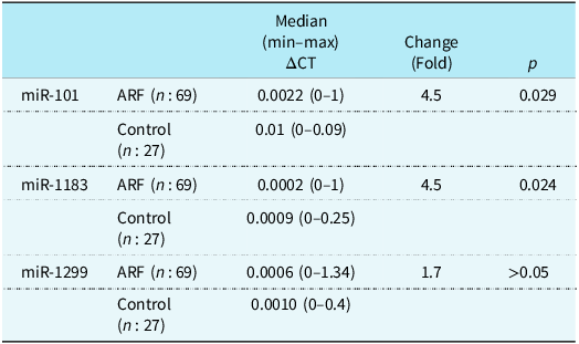
RT-PCR results presented in Table 2 revealed that median ΔCT for miR-101 and miR-1183 was 0.0022 and 0.0002 in the acute rheumatic fever group, and both miRNAs were significantly downregulated 4.5-fold in the patients compared to the controls (p : 0.029 and p : 0.024, respectively) (Table 2).
miRNA expressions in ARF patients and controls

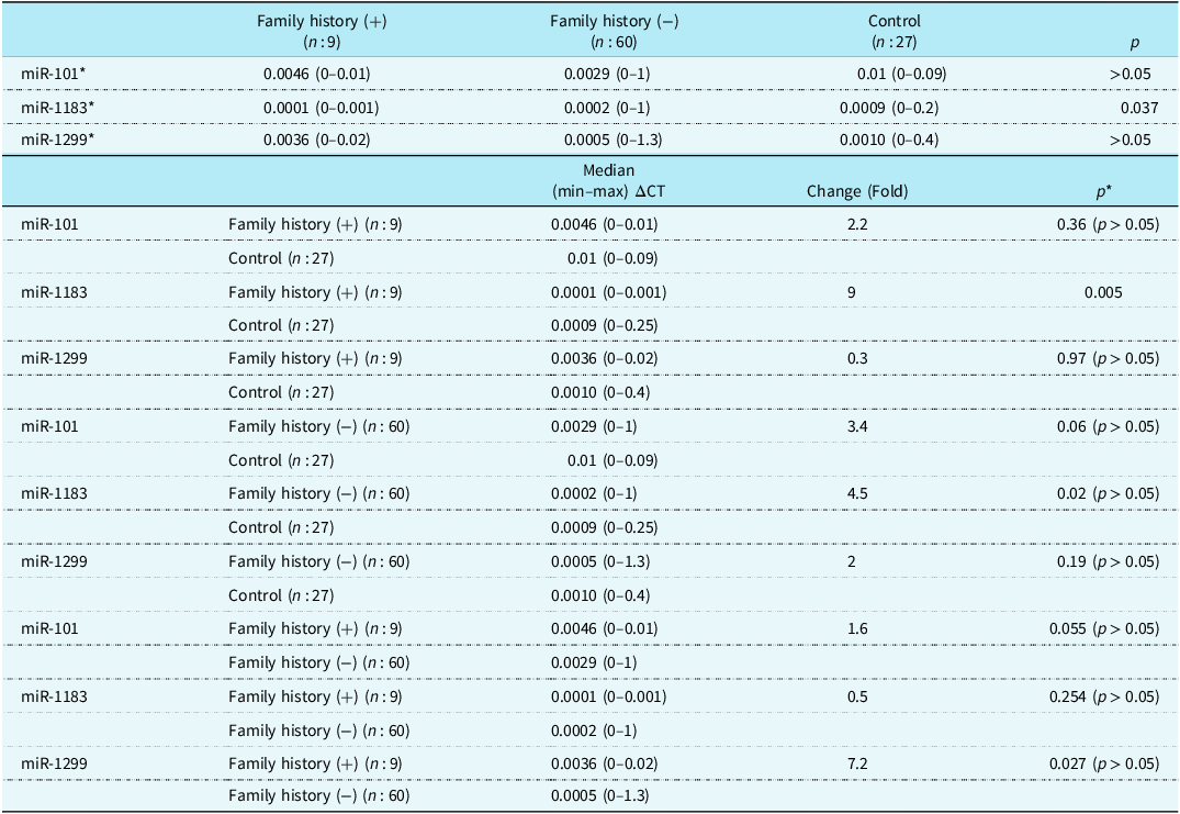
The expression of miR-1183 in positive family history patients was significantly reduced (median ΔCT: 0.0001) compared to those of the patients with negative family history and the controls (p : 0.037) and downregulated 9-fold in the acute rheumatic fever patients who had a positive family history compared to controls (p : 0.005). No significant relationship was observed when the expression of other miRNAs in our study was compared between negative family history patients and controls or between positive and negative family history patients (Table 3).
miRNA expression in ARF patients with positive and negative family history

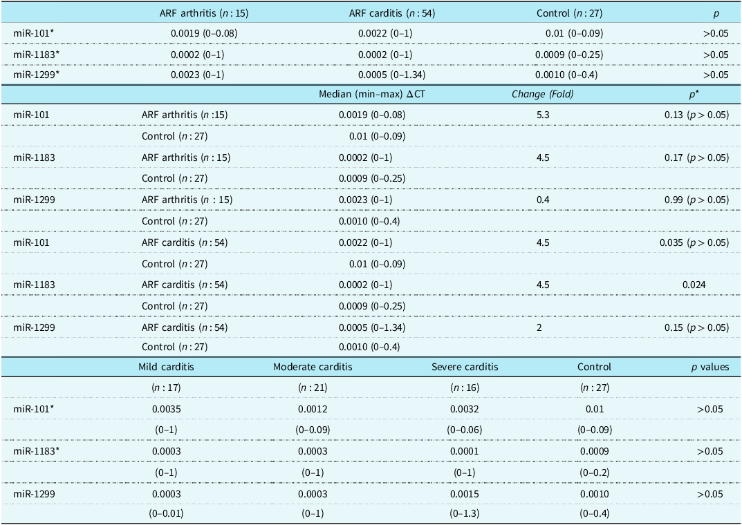
The expressions of miR-101, miR-1183, and miR-1299 were compared among acute rheumatic fever patients with isolated arthritis, with carditis and controls revealed no significant difference. When the statistical analysis was performed to compare the miRNA expressions in acute rheumatic fever carditis patients and controls, we observed that the miR-1183 expression was significantly downregulated 4.5-fold in the acute rheumatic fever carditis patients (p : 0.024) (Table 4).
miRNA expressions in ARF patients with isolated arthritis and carditis

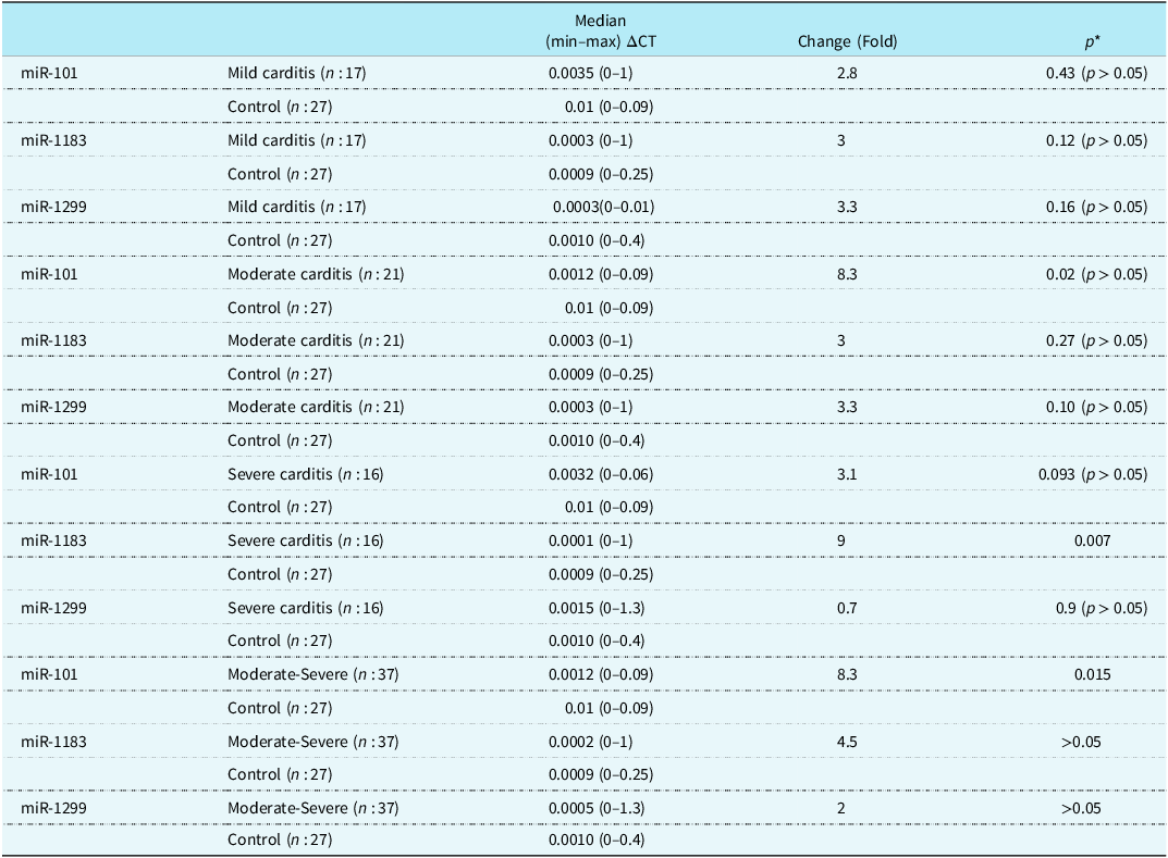
The miRNA expression results among patients with mild, moderate, and severe carditis are shown in Table 4. No statistical difference in miR-101, miR-1183, and miR-1299 expressions was observed among patient groups. The relationships between the expressions of miRNAs and patients with mild, moderate, and severe carditis were further investigated on an individual basis. We observed a significant 9-fold downregulation of miR-1183 in the acute rheumatic fever patients with severe carditis (p : 0.007). Next, we grouped the patients with moderate and severe carditis together (n : 37), and the results showed that the expression of miR-101 in the patients was significantly downregulated 8.3-fold than that in the controls (p : 0.015) (Table 5).
miRNA expressions related to the severity of carditis

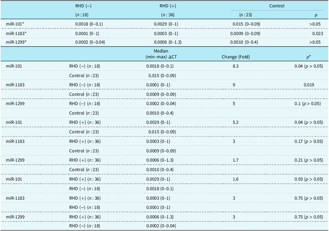
When the miRNA expressions were compared among patients who developed rheumatic heart disease, who did not develop rheumatic heart disease and the controls, we observed a significant difference in the expression of miR-1183 among groups (p : 0.023). When the expression of miR-1183 was compared between patients without rheumatic heart disease and controls, we found a significant 9-fold downregulation (p : 0.019) (Table 6).
miRNA expression related to RHD developing

The AUC value for miR-101 and miR-1183 in ROC analysis was 0.64 and statistically significant for acute rheumatic fever diagnosis for both miRNAs (p < 0.05). In rheumatic heart disease patients, ROC analysis did not indicate any significance for miR-101 (AUC: 0.63) and miR-1183 (AUC: 0.61) expressions (p > 0.05).
Discussion
The late autoimmune response to Group A haemolytic streptococcus infection in the pathogenesis of acute rheumatic fever and rheumatic heart disease consists of a complex network of genes and regulatory elements. Recent advances in immunogenetics research indicated the inhibitory role of miRNAs on their target genes like TGFβR1, TLR2, ZNF 708, ZNF101, TAOK1, IL-1β, IL1R1.Reference Lu, Sun and Duan15,Reference Dong, Sun and Shan17,Reference Zhao, Wang and Liao20,Reference Balashanmugam, Shivanandappa and Nagarethinam21 The presence of miRNAs in the circulation suggests their use as diagnostic, prognostic, and therapeutic biomarkers.Reference Balashanmugam, Shivanandappa and Nagarethinam21,Reference Pokhrel and Guotian22 Despite acute rheumatic fever primarily affecting children, with the risk of rheumatic heart disease later in life, research on miRNA roles in the pathogenesis of acute rheumatic fever and rheumatic heart disease in children is scant. The limited literature prompted us to investigate the expressions of miR-101, miR-1183, and miR-1299 in children with acute rheumatic fever and who developed rheumatic heart disease later.
The control groups were age and gender-matched to the patients as confirmed by no statistically significant difference in age and gender between study groups. Echocardiography was repeated to determine the current status of cardiac involvement and diagnose the presence of rheumatic heart disease. The echocardiographic measurements in 36 patients compared to those of the controls showed significant differences indicating the presence of left-sided heart involvement. As recently reported, the mitral valve was observed to be affected in almost all rheumatic heart disease patients, consistent with our study findings.Reference Arvind and Ramakrishnan1,Reference Bright, Mayosi and Martin5 Although the sequence from Group A haemolytic streptococcus pharyngitis to acute rheumatic fever to rheumatic heart disease progression is accepted, diagnosing rheumatic heart disease cases without acute rheumatic fever history is not uncommon.Reference Bright, Mayosi and Martin5 Hence, our study includes the acute rheumatic fever cases that progressed to rheumatic heart disease and those that did not and compared the expression levels of miRNAs between these patients and the controls. Oury et al. noted that research on the role of miRNAs in valvular heart diseases was in its infancy, with most data originating from studies primarily focused on aortic stenosis, one of the most common valvular diseases in developed countries.Reference Oury, Servais and Bouznad23 This statement partly elucidates the dearth of data on acute rheumatic fever-associated rheumatic heart disease, which predominantly affects the mitral valve and is more prevalent in developing countries compared to developed ones. Oury et al. also emphasised the significance of miRNA profiling in biomarker detection to assess the degree of valve impairment and early left ventricular dysfunction.Reference Oury, Servais and Bouznad23 A study on paediatric atrial septal defect patients demonstrated upregulation of hsa-let-7a, hsa-let-7b, and hsa-miR-486. The authors concluded that circulating miRNAs were extremely valuable as diagnostic biomarkers and risk-predictive tools for subsequent generations.Reference Song, Higgins and Guo11 In a recent study, inhibition of cardiac expression of miR-155-5p, using an adeno-associated viral vector in a murine model of rheumatic heart disease, caused limitation of the valvular damage by upregulating miR-155-associated S1PR1, SOCS1, and STAT3 pathways.Reference Chen, Wen and Lu14 In a recent miRNA microarray study, among the more than 1000 miRNAs identified to be expressed in aortic valve tissues, miR-145-5p, miR-199a-5p, and miR-5701 were found to be significantly elevated in rheumatic valves compared to calcified valves.Reference Asulin, Volinsky and Grosman-Rimon9 Previously, the roles of IL-1β and IL1R1 genes in rheumatic heart disease progression were determined by reduced levels of serum hsa-miR-205-3p and hsa-miR-3909.Reference Lu, Sun and Duan15 The downregulation of miR-16-5p, miR-223-3p, and miR-92a-3p was significantly associated with rheumatic carditis in children.Reference Gumus, Giray and Bobusoglu16 Although children similar to those in our study were studied by Gumus et al., the investigated miRNAs differed between the two studies, making direct comparison of outcomes impossible. We observed that the expressions of miR-101 and miR-1183 in acute rheumatic fever children were significantly 4.5-fold lower than the controls. MiR-1299 expression in acute rheumatic fever children was also lower than in controls; however, it did not reach the statistical significance threshold.
MiR-101 targets multiple genes and participates in a wide spectrum of cellular processes, ranging from IL-1β-induced degradation of the extracellular matrix of chondrocytesReference Dai, Zhang and Hu24 to impaired apoptosis observed in cancers.Reference Chen, Ponnusamy and Liu25,Reference Sayed and Abdellatif26 Different target genes for miR-101, such as TGFBR1, FDZ4, FDZ6, MAP3K13, and MAP3K4 were found to have potential roles in valvular heart disease.Reference Wang, Rao and Liu27 MiR-101a was found to play a limiting role in the hypoxia-induced proliferation of cardiac fibroblasts by suppression of the TGFβR1.Reference Zhao, Wang and Liao20 An interesting study has recently revealed that a herbal product upregulated miR-101, subsequently suppressing TGFBR1 and TLR expressions in cardiomyocytes injured by hypoxia/reoxygenation.Reference Wu, Fan and Chen28 Similarly, the authors of another study suggested that miR-101a might have the potential to reduce the speed of chronic renal fibrosis by acting on KDM3A and leading to the inhibition of the YAP-TGF-β-Smad signalling pathway.Reference Ding, Xu and Jiang29 The impaired expression of TLR2, which was proposed as a factor in rheumatic heart disease pathogenesis, was previously associated with miR-101 downregulation. The expression of miR-101 in rheumatic heart disease patients who underwent valve replacement was reported to be 1.7-fold lower compared to that of patients who underwent valve replacements due to other causes. The reduced expression of miR-101 in cardiac tissue had been associated with an increase in inflammatory cytokines such as TNF-α, IL-1β, and IL-6. Conclusively, the authors reported that miR-101 impairs the immune response to streptococcus infection by suppressing the expression of TLR2.Reference Dong, Sun and Shan17 Consistently, we observed 4.5-fold and 5.2-fold reduction in the expression of miR-101 in children with acute rheumatic fever and rheumatic heart disease, respectively. We found that the miR-101 expression in children who had carditis during the active state of acute rheumatic fever and developed rheumatic heart disease later had been lower than in normal cases, although it did not reach statistical significance. This could be attributed to either the limited number of cases or the categorisation of cases into three subgroups. Therefore, we assessed the moderate and severe carditis cases together and observed a significant 8.3-fold reduction in miR-101 expression compared to controls. We hypothesised that there might be an association between miR-101 and the severity of carditis in acute rheumatic fever. The ROC curve analysis for miR-101 expression was statistically significant indicating that miR-101 could be used as a biomarker for acute rheumatic fever diagnosis. The significantly reduced expression of miR-101 in our patients was consistent with the findings of a previous study that demonstrated significant downregulation of tissue miR-101 in acute rheumatic fever and rheumatic heart disease patients.Reference Dong, Sun and Shan17
A recent study demonstrated a 4-fold upregulation of miR-1183 in cardiac stromal cells of patients with arrhythmogenic cardiomyopathy, with its target gene identified as SP4.Reference Rainer, Meraviglia and Blankenburg30 In a microarray study of miRNAs and their target genes in coronary artery disease, upregulated miR-1183 interacting with HMGB1 was predicted as a novel prognostic and diagnostic biomarker.Reference Balashanmugam, Shivanandappa and Nagarethinam21 Recently, miRNA profiling in rheumatic heart disease patients revealed dysregulation of miR-1299 in the right atrium.Reference Yan, Shi and Yu31 In a study similar to ours, Li et al. investigated the expression of miR-1183 and miR-1299 in both the plasma and cardiac tissue of adult patients with rheumatic heart disease. They found significant upregulations of both miR-1183 and miR-1299, suggesting a potential role for miR-1299 in cardiac apoptosis.Reference Li, Lian and Zhao18
On the contrary, our results indicated reductions in the expressions of miR-1183 and miR-1299 in children with. There could be several reasons for the different outcomes between our study and the study by Li et al. First, the age difference of the studied populations could account for the contradictory outcome. Second, temporal and spatial differences in the expressions of miRNAs, as observed in other regulatory nucleic acids across the human genome. Moreover, considering the intricate network of genes and pathways influenced by most miRNAs, it is plausible that the pathways and target genes affected by the downregulations of miR-1183 and miR-1299, related to acute rheumatic fever in children in our study, could differ from those in the study by Li et al. Notably, EGFR has been identified as one of the target genes for miR-1299.Reference Cao, Li, Li and Zhao32 Additionally, dysregulated EGFR expression has been implicated in inflammationReference Li, Fang and Zhong33 and cardiac valve pathology.Reference Barrick, Roberts and Rojas34 Therefore, one might consider that the downregulation of miR-1299 could be involved in acute rheumatic fever and related valvulopathy in rheumatic heart disease by leading to the overexpression of EGFR. In our study, family history positivity for acute rheumatic fever was significantly associated with the downregulation of miR-1183 in acute rheumatic fever patients. To the best of our knowledge, no study assessed the association of family history and expression of miRNAs in acute rheumatic fever, making direct comparisons unavailable. The ROC curve analysis for miR-1183 expression was statistically significant and indicated that miR-1183 could be used as a diagnostic biomarker for acute rheumatic fever.
There are inherent limitations and strengths in our study. The paediatric age group of patients with acute rheumatic fever who were followed up for rheumatic heart disease development later was an advantage of the current study, as it allowed us to compare the expressions of miRNAs in the same individuals across different clinical settings of related diseases. The results of this study could not be compared directly with others due to the absence of studies sharing the exact same design as ours regarding patient population and the selected miRNAs for investigation. The primary aim of the current study, which was to determine the potential of miR-101, miR-1183, and miR-1299 as biomarkers for acute rheumatic fever/rheumatic heart disease, naturally encounters challenges inherent in using circulating miRNAs as biomarkers as recently reported.Reference Felekkis and Papaneophytou35
In conclusion, we observed significantly lower plasma expressions of miR-101 and miR-1183 in children with acute rheumatic fever, especially marked in children with moderate-severe carditis. Our findings, which indicated the roles of miR-101 and miR-1183 in the pathogenesis of acute rheumatic fever and rheumatic heart disease, support the use of these miRNAs as diagnostic and prognostic biomarkers. miRNAs may also be used in the diagnosis and treatment of acute rheumatic fever and rheumatic heart disease in the near future. We suggest further studies with larger patient populations should be conducted to delineate the exact roles of miR-101, miR-1183, and miR-1299 in acute rheumatic fever and rheumatic heart disease pathogenesis as well as to reach a consensus on their use as biomarkers.