




No CrossRef data available.
Article contents
A novel technique for creation of a systemic-to-pulmonary arterial shunt
Published online by Cambridge University Press: 14 April 2005
Abstract
A systemic-to-pulmonary arterial shunt is still widely used for palliation of some neonates with cyanotic congenital cardiac lesions. This procedure, however, is well known to be associated with some degree of morbidity and mortality. To reduce the incidence of iatrogenic pulmonary arterial deformities, we have devised a new and simple technique to create the shunt using a partial sternotomy, and have used our technique in 10 neonates with cyanotic cardiac malforamations. All but one of our patients survived, and an early reoperation was needed in only one further patient for revision of the shunt. Successful bidirectional Glenn procedures were performed in five of the nine surviving patients within 18 months of the initial procedure. We believe that our technique provides superior palliation by permitting equal enlargement of the right and left pulmonary arteries, and thus facilitates subsequent completion of the Fontan circulation.
Information
- Type
- Original Article
- Information
- Copyright
- 2005 Cambridge University Press
Palliative shunts are still used widely in congenital cardiac surgery, the classical and modified variants of the Blalock–Taussig shunt being the most common. The optimal technique for creating systemic-to-pulmonary arterial anastomoses, however, remains debatable. Some authors recommend thoracotomy, whereas others prefer median sternotomy, which has gained popularity in recent years.1 Shunts to the right or left branches of the pulmonary trunk using either approach may cause complications, such as deformity at the anastomotic site, or excessive flow of blood in one pulmonary artery.2, 3 Here, we present a new technique for interposition of a graft between the brachiocephalic artery and the pulmonary trunk or its bifurcation, reviewing our initial experience in 10 neonates.
Materials and methods
The systemic-to-pulmonary arterial shunts were performed via a partial midline sternotomy in 10 neonates with complex cyanotic cardiac disease between January 2001 and September 2003. This group of patients constitutes a subset of those previously mentioned in published correspondence.4 The age of the patients ranged between two and thirty days, with a mean of 16 days. Their other pertinent characteristics are summarized in Table 1. All patients with functionally univentricular pathology were palliated with a view to subsequent construction of a bidirectional cavopulmonary anastomosis, or an extracardiac Fontan operation. We postulated that, by placing our shunt to the pulmonary trunk or its bifurcation, the flow of blood would be distributed uniformly into the right and left pulmonary arteries, permitting uniform growth of both arteries.
Table 1. Preoperative and operative findings.

Surgical technique.
We performed a partial median sternotomy from the suprasternal notch to the level of the third intercostal space. Only the upper third of the pericardium was opened, and the aorta and the pulmonary trunk were minimally dissected. The pleural cavities were kept intact, the right lobe of the thymus gland was completely removed, and the brachiocephalic artery and vein were mobilized. After intravenous administration of heparin at 100 international units per kilogram, a partially occluding clamp was placed on the pulmonary trunk. Following a longitudinal incision, we made an end-to-side anastomosis with a 3.5 millimetre Gore-tex graft for patients weighing from 2.5 to 3.5 kilograms, or a 4 millimetre graft for patients weighing from 3.4 to 4.0 kilograms (Fig. 1). The other end of the graft was taken in front of the aorta, and anastomosed to the brachiocephalic artery, again using a partially occluding clamp. We used continuous polypropylene sutures of 7/0 dimensions for both anastomoses. In two patients, in whom the pulmonary arteries lay posterior to the aorta, the pulmonary side of the anastomosis was placed on the retroaortic pulmonary bifurcation. Having secured haemostasis, the lower half of the opened pericardium was loosely closed. A drain was placed into the mediastinum, and the partial sternotomy was closed in conventional fashion.

Figure 1. The completed systemic-to-pulmonary arterial anastomosis between the brachiocephalic artery (IA) and the pulmonary trunk (PA). AO: aorta; RPA: right pulmonary artery; RA: right atrium; SVC: superior caval vein.
Results
In one patient, with pulmonary atresia with intact ventricular septum, pneumonia and septic shock developed on the 15th postoperative day, and the patient died (Table 2). Early surgical reintervention was performed in another patient because of occlusion of the shunt, arterial saturations of oxygen being below 70 percent. At re-operation, the graft was noted to be stretched and compressed by the aorta. It was successfully treated by interposition of a longer prosthesis. We did not observe any signs of excessive flow of blood to the lungs in any patient. Minimal inflammation of the wound was observed postoperatively on three occasions. All surviving patients were followed up by echocardiographic examination, and control angiography was performed for one patient (Fig. 2).
Table 2. Postoperative course.


Figure 2. Lateral view of a postoperative aortogram, showing a patent shunt, which uniformly fills both pulmonary artery branches. LPA: left pulmonary artery; MPA: pulmonary trunk; RPA: right pulmonary artery.
A bidirectional Glenn procedure has been performed in five of the surviving patients within 18 months of the initial operation. All five patients survived, and pressures in the superior caval vein ranged from 13 to 16 millimetres of mercury. Prolonged pleural drainage was needed in one of these patients. We did not encounter dense adhesions during preparations of the right pulmonary artery and superior caval vein at resternotomy. In two cases, the graft had become adherent to the left sternal border, but there was no vascular damage. It was our impression that keeping intact the right pulmonary artery at the first intervention had facilitated the subsequent construction of the bidirectional cavopulmonary anastomosis.
Comment
The ideal approach for creation of a systemic-to-pulmonary arterial anastomosis in neonates remains controversial. When shunts are created via thoracotomy incisions, the subsequent flow may not enlarge both pulmonary arteries equally, and may result in occlusion or iatrogenic distortion of small vessels. Alkhulaifi et al.1 reported on 33 neonatal right modified Blalock–Taussig shunts performed via a median sternotomy, while Potapov et al.2 reported optimal enlargement of the left and right pulmonary arteries following shunts placed centrally to the pulmonary trunk. In the latter technique, however, there is a risk of pulmonary overperfusion, because the ascending aorta is the site of the proximal anatomosis, while in the former, anatomoses to the right pulmonary artery many compromise the site of a future cavopulmonary anastomosis. Our technique minimizes both of these risks, as the shunt is placed between the brachiocephalic trunk and the pulmonary trunk.4 Partial occlusion of the pulmonary trunk did not compromise the haemodynamics in those patients who were ductally dependent, and cardiopulmonary bypass was not needed in any patient.
We selected for our initial experience the patients with functionally univentricular pathology for whom a Glenn or Fontan procedure was planned as a second operation, and also those with well-developed pulmonary arteries. Successful growth of the pulmonary arteries has also been achieved by creating an aorto-pulmonary window in patients with pulmonary atresia and hypoplastic pulmonary arteries.5 We believe that our operation may also offer particular advantages for patients with extremely small pulmonary arteries.
Our results were unsatisfactory in two patients (20 percent), one of whom died of sepsis, and another who required early revision of the shunt. Considering the small number of cases, this compares not unfavourably with the results of Alkhulaifi et al.,1 who reported a mortality of 4 percent, and 9 percent incidence of complications. Moreover, our only death was not related to the surgical technique we employed. Median sternotomy is considered as a risk factor for subsequent operations, due to adhesions, and this may be particularly relevant with a prosthetic graft lying anterior to the aorta. In our suggested technique, the risk of complications is reduced as a result of the partial median sternotomy and pericardiotomy at the initial palliation, and closure of the partially opened pericardium.
We conclude, therefore, that a modified Blalock–Taussig shunt placed between the pulmonary trunk and the brachiocephalic artery, using partial sternotomy, is a simple and practical technique without complications. This method provides equal enlargement of both branches of the pulmonary trunk, and may be advantageous for subsequent operations, since it leaved the right pulmonary artery intact, and causes fewer adhesions.
References
Alkhulaifi AM, Lacour-Gayet F, Serraf A, Belli E, Planche C. Systemic pulmonary shunts in neonates: Early clinical outcome and choice of surgical approach. Ann Thorac Surg 2000; 69: 1499–1504.Google Scholar
Potapov EV, Alexi-Meskishvili VV, Dahnert I, et al. Development of pulmonary arteries after central aortopulmonary shunt in newborns. Ann Thorac Surg 2001; 71: 899–906.Google Scholar
Tamisier D, Vouhe PR, Vernant F, et al. Modified Blalock–Taussig shunts: Results in infants less than 3 months of ages. Ann Thorac Surg 1990; 49: 797–801.Google Scholar
Tireli E, Basaran M. Aortopulmonary shunts in neonates. Ann Thorac Surg 2001; 72: 2187–2188.Google Scholar
Rodefeld MD, Reddy VM, Thompson LD, et al. Surgical creation of aortapulmonary window in selected patients with pulmonary atresia with poorly developed aortapulmonary collaterals and hypoplastic pulmonary arteries. J Thorac Cardiovasc Surg 2002; 123: 1147–1154.Google Scholar
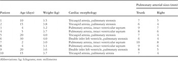
Table 1.

The completed systemic-to-pulmonary arterial anastomosis between the brachiocephalic artery (IA) and the pulmonary trunk (PA). AO: aorta; RPA: right pulmonary artery; RA: right atrium; SVC: superior caval vein.
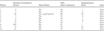
Table 2.

Lateral view of a postoperative aortogram, showing a patent shunt, which uniformly fills both pulmonary artery branches. LPA: left pulmonary artery; MPA: pulmonary trunk; RPA: right pulmonary artery.
 You have
Access
- 1
- Cited by