Implications
Dietary turmeric powder increases rabbit viability and fecundity (the number of liveborn and weaned pups), as well as the production and growth of ovarian follicles by altering both the release of ovarian hormones and the response of ovaries to gonadotropin.
Introduction
Turmeric (Curcuma longa L.) is widely used in both folk medicine and cooking, and along with other spices, is an important component of curry powder. The species’ rhizome contains several biologically active curcurminoids, of which the most prominent is polyphenol curcumin – 1,7-bis(4-hydroxy-3-methoxyphenyl)-1,6-eptadiene-3,5-dione (Lestari and Indrayanto, Reference Lestari and Indrayanto2014), and its antioxidant, antiinflammatory, antimicrobial, antiangiogenic and anticancer properties have been widely reported (Fan et al., Reference Fan, Zhang, Liu, Yan and Liang2013; Kunnumakkara et al., Reference Kunnumakkara, Bordoloi, Padmavathi, Monisha, Roy, Prasad and Aggarwal2016; Stanić, Reference Stanić2017). For example, the ability of curcumin to suppress the proliferation of epithelial cancer cells and to promote apoptosis has been well documented (Inano et al., Reference Inano, Onoda, Inafuku, Kubota, Kamada, Osawa, Kobayashi and Wakabayashi2000; Terlikowska et al., Reference Terlikowska, Witkowska, Zujko, Dobrzycka and Terlikowski2014; Bondì et al., Reference Bondì, Emma, Botto, Augello, Azzolina, Di Gaudio, Craparo, Cavallaro, Bachvarov and Cervello2017), and curcumin has also been reported to protect mouse ovarian cells from the pro-apoptotic, pro-necrotic, antiproliferative, and anti-oogenic actions of stresses induced by the downregulation of FSH receptors (Tiwari-Pandey and Ram Sairam, Reference Tiwari-Pandey and Ram Sairam2009), immunological shock (Voznesens’ka et al., Reference Voznesens’ka, Bryzhina, Sukhina, Makohon and Aleksieieva2010), hypoxia (Sak et al., Reference Sak, Soydinc, Sak, Evsen, Alabalik, Akdemir and Gul2013) and ionizing irradiation (Aktas et al., Reference Aktas, Kanter and Kocak2012). Furthermore, the dietary administration of curcumin does not affect the ovarian weight or litter size of rats subjected to γ irradiation (Inano et al., Reference Inano, Onoda, Inafuku, Kubota, Kamada, Osawa, Kobayashi and Wakabayashi2000), whereas an artificial pegylated curcumin analogue reportedly suppresses rat ovarian folliculogenesis, sexual maturation and fecundity (Murphy et al., Reference Murphy, Tang, Van Kirk, Shen and Murdoch2012), thereby suggesting that curcumin affects healthy reproductive systems. However, despite widespread medicinal and nutritional application, the effects of turmeric and its constituent compounds on reproductive processes and healthy ovaries have yet to be examined. Such studies could help identify cheap and natural plant compounds that stimulate mammalian reproduction, with important applications in farm animal production, and rabbits, which are small farm animals with high reproductive potential and large variation in reproductive efficiency, could be used as a sensitive and economically important model system.
The mechanisms that underlie turmeric’s effects on reproduction remain unknown. However, it is generally accepted that fecundity depends on the growth, development and atresia of ovarian follicles, and these processes are regulated by gonadotropins (LH and FSH), ovarian steroids (progestogens, androgens and estrogens), and peptide hormones, including leptin (Sirotkin, Reference Sirotkin2011 and Reference Sirotkin and Harrath2014; Sirotkin et al., 2014). Curcumin is a phytoestrogen (Bachmeier et al., Reference Bachmeier, Mirisola, Romeo, Generoso, Esposito, Dell’eva, Blengio, Killian, Albini and Pfeffer2010) and can, therefore, interact with the endocrine system, affect the hypothalamo–hypophysial–ovarian axis and treat its disorders (Sirotkin and Harrath, Reference Sirotkin, Chrenek, Kolesarová, Parillo, Zerani and Boiti2014). For example, in mouse ovaries, curcumin has been shown to downregulate androgen receptors and upregulate 3-β-hydroxysteroid dehydrogenase (Tiwari-Pandey and Ram Sairam, Reference Tiwari-Pandey and Ram Sairam2009), although it had no effect on aromatase (Valentine et al., Reference Valentine, Le Nedelec, Menzies, Scandlyn, Goodin and Rosengren2006), and curcumin also failed to affect plasma levels of progesterone, estradiol and FSH in γ irradiated rats, even though it increased levels of LH and decreased levels of prolactin (Inano et al., Reference Inano, Onoda, Inafuku, Kubota, Kamada, Osawa, Kobayashi and Wakabayashi1999 and Reference Inano, Onoda, Inafuku, Kubota, Kamada, Osawa, Kobayashi and Wakabayashi2000). Yet, to the best of our knowledge, no studies have investigated the action of turmeric on endocrine regulators of reproduction. Therefore, the aim of the present study was to examine the effect of dietary turmeric on the growth, viability, reproduction, ovary development, hormone release and the release of leptin in response an upstream hormonal stimulator, LH.
Material and methods
In vivo experiments
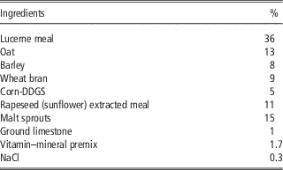
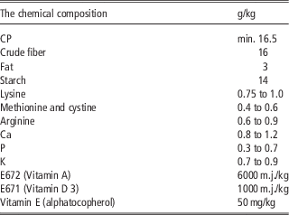
We obtained 60-day-old, clinically healthy female New Zealand White rabbits (Oryctolagus cuniculus; n=46) from the farm of the Research Institute for Animal Production, Nitra, Slovakia. Throughout the experiment, the animals were housed in individual cages (103×55×51 cm) under a light cycle of 16 h light and 8 h dark and were given ad libitum access to both a commercially available feed mixture (KV; Tekro, s.r.o., Párovské Háje, Nitra, Slovakia; Tables 1 and 2) and water (with nipple drinkers). The rabbits were divided into three groups, based on the amount of turmeric powder (Mäspoma s.r.o., Zvolen, SlovakRepublic) added to their feed: 0, 5 and 20 g per 100 kg feed for the control group (n=15) and two experimental groups (E1, n=16; E2, n=15), respectively. The turmeric and feed were combined to form 6 mm pellets, and the corresponding diets were provided to the animals for the duration of the experiment (i.e. 235 days, from the 60th to 295th day of life, after weaning pups in the second reproductive cycle).
Ingredients of the pelleted rabbit feed used in the experiments (data provided by Tekro, s.r.o., Párovské Háje, Nitra, Slovakia)

Chemical composition of the pelleted rabbit feed used in the experiments (data provided by Tekro, s.r.o., Párovské Háje, Nitra, Slovakia)

For artificial insemination, ejaculates were collected from tested bucks using an artificial vagina, and semen samples from three to four bucks were mixed and, thereafter, diluted (1 : 6) with an insemination diluent (DMRS; MiniTüb, Tiefenbach, Germany) to a final concentration of ~100 million spermatozoa/ml, which was determined using a computer-assisted sperm analysis system (Sperm Vision, MiniTüb, Tiefenbach, Germany). At 48 h after treatment with pregnant mare serum gonadotropin (intramuscular injection of 25 IU in 1 ml PBS; Sergon, Bioveta s.r.o., Ivanovice to Hané, Czech Republic), each doe was artificially inseminated with 0.5 ml diluted semen, using a plastic insemination pipette, and injected intramuscularly with 100 μg synthetic gonadotropin-releasing hormone (Supergestran 36; Ferring Pharmaceuticals s.r.o., Jesenice, Czech Republic), in order to induce ovulation.
The conception rate of the inseminated does (i.e. proportion becoming pregnant) was determined by abdominal palpation at 15th day after insemination, and the kindling rate, as well as the numbers of liveborn and stillborn pups, were recorded at 31 days after insemination. The first artificial insemination of the rabbit does was performed after 56 days of experimental feeding (day 110 of life), and artificial insemination was repeated for two reproductive cycles, both during the 235-day feeding period. The second cycle was initiated following the methods of the first cycle, and the second insemination was performed after 166 days of experimental feeding (day 220 of life).
After the pups from the second litter had weaned (day 295 of the experiment), the does were killed via decapitation and were dissected, in order to collect their ovaries. The extracted organs were weighed and measured, and five of the left-hand ovaries from each group were subjected to histological analysis, whereas the remaining ovaries were used for subsequent in vitro experiments. The study’s treatment of animals was approved by the Ministry of Agriculture and Rural Development of the Slovak Republic, No. SK P 28004 and Ro 1488/06-221/3a.
In vitro experiments
The collected ovaries were washed in phosphate buffered saline (PBS) with 1% antibiotic-antimycotic solution (Sigma, St. Louis, IL, USA), placed onto 100 mm diameter culture dishes (Gama, České Budejovice, Czech Republic), and dissected using a blade knife. The resulting 1/8th of the ovaries were washed thrice in PBS. Two randomly selected ovarian fragments were cultured in 24-well Falcon plates (Becton Dickinson, Lincoln Park, USA), each with 2 ml sterile incubation medium (DMEM/F12 1 : 1) that was supplemented with 10% fetal calf serum (BioWhittaker, Verviers, Belgium) and 1% antibiotic-antimycotic solution (BioWhittaker). To determine the effect of dietary turmeric on the basic release of ovarian hormones, fragments from the ovaries of each group were cultured without the addition of hormones, and to determine the effect of dietary turmeric on the response of ovaries to LH, fragments from the ovaries of each group were cultured in the presence of biological-grade pLH (Sigma), which dissolved immediately before the experiments at doses of 0, 1, 10 or 100 ng/ml incubation medium. A blank control included the culture medium but no follicular fragments. After 2 days, the medium from plate wells was aspirated and frozen at −18°C until analyzed via radioimmunoassay (RIA).
Histology
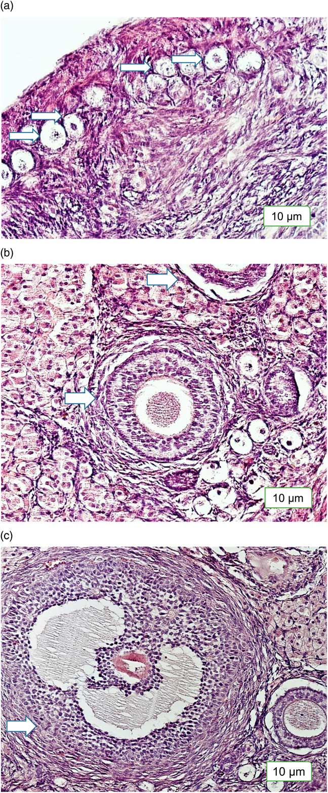
After isolation, the five left-hand ovaries collected from each group were immediately fixed in 10% formalin for 24 h; washed in flowing tap water for 2 h; dehydrated in 70, 96 and 100% ethanol (2 h each); and embedded in paraffin (Heraeus Kulzer GmbH&CoKG, Wehrheim/Ts., Germany). Afterward, the ovaries were serially sectioned using a standard rotation microtome at 5 μm thickness, and every tenth section was transferred to a glass slide (Bamed s.r.o., Ceske Budejovice, Czech Republic), dewaxed, rehydrated, stained with eosin-hematoxylin (Diapath S.p.A., Martinengo, Italy), and embedded in entellan (Merck, Darmstadt, Germany). All histological sections were examined and photographed using an Eclipse E600 light microscope (Nikon spol s.r.o., Prague, Czech Republic) and analyzed using the NIS-Elements image-analysis software (version 3.0; Nikon). For each slide, 20 representative areas were analyzed, and both the diameters of visible primary, secondary, and tertiary follicles were measured using an ocular-micrometer in the area of oocyte localization (Figure 1).
(Colour online) Histological slices of (a) primary (E2 group), (b) secondary (control group) and (c) tertiary (E1 group) ovarian follicles from rabbits. Magnification ×200, staining with eosin–hematoxylin. Control (no dietary turmeric), E1–5 g turmeric per 100 kg food, E2–20 g turmeric per 100 kg food. Scales are included at the bottom of each image.

Immunoassay
The concentrations of progesterone, testosterone, estradiol and leptin in the incubation medium samples from the in vitro experiment were measured using 25 μl aliquots of each incubation medium, RIA kits for progesterone and estradiol (DSL, Webster, TX, USA) and testosterone (Immunotech, Marseille, France) and a multispecies leptin RIA kit from Linco (St. Charles, USA), according to the manufacturers’ instructions. The RIAs were validated for use in culture medium samples. The anti-progesterone antiserum exhibited <0.001% cross-reactivity to cortisol, corticosterone, cortisol, androstenediol, pregnenolone, estradiol and testosterone. The sensitivity of this assay was 0.12 ng/ml, and the intra- and inter-assay coefficients of variation did not exceed 13.1% or 8.0%, respectively. The antitestosterone antiserum exhibited <0.001% cross-reactivity with dehydroepiandrosterone (DHEA), progesterone, estradiol and other related molecules. The sensitivity of this assay was 0.025 ng/ml, and the intra- and inter-assay coefficients of variation did not exceed 14.8% or 15.0%, respectively. The anti-estradiol antiserum exhibited <0.01% cross/reactivity to DHEA, progesterone, testosterone, cortisol, androsterone, corticosterone and cortisone. The sensitivity of this assay was 6.5 ng/ml, and the maximal intra- and inter-assay coefficients were 19.2% and 9.4%, respectively. The anti-leptin antiserum exhibited 100%, 67%, 61%, 73% and 3% cross-reactivity to human, porcine, rat, mouse and canine leptin, respectively, and <0.001% cross-reactivity to human insulin, proinsulin, and C-peptide and rat insulin, glucagon, and IGF-I. The sensitivity of this assay was 0.801 ng/ml, and the intra- and inter-assay coefficients of variation did not exceed 3.6% or 8.7%, respectively.
Statistical analysis
The in vivo experiment involved 46 animals, which were studied over the course of two consecutive reproductive cycles, and all the animals were sacrificed at the end of the experiment. Histological analysis was performed on ovaries from five animals per group, and the histological data represent ⩾20 slices per sample. In the in vitro experiments, each experimental group was represented by six culture wells (two ovarian fragments per well), and hormone release was calculated per milligram tissue per day by subtracting the measured values from the non-specific background (medium cultured without follicular fragments, less than 10%). Differences between the experimental groups were evaluated using one- or two-way ANOVA, followed by either χ 2 or Wilcoxon–Mann–Whitney tests to compare percentages or numbers, weights and sizes, respectively. The analyses were performed using the statistical software Sigma Plot 11.0 (Systat Software, GmbH, Erkrath, Germany), and differences between experimental and control groups were considered significant at P<0.05.
Results
In vivo experiments
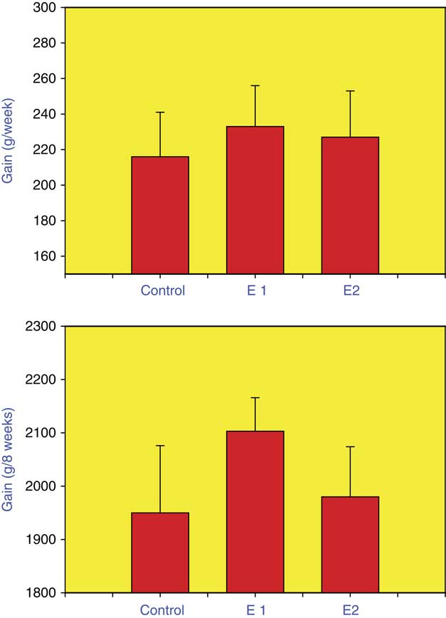
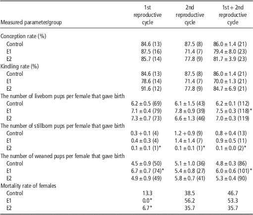
The initial BW of the does was 680±20 g, and by the end of the experiment, the median BW of the does was 2980±120 g. Weekly weight gain varied from 69.3 g in the 8th week of the experiment to 327.1 g in the 3rd week. After the 8th week, the weight gain was insignificant, so we did not analyze it further. Dietary turmeric did not affect the median weekly or median total BW gain of the pups (Figure 2). Weekly measurement of weight gain did not indicate significant differences between the groups either. There were no significant differences in the conception of kindling rates of the three treatment groups (C, E1 and E2). However, group E1 produced significantly more liveborn (21.0%) and weaned (25.0%) pups than group C, and E2 produced 87.5% fewer stillborn pups. Dietary turmeric also decreased doe mortality during the first reproductive cycle, from 13.3 in group C to 0% in group E1 and 6.7% in group E2, but not during the second cycle. For most variables, the effect of dietary turmeric was evident during both the first and second reproductive cycles, although this was not the case for the effect of turmeric on the number of weaned pups and doe mortality, which was only significant during the first reproductive cycle (Table 3).
Effect of dietary turmeric on the BW gain of rabbits. Top – BW gain per week; bottom – BW gain during entire experiment (8 weeks). Control (no dietary turmeric), E1–5 g turmeric per 100 kg food, E2–20 g turmeric per 100 kg food. Values and error bars represent means±SEM.

Effect of dietary turmeric on the reproduction and vitality indices of female rabbits during two consecutive reproductive cycles

Control (no dietary turmeric), E1–5 g turmeric per 100 kg food, E2–20 g turmeric per 100 kg food. Values and error bars represent means±SEM.
*Significant difference (P<0.05) from the control group. Numbers in brackets indicate sample numbers.
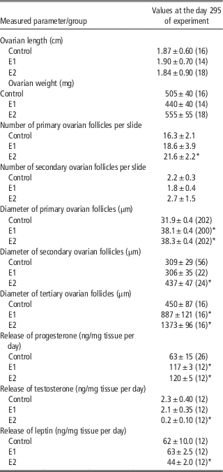
Dietary turmeric did not affect ovarian length or weight. However, higher doses (i.e. group E2) significantly increased the number of primary follicles by 32.5%. Turmeric also significantly increased the diameters of primary (E1: +19.4%; E2: +21.1%), secondary (E1: no significant difference; E2: +41.4%,), and tertiary (E1: +97.1%; E2: +205.1%) follicles (Table 4). The number of tertiary follicles per photo was not calculated, owing to their rarity (i.e. most photos lacked them). No other visible differences were observed in the state of the follicles, which indicated either degenerative or reparative processes.
Effect of dietary turmeric on the morphometric and endocrine indices of female rabbits during two consecutive reproductive cycles

Control (no dietary turmeric), E1–5 g turmeric per 100 kg food, E2–20 g turmeric per 100 kg food. Values and error bars represent means±SEM.
*Significant difference (P<0.05) from the control group. Numbers in brackets indicate sample numbers.
In vitro experiments
In the first in vitro experiment, the ovarian fragments from groups E1 and E2 released 97.1 and 205.1% more progesterone than group C, respectively, and group E2 also produced 87.0% less testosterone and 29% less leptin (Table 4). However, the concentration of estradiol in the incubation medium was below the detection limit of RIA.
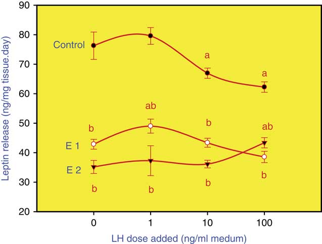
In the second in vitro experiment, the ovaries isolated from groups E1 and E2 released significantly less leptin in the absence of LH than those isolated from group C (Figure 3), and the leptin output of group C was reduced by the 10 and 100 ng/ml LH treatments. However, the release of leptin from the ovaries isolated from groups E1 and E2 was promoted by the addition of 1 and 100 ng/ml LH, respectively (Figure 3). Meanwhile, as in the first experiment, the concentration of estradiol in the incubation medium was below the detection limit.
Effect of turmeric on the release of leptin by fragments of rabbit ovaries. Fragments were either cultured with LH (1, 10 or 100 ng/ml medium) or without LH (0 ng/ml), and leptin was measured after 295 days of turmeric administration (after completion of the second reproductive cycle). (a) Effect of LH. (b) Effect of dietary turmeric. Control (no dietary turmeric), E1–5 g turmeric per 100 kg food, E2–20 g turmeric per 100 kg food. Values and error bars represent means±SEM.

Discussion
Effects of turmeric on female reproduction
In the in vivo experiment, dietary turmeric increased the number of primary follicles, as well as the diameter of primary, secondary and tertiary follicles, which suggests that turmeric promotes rabbit fecundity in two ways: by promoting the production of primary ovarian follicles and by stimulating follicle growth throughout folliculogenesis. To the best of our knowledge, this is the first report of turmeric’s promoting activity on the folliculogenesis of healthy animals. However, our findings are in line with those of previous studies that have described the influence of curcumin on stressed mouse ovaries, namely that curcumin helps preserve the ovarian functions of mice subject to FSH receptor downregulation (Tiwari-Pandey and Ram Sairam, Reference Tiwari-Pandey and Ram Sairam2009), immunological shock (Voznesens’ka et al., Reference Voznesens’ka, Bryzhina, Sukhina, Makohon and Aleksieieva2010), hypoxia (Sak et al., Reference Sak, Soydinc, Sak, Evsen, Alabalik, Akdemir and Gul2013) and γ irradiation (Aktas et al., Reference Aktas, Kanter and Kocak2012). Because the previous studies determined that curcumin can prevent stress-induced reductions in ovarian cell proliferation and can promote ovarian apoptotic and necrotic activities, we suggest that the generation and growth of rabbit follicles in the present study were caused by the action of curcumin on ovarian cell proliferation: death rate. The mechanism underlying such activity requires further elucidation, although it is possible that curcumin reduces ovarian cell death and causes ovarian follicle atresia through its anti-oxidative properties in both ovarian (Sak et al., Reference Sak, Soydinc, Sak, Evsen, Alabalik, Akdemir and Gul2013) and non-ovarian (Kunnumakkara et al., Reference Kunnumakkara, Bordoloi, Padmavathi, Monisha, Roy, Prasad and Aggarwal2016) tissues.
The ability of curcumin to prevent the stress-induced degeneration of rat oocyte zona pellucida (Tiwari-Pandey and Ram Sairam, Reference Tiwari-Pandey and Ram Sairam2009) and stress-induced suppression of murine oocyte nuclear maturation (Voznesens’ka et al., Reference Voznesens’ka, Bryzhina, Sukhina, Makohon and Aleksieieva2010) suggest that turmeric provides protective effects during both ovarian folliculogenesis and oogenesis, which together determine fecundity. However, in the present study, dietary turmeric had no effect on the number of either liveborn or weaned pups. To our knowledge, this is the first report that turmeric can function as a biostimulator of fecundity in healthy animals. Dietary turmeric also failed to affect rabbit conception and kindling rate but did improve pup viability, which indicates that turmeric promotes rabbit reproductive efficiency by improving pup viability, rather than by its action on the number of pups. Turmeric could also improve the viability of adult does. However, in the present study, such a positive influence was only observed in a limited number of animals and only during the first reproductive cycle.
Potential mechanisms of turmeric’s activity
Steroid hormones
Even though the mechanisms of turmeric’s action on rabbit reproduction require further elucidation, ovarian functions, including rabbit reproduction, are typically regulated by phytoestrogens (Sirotkin and Harrath, Reference Sirotkin, Chrenek, Kolesarová, Parillo, Zerani and Boiti2014), gonadotropins, and other steroid and peptide hormones (Sirotkin et al., 2014; Sirotkin, Reference Sirotkin and Harrath2014). Owing to curcumin’s strong phytoestrogenic properties (Bachmeier et al., Reference Bachmeier, Mirisola, Romeo, Generoso, Esposito, Dell’eva, Blengio, Killian, Albini and Pfeffer2010) and its effects on steroid hormone synthesis and reception in the ovaries of stressed mice (Tiwari-Pandey and Ram Sairam, Reference Tiwari-Pandey and Ram Sairam2009), it is likely that the action of turmeric on the reproductive system is mediated by ovarian steroids and/or steroid receptors. However, some authors have reported that curcumin fails to affect levels of aromatase (mice: Valentine et al., Reference Valentine, Le Nedelec, Menzies, Scandlyn, Goodin and Rosengren2006) or plasma levels of progesterone and estradiol (rat: Inano et al., Reference Inano, Onoda, Inafuku, Kubota, Kamada, Osawa, Kobayashi and Wakabayashi1999 and Reference Inano, Onoda, Inafuku, Kubota, Kamada, Osawa, Kobayashi and Wakabayashi2000).
In the present study, rabbit ovaries released high levels of progesterone but not estradiol, which is characteristic of ovarian tissue luteinization (Sirotkin, Reference Sirotkin and Harrath2014), and the ovaries of rabbits fed high doses of turmeric (group E2) released more progesterone and less testosterone in vitro than the ovaries of control does (group C). Given the key regulatory roles of progestogens and androgens (Sirotkin, Reference Sirotkin and Harrath2014), our observations suggest that ovarian folliculogenesis, oogenesis and fertility are affected by turmeric through changes in the output of ovarian steroid hormones. However, not all changes in the release of steroid hormones were associated with changes in ovarian folliculogenesis or fecundity and vice versa, which indicates that steroid hormones are not the only mediators of turmeric’s action.
Leptin
Our in vitro experiments provided the first evidence that turmeric can downregulate ovarian leptin release. Leptin is a potent regulator of ovarian function in a variety of mammals (Sirotkin, Reference Sirotkin2011), including rabbits (Sirotkin et al., 2014). Therefore, leptin is an additional candidate hormone mediator of turmeric’s effect on rabbit reproduction.
Luteinizing hormone
It is also possible that pituitary hormones could mediate the effects of turmeric. For example, Inano et al. (Reference Inano, Onoda, Inafuku, Kubota, Kamada, Osawa, Kobayashi and Wakabayashi1999 and Reference Inano, Onoda, Inafuku, Kubota, Kamada, Osawa, Kobayashi and Wakabayashi2000) reported that curcumin increases LH (but not FSH) in the plasma of rats subjected to γ irradiation. The present study provides the first evidence that LH affects the release of leptin in rabbits and confirms previous reports that LH is involved in the regulation of rabbit ovarian functions (Sirotkin et al., Reference Stanić2014) and mediates the action of turmeric on ovarian functions (Inano et al., Reference Inano, Onoda, Inafuku, Kubota, Kamada, Osawa, Kobayashi and Wakabayashi2000). In addition, our observation that LH suppresses the release of ovarian leptin in control animals (at doses of 10 and 100 ng/ml) and promoted it in turmeric-fed does (at 1 and 100 ng/ml in groups E1 and E2, respectively). This suggests that turmeric can stimulate ovarian functions not only by promoting the release of leptin but also by modulating the response of ovaries to LH. We suggest that dietary turmeric may affect ovarian LH receptors, with lower doses being more effective than higher doses. It is also possible that turmeric promotes rabbit ovarian function and fecundity by respectively reducing and inducing the inhibitory and stimulatory actions of LH on ovarian leptin. Therefore, we hypothesize that turmeric stimulates rabbit folliculogenesis and fecundity altering the response of ovarian cells to LH, probably by modulating LH receptors, that some of the effects of LH are mediated by leptin, and that both LH and leptin affect ovarian processes via steroid hormones. On the other hand, steroid hormones could affect LH, LH receptors and leptin through feedback mechanisms, and finally, turmeric could affect both ovaries and LH through the phytoestrogenic effects of curcumin on steroid hormone receptors.
It is important to note that turmeric-induced changes in regulatory molecules do not necessarily indicate that these molecules are functional mediators of turmeric action. The effect of turmeric on some parameters was not dose-dependent, which could be due to negative feedback mechanisms or multiple mechanisms of action on different sites. Furthermore, the effects of turmeric on some parameters were statistically significant during the first, but not the second, reproductive cycle. This indicates the lack of a cumulative effect but, rather, a decrease in turmeric action during its application, which is to be considered by its long lasting use. Therefore, functional interrelationships between the ovarian indices, processes, and molecules affected by turmeric require further clarification, and additional basic studies are needed to elucidate the potential roles of other hormonal and intracellular mediators, both in order to validate our findings and to identify the specific molecules responsible for turmeric’s effects. Indeed, most reports of turmeric’s action are based on only one of its molecular components, namely curcumin. However, curcumin is rarely consumed alone, and both turmeric and turmeric-containing food contain a number of other biologically active molecules, as well. Therefore, from a practical point of view, our observations suggest that turmeric can be used as an effective, natural, cheap, and efficient biostimulator of rabbit viability and reproduction, with important applications for commercial rabbit production. We recommend a dose of 5 g turmeric per 100 kg of feed mixture for application on rabbit farms since this treatment significantly improved the numbers of liveborn and weaned pups and was the most effective in improving the viability of does during the first reproductive cycle. Further studies will contribute to further improving the regime (dose, duration, etc.) of turmeric application in rabbit breeding. Furthermore, the abilities of turmeric to promote rabbit ovarian folliculogenesis and fecundity suggest that the plant may also be useful for improving the reproductive efficiency of other economically important farm animals. Finally, future studies should also determine whether turmeric can affect the reproductive processes or humans and whether it can be used to regulate human reproduction or to treat reproductive disorders.
Even though we are just beginning to understand the effects of turmeric on mammalian reproduction, the present study provides clear evidence that turmeric stimulates rabbit viability, ovarian folliculogenesis, and fecundity and that these effects could be related to the release of ovarian hormones and ovarian response to LH.
Acknowledgements
The authors express their gratitude to Ms K. Tothová and Ž. Kuklová for technical assistance. This work was supported by the Slovak Research and Development Agency (APVV), under contract numbers APVV-0854-11 and APVV-15-0296; Slovak Grant Agency of the Ministry of Education, Science and Sport; and Slovak Academy of Science (VEGA), project VEGA 1/0392/17. The authors would also like to extend their sincere appreciation to the Deanship of Scientific Research at King Saud University for funding this research RGP-164.