Introduction
Several surgical reconstructive techniques have been used in patients with tracheal lesions of diverse causes. However, none of these techniques are completely effective.Reference Delgado-Pecellin, González-Valencia, Machuca-Contreras and Pineda-Mantecón1, Reference Krajc, Janik, Benej, Lucenic, Majer and Demian2 Some reports have described favourable outcomes of tracheal prostheses in adult patients.Reference Jungebluth, Alici, Baiguera, Le Blanc, Blomberg and Bozóky3 Children usually experience rapid growth of all organs, including the trachea, during early life.Reference Griscom and Wohl4 Therefore, tracheal prostheses implanted in children, particularly during infancy, need to have sufficient malleability to allow continued tracheal growth, without imposing significant structural changes or functional limitations during growth.
In a previous study, implantation of a mersylene-coated homologous collagen prosthesis in adult rats, to repair a partial non-circular defect made on the tracheal cervical wall, had good outcomes.Reference Villegas-Álvarez, González-Zamora, González-Maciel, Soriano-Rosales, Pérez-Guille and Padilla-Sánchez5 The present study aimed to examine the clinical and histological properties of the trachea after partial tracheal wall substitution with this prosthesis, and to assess the effects of this prosthesis on the growth of the tracheal lumen during the first year of life in dogs.
Materials and methods
The study was reviewed and approved by the Research and Ethic Committees for Laboratory Animal Care and Use, of the Instituto Nacional de Pediatría. All animal procedures were conducted in accordance with Mexican Official Norms (NOM-082-ZOO), Internal Facility Guidelines of the Instituto Nacional de Pediatría, and the “Guide for the Care and Use of Laboratory Animals” for the National Research Council of the National Academies U.S.A.6
A total of 16 puppies (9 males and 7 females) from 4 deliveries by 2 female dogs crossed with the same male (born at the Instituto Nacional de Pediatría) were used in this study. The puppies were consecutively assigned a number and allocated to an experimental group or a control group. The biological father of the puppies was used as the source of the collagen employed to cover the prosthesis.
Implantation and surgical procedures
The prosthesis creation process, implantation procedure, surgical techniques and histological analyses were performed as described previously.Reference Villegas-Álvarez, González-Zamora, González-Maciel, Soriano-Rosales, Pérez-Guille and Padilla-Sánchez5 Briefly, a 40 denier polyester XRZ4 mesh (Celanese, Dallas, Texas, USA) was used to wrap two Silastic tubes (1 cm diameter, 5 cm long; Dow Corning, Midland, Michigan, USA). The wrapped tubes were sterilised with ethylene oxide at 80 per cent for 24 hours, and were subcutaneously installed on the back of the donor dog using antiseptic surgical techniques. The prostheses were extracted 4 weeks later and soaked in 0.5 per cent Hank solution for 15 days, after which the tubes were removed and washed in saline solution for 45 minutes. The prostheses were then split into 1 × 1 cm segments, yielding five cylindrical prostheses coated with fibrocollagen. A Silastic tube (1.5 cm long, 1 cm diameter) was introduced into each tube to maintain their shape. The remaining prosthetic fragment was sent to the Faculty of Chemistry, Universidad Nacional Autónoma de México, for fibrocollagen component analyses.
The experimental group underwent surgical implantation at 29–32 days of age. The objective of the procedure was to generate adhesion and promote angiogenesis on the prostheses, as in our previous study.Reference Villegas-Álvarez, González-Zamora, González-Maciel, Soriano-Rosales, Pérez-Guille and Padilla-Sánchez5 The prosthesis was placed on the external wall of the trachea immediately below the second cervical cartilage, and was fixed with a single-point suture (polyglactin 6-0; Ethicon, Somerville, New Jersey, USA) and covered with cervical muscle tissue. The incisions were closed, and once the puppies had recovered from the residual effects of anaesthesia, they were re-integrated with their mothers and kept under controlled temperature and light conditions.
Four weeks later, the puppies in the experimental group underwent sterile surgery to expose the anterior surface of the prosthesis. By using an anterior incision and avoiding separating the prosthesis from the muscle mass, approximately 30–40 per cent of the total tracheal cartilaginous wall was removed, from tracheal rings C4–C7. The resulting defect was covered by the concave face of the prosthesis, and the Silastic tube and fibrocollagen cylinder remnants were removed. The tissue was sutured using polypropylene 6-0 sutures (Meadox Medical, Oakland, New Jersey, USA) to ensure the edges of the cartilage coincided with the edges of the prosthesis. The muscle layers were sutured with 4-0 polyglactin 9101 sutures (Ethicon) and the skin was sutured with polypropylene 4-0 sutures (Meadox Medical).
Post-operative management and endoscopic evaluation
Following the surgical procedure, the experimental animals underwent laryngotracheoscopy using a rigid bronchoscope (Karl Stortz, Tuttlingen, Germany). The implanted zone was localised and filmed. Endoscopy was repeated every month for the first 6 months and then every 60 days for 8 months. Tomographic measurements were obtained at the same times.
After recovery from anaesthesia, the puppies were housed individually in controlled-temperature conditions. Water and food (Pedigree Cachorros; Effem, Querétaro, Mexico) were given to the puppies on demand; the amount of food given depended on their weight. The dogs received nebulisation with budesonide 250 mg/2 ml (Pulmicort; AstraZeneca, London, UK) per 12 hours twice in the immediate post-operative period. Flunixin meglumine 0.04 mg/kg (Napzin; Pisa, Hidalgo, Mexico) was also administered intramuscularly every 12 hours for 2 days. Enrofloxacin (Baytril; Bayer Healthcare Animal Health Division, Berlin, Germany) was administered intramuscularly at a dose of 5 mg/kg every 24 hours for 5 consecutive days.
Tomographic measurements
Tomographic measurements were performed by the Helicoidal Multi-cut Tomography Service of the Imaging Department of Instituto Nacional de Pediatría. Measurements were taken after determining body weight, whilst the dogs were under non-intubated general anaesthesia. These measurements were recorded in both groups of dogs every 30 or 60 days for 14 consecutive months, starting from 4 weeks of age. A helicoidal multi-cut tomographic system (Somaton Sensation 4; Siemens, Munich, Germany) was used, with a voltage of 100 kV, current of 50 mAs, collimation of 1 mm and reconstruction window of 0.75 mm.
For measurements, the dogs were placed in a supine position (using lateral positioners) on the topographic table, at a height of 1.35 m (on soft pillows). The laser was applied for 6–12 seconds depending on the thoracic region, from above the larynx to below the diaphragmatic dome.
Tracheal fore–back diameter and transversal diameter were measured in three sites: (1) immediately above the border of the implant; (2) in a region corresponding to the middle of the implant; and (3) at the inferior edge of the implant. The adjacent vertebral body was used as a reference point to localise the target sites in the control group. The area and circumference values of the tracheal lumen were analysed using software provided by the manufacturer (Siemens). The images, taken at slice thicknesses of 1–5 mm, were saved onto a compact disc and were analysed at the end of the study period.
Anaesthetic procedure
All surgical, tomographic and endoscopic procedures were carried out under non-intubated general anaesthesia, using a mixture of 7 mg/kg xylacine (Rompun; Bayer Healthcare, Leverkusen, Germany), tiletamine plus zolazepam 10 mg/kg (Zoletil; Virbac, Carros, France) and intramuscular administration of 0.05 mg/kg atropine (Atropisa; Pisa, Guadalajara, Mexico).
Clinical evaluation
Respiratory frequency was measured daily for the first four weeks after implantation. Spontaneous activity of the animals was also recorded, as well as any evidence of wheezing, intercostal retraction or cyanosis. The body weight of the dogs in both groups was measured every 30 days until the end of the study. At the end of the study, the dogs in the experimental group were euthanised with an overdose of sodium pentobarbital. The tracheas were removed and sent to the microscopic and immunochemical laboratories of the Instituto Nacional de Pediatría and Universidad Nacional Autónoma de México, respectively, for analysis.
Statistical analysis
The independent samples Student's t-test was used to compare body weight and the tracheal variables at baseline between the two groups. Growth curve analysis was conducted to examine changes in tracheal dimensions over time in both groups; specifically, a linear mixed model with random effectsReference Brown and Prescott7 was applied to each tracheal parameter. Mixed models are suitable when repeated observations are made on the same animal at fixed but unequally spaced times. The treatment group and observation time were included in the analysis as fixed effects (i.e. as independent variables). Where necessary, we also included a quadratic effect for time, and an interaction between time and treatment group. The lack of independence among observations in the same dog was taken into account by using the random effects model. A mixed model analysis was also used to examine changes in body weight over time in both groups of dogs. An analysis of residual variance was performed to confirm the assumptions of the models. All analyses were conducted using R2.10.1 software.8, Reference Pinheiro, Bates, DebRoy and Sakar9
Results
General characteristics
None of the dogs died during the study. In the immediate post-operative period, the dogs showed moderate tachypnoea and limited spontaneous activity, which normalised by day 3–4, except in dog number 8, which suffered from moderate wheezing for 2 consecutive weeks. In that dog, the signs progressively decreased and disappeared by the end of week three.
Endoscopic analysis
Endoscopy conducted after implantation in seven of the eight dogs in the experimental group did not reveal any significant alterations except for changes to the tracheal contour caused by the prosthesis, as the outline of the mesh could be clearly seen through the collagen coat (Figure 1a). By one month after implantation, the shiny surface of the implant had been replaced by a dense amber-coloured region covered by hyaline mucous secretion that was easy to remove by aspiration (Figure 1b). At two months, the surface of the trachea at the site of the implant had a similar appearance to the rest of the tracheal wall. However, this region did not have a circular shape, unlike the rest of the trachea, a feature that was apparent until the end of the study (Figure 1c).
Endoscopic view in the experimental group. (a) The recently implanted prosthesis is surrounded by ecchymosis at its junction with the tracheal wall. (b) One month later, the implant slightly protrudes into the lumen; it has a more friable appearance and is surrounded by hyaline mucus. (c) At the end of the study, the surface of the implant is smooth and is similar to the normal tracheal wall; it no longer projects into the lumen, but the contour of the trachea is still not circular in shape. (d) In dog number eight, part of the prosthetic lip, which initially protruded during implantation, exhibits a polyp appearance; it is similar in colour to the rest of the tracheal wall and was unchanged at the end of the study.

In dog number eight, which suffered from wheezing for two weeks, the implant was found to protrude into the lumen along one border of the prosthesis–tracheal connection; this was observed during the first post-operative endoscopic evaluation. At the end of the study, this protruding edge formed a polypoid shape with an irregular smooth surface across the implant site, showing no changes during the course of the study (Figure 1d).
Tomographic analysis
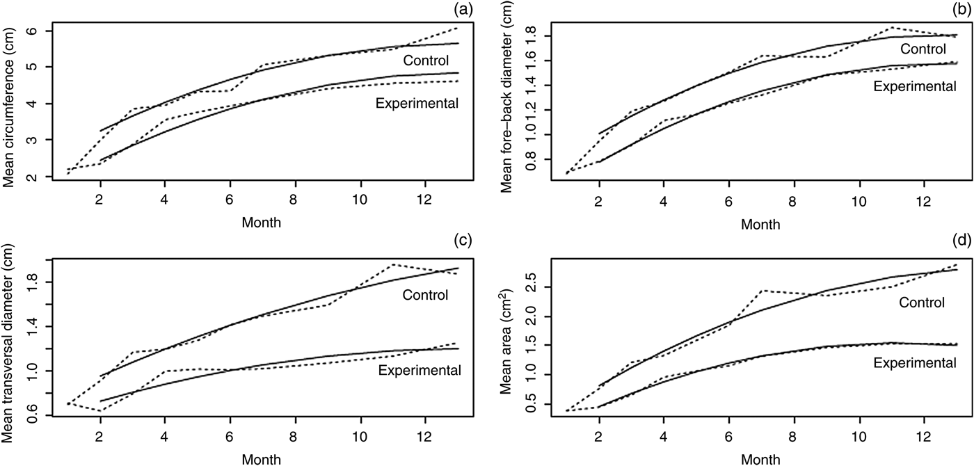
At baseline, body weight and the four tracheal parameters were similar in both groups of dogs (Table I). Plots were constructed to visualise the changes in each parameter for each dog (data not shown). As would be expected, body weight and the four tracheal parameters tended to increase as the dogs grew, with a quadratic trend in some parameters and linear trends in others. Although the growth curves in the experimental group were generally similar in the eight dogs during the study period, some outliers were observed. For example, the tracheal circumference in one dog at 180 days after birth was noticeably smaller than the previous and subsequent measurements. The growth curves for the four tracheal parameters in both groups are shown in Figure 2. The curves generally showed a quadratic trend, and the upper curves usually reflected the control group.
Growth curves for the four tracheal structural characteristics, for both groups (starting at 30 days of life): (a) circumference, (b) fore–back diameter, (c) transversal diameter and (d) area. These show the observed mean values (dashed lines) and predicted mean values (solid lines). Graphically, the absence of an interaction between group and time produces two parallel curves, while the presence of an interaction produces two diverging curves.
Relative group differences in baseline tomographic characteristics and body weight
A two-way analysis of variance with a block effect for the matched pairs of dogs revealed no significant differences between the two groups in terms of baseline body weight or tracheal characteristics. *Final value reported as a ratio of the baseline value.
The results of the statistical models are shown in Table II. Baseline measurements were not included as a covariate in the model because they did not predict subsequent observations. All four tracheal parameters were significantly smaller in the experimental group than in the control group, whereas there were no differences in body weight between the two groups. All of the mixed models included linear and quadratic effects of time. The interaction between group and time was only significant in the models for tracheal area and transversal diameter. These results imply that the area and circumference of the trachea increased at different rates in each group. The variance explained by the random effects model ranged from 0.23 to 0.75, and was significantly greater than 0 in all of the models. Residual analyses showed that the data followed a normal distribution and had constant variance, despite the presence of a few outliers (Table III).
Residual maximum likelihood estimates for fixed effects*
Values in parentheses are standard errors.
* For linear mixed models. Quad = quadratic
Residual maximum likelihood estimates for residual terms and random effects
Values in parentheses are 95 per cent confidence intervals. Variability in repeated observations was greater within animals (residual term) than between animals (random effect) for the four tracheal characteristics. The variability among animals explained between 23 and 46 per cent of the total variance in the tracheal characteristics. The variance between animals explained 75 per cent of the total variance in log body weight.
Using these models, the mean tracheal circumference in the control group was 3.25 cm on day 60 after birth and 5.65 cm at the end of the study. By comparison, the mean tracheal circumference was 0.81 cm shorter in the experimental group than in the control group at both time-points, corresponding to decreases of 24.9 per cent (0.81 out of 3.25) and 14.3 per cent (0.81 out of 5.65), respectively.
The mean estimated fore–back diameter ranged from 1.01 to 1.81 cm between days 60 and 390 after birth in the control group. The fore–back diameter was 0.23 cm smaller in the experimental group, corresponding to reductions of 22.8 per cent and 12.7 per cent at these times.
At 60 days after birth, the mean transversal diameter of the trachea was 0.96 cm in the control group and 0.73 cm in the experimental group. This difference of 0.23 cm represents a 23.95 per cent reduction in the experimental group. At the last measurement, the mean transversal diameters were 1.93 and 1.20 cm in the control and experimental groups respectively, yielding a difference of 0.73 cm (37.7 per cent).
The mean tracheal area at day 60 after birth was 0.81 cm² in the control group and 0.45 cm² in the experimental group, resulting in a difference of 0.36 cm² (44.3 per cent). The mean tracheal area at the end of the study was 2.78 cm² in the control group and 1.50 cm² in the experimental group, revealing a difference of 1.30 cm² (46.5 per cent).
Although the sample sizes were relatively small (eight dogs per group), the analyses were powerful enough to detect significant differences between the two groups in all four tracheal parameters. Power is only an issue when the null hypothesis of no difference in mean effects between groups is not rejected.Reference Spybrook, Raudenbush, Liu, Congdon and Martinez10
Light microscopy
This revealed that the subcutaneously implanted mesh from the donor was surrounded by dense connective tissue characterised by the presence of thick bundles of type I collagen fibres and fine type V collagen fibres. Type I collagen fibres were organised as lamellae of parallel fibres sited on the inner layer of the tubular mesh. By contrast, type V collagen fibres were localised on the external layer of the implant (Figure 3a,b).
Immunohistochemistry of the polyester mesh before implantation: (a) type I collagen (brown) is widely disseminated throughout the tissue, although its density is greatest around the polyester mesh; (b) type V collagen (purple) is also present, particularly under the mesh. (ABC Kit (Vector Laboratories, Burlingame, California, USA) and 3′3′diaminobenzidine chromogen (Sigma Chemical, St Louis, Missouri, USA); ×400)
Microscopic analysis showed that the prosthesis had fully adhered to the host's trachea without any granulation. The thickness of the implanted tracheal wall was about 20–30 per cent greater than that of the corresponding region in control dogs at the end of the study, but was 40 per cent smaller at the start of the study (Figure 4c). The material that formed the basis of the prosthesis was surrounded by, and was continuous with, connective and muscular tissue. We found vascular neoformation, blood vessels, arterioles, capillaries and venules in the region of the prosthesis. Vessels were scattered throughout the connective tissue and around the prosthesis. However, there were a larger number of capillaries immediately below the mucosa (Figure 4a,b). We also noted regeneration of pseudostratified ciliated epithelium on the surface of the implant (Figure 5a,b). Only one dog (number eight) had granulation tissue. The trachea at the site of the implant was folded and thickened, with three types of tissue, namely squamous epithelium, stratified epithelium with ciliated cells, and pseudostratified ciliated epithelium similar to normal tracheal tissue (Figure 6a–d).
Thickness of: (a) normal tracheal wall, (b) the prosthesis 12 months after implantation and (c) the prosthesis before implantation. The lines indicate the thickness from the mucosa (muc) to the adventitia (adv). As seen in part (b), the increase in prosthesis thickness seems to be at the expense of the extracellular matrix (collagen and glycosaminoglycans) and neovascularisation (nv). Some areas of submucosal tissue appear to be more compact (arrows) compared with the rest of the tracheal wall. Under the mucosa, there are many capillaries proximal to the prosthesis (arrowheads), as well as arteries and arterioles. As shown in part (c), type I collagen is expressed around the mesh, while the mucosa and adventitia are absent. (Masson's trichrome stain; ×50)
At the end of the study, respiratory epithelium covers the surface of the trachea and implant in both the control (a) and experimental (b) groups. Ciliated epithelium covers the regenerated mucosa and is similar to that in the control trachea. Neovascular formation is evident under the mucosa (arrows). The arrowheads indicate regions of fibrocollagen covering the polyester mesh. Sub = submucosa; muc = mucosa; bv = blood vessels. (Toluidine blue; ×10, inset ×100)
Features of dog number eight, showing sections of the trachea where the prosthesis was folded: a dense area of tissue protruding towards the lumen (a); the luminal surface covered with stratified ciliated cells (b) and pseudostratified cells (c); and between the prosthesis (arrowheads) and the connective tissue, where there are numerous blood vessels, mononuclear infiltrates and groups of macrophages (d). Bv = blood vessels. (Toluidine blue; ×5 in (a), ×20 in (b–d))
Transmission electron microscopy
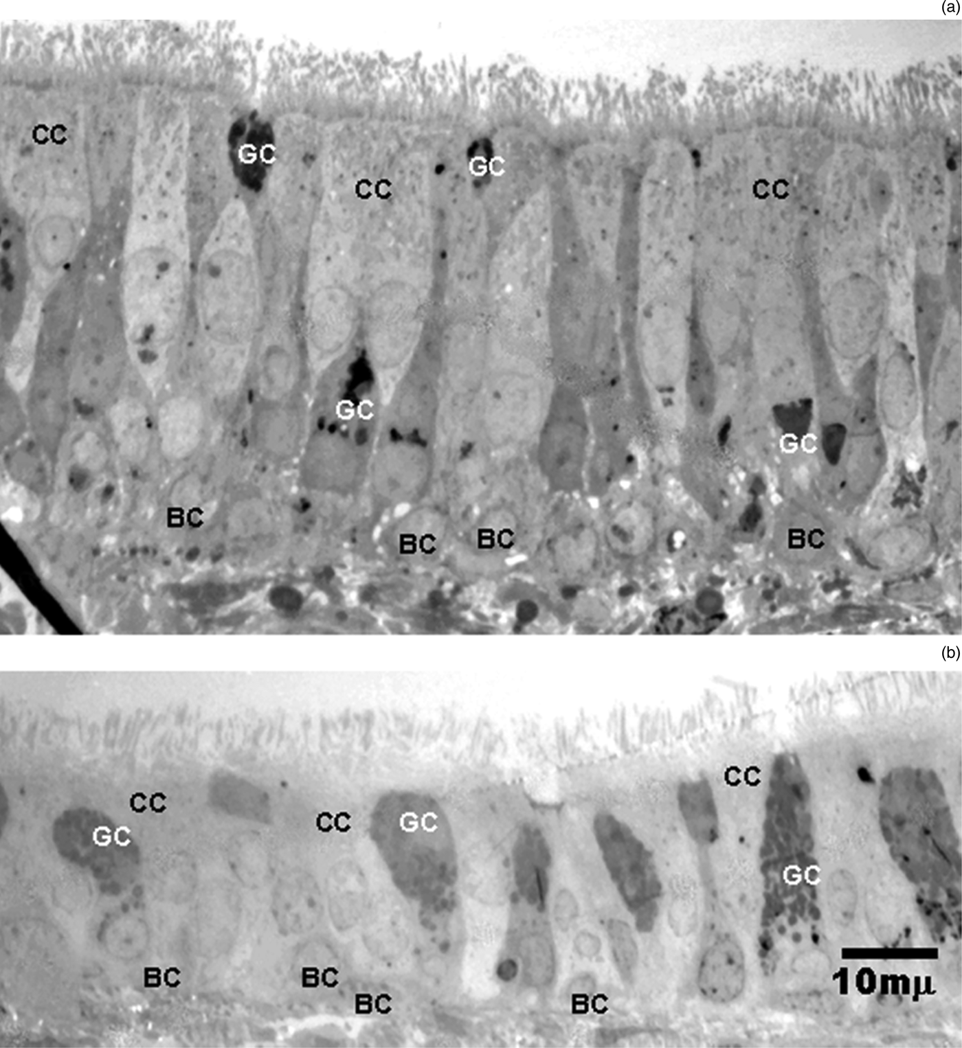
Transmission electron microscopy of tracheal mucosa showed the presence of the same types of cells in both groups, including goblet cells, ciliated cells and basal cells. In the experimental group, regeneration of the mucosa was apparent and these cells were detected 12 months after implantation (Figure 7a,b).
Electron microscopy of the trachea, showing respiratory epithelium formed in the trachea (a) and over the implant (b). The same types of cells are present in both groups. The features of cilia in the normal trachea and in the implant are similar. CC = ciliated cells; GC = goblet cells; BC = basal cells
Discussion
Some experimental models have proposed the use of collagen-coated prosthetic materials to repair a lesion in a circular segment of the trachea.Reference Kuriloff and Fayad11–Reference Gilbert, Gilbert, Madden, Reynolds and Badylak14 Moreover, there is some evidence, albeit limited, showing the success of these models in clinical applications.Reference Omori, Nakamura, Kanemaru, Asato, Yamashita and Tanaka15 In the present study, in addition to describing the macroscopic and microscopic changes, we have revealed some changes that could accompany the implantation of a homologous collagen-coated mersylene prosthesis into the trachea during the growth of the host. These changes were visualised using direct endoscopy and computerised axial tomography, as these allow higher precision and versatility than standard radiological methods.Reference Griscom and Wohl4, Reference Kara, Turan, Dabanoglu and Ocal16
Distortion of the tracheal lumen at the site of implantation was evident as early as the first post-operative endoscopic evaluation. This distortion was likely caused by the loss of integrity of the trachea, and traction between the prosthesis and the attached cartilaginous remnants. Using tomography, we detected reductions in fore–back diameter, transversal diameter, circumference and area of the trachea in the experimental group compared with the control group, but no collapse occurred. There was a loss of tracheal contour at the site of implantation, which remained for the entire duration of the study. Although this did not markedly impair tracheal growth, the prosthesis did limit tracheal size compared with that in control dogs. At the end of the experiment, transversal diameter, fore–back diameter, circumference and area of the trachea at the site of implantation were 37, 12, 14 and 44 per cent smaller, respectively, in the experimental group than in the control group.
Despite these apparent deficits, the dogs exhibited no abnormal signs, except in the immediate post-operative period. All of the dogs in the experimental group showed an increase in body weight, similar to that in the control group. Clinically, they did not show signs of impaired ventilatory function, except in one dog that suffered a technical defect in the prosthesis–tracheal ring joint. That dog suffered from moderate wheezing that lasted for two weeks. This technical defect culminated in a small protrusion of the implant into the tracheal lumen, which was later covered with polypoid material. Nevertheless, the dog remained asymptomatic for the remainder of the study.
Histological and immunochemical analysis of the prosthesis before implantation revealed that it was coated with types I and V collagen, and that its thickness was only 31 per cent of that of the trachea. The thickness of the prosthesis had increased four-fold at one year after implantation as a partial cervical tracheal substitute. This growth was not projected towards the lumen. Thickening occurred through the addition of key structural components, and by one year the epithelial tissue covering the prosthesis resembled that of the neighbouring tracheal wall. These morphological changes could be attributed to the migration of cells, particularly epithelial, secretory and squamous cells, to cover the scaffold provided by the prosthesis, followed by metaplasia of pseudostratified columnar epithelium and differentiation of ciliated respiratory epithelium. The rest of the tissue was covered by abundant collagen fibres, principally type I collagen, which is the principal promoter of angiogenesis and is essential to maintain the viability of the epithelial cells deposited on the implant surface. As in an earlier study,Reference Villegas-Álvarez, González-Zamora, González-Maciel, Soriano-Rosales, Pérez-Guille and Padilla-Sánchez5 vascular proliferation occurred even though we did not administer any stimulants, such as vascular endothelial growth factor, vascular permeability factor or basic fibroblast growth factor.Reference Nakanishi, Hashimoto and Yasumoto17–Reference Sweeney, DiLullo, Slater, Martinez, Iozzo and Lauer-Fields20 Another element underlying the increase in thickness of the prosthesis was that muscle fibres started to adhere to it. These changes were observed in all of the prostheses implanted. However, the inward fold of the prosthesis into the lumen in one dog likely hindered regeneration of the epithelium, favouring focal development of granulation tissue with deposition of fibroblasts, inflammatory cells and interstitial fluid. In that dog, in regions of the trachea unaffected by the fold, we observed normal respiratory epithelium.
• Evidence for the long-term efficacy or safety of tracheal wall implants in young people is limited
• In this pilot study, 30–40 per cent of the total tracheal cartilaginous wall was removed in dogs, from tracheal rings C4–C7, and replaced with a fibrocollagen-coated prosthesis
• The tracheal area grew following implantation, but growth was less than that in the control group
• Once the prosthesis had fully integrated into the segmented tracheal wall, there were no group differences in growth or development
Tracheal reconstruction in children with acquired severe or congenital stenosis is now a major focus of research.Reference Grillo21 Homogeneous graftsReference Jacobs, Elliott, Haw, Bailey and Herberhold22 and tracheoplastyReference Grillo, Wright, Vlahakes and MacGillivray23 have been proposed for the treatment of these patients. One experimental study showed that tracheal resection with extension ranging from 35 to 38 per cent, and posterior anastomosis with tension not exceeding 1450 g at the suture site, did not impair local healing in dogs.Reference Blanchard, Brochu, Bensoussan, Lagacé and Khan24 However, by adulthood, the trachea had lost its circular contour, an outcome that was coupled with enlargement of the intercartilaginous ligament. The trachea was also shorter than that in dogs of a similar age that were not subjected to resection; this persisted for the duration of the study.Reference Blanchard, Brochu, Bensoussan, Lagacé and Khan24 Despite these morphological differences, the dogs in that study did not exhibit functional defects.
Thus far, clinical studies have not yet been conducted in children with equivalent resections. Nevertheless, several procedures developed to correct severe stenosis in children have proven successful in isolated cases.Reference Othersen, Hebra and Tagle25 Indeed, some authors have suggested that slide tracheoplasty has good short- and medium-term outcomes, with immediate increases in the tracheal luminal size and reductions in tracheal length.Reference Grillo, Wright, Vlahakes and MacGillivray23, Reference Koopman, Bogers, Witsenburg, Lequin, Tibboel and Hoeve26
The use of autologous pericardial patches is associated with some complications and is not recommended in all patients.Reference Cheng, Backer, Holinger, Dunham, Mavroudis and Gonzalez-Crussi27 Autogenous tissues (e.g. from the carotid artery) have been used to enlarge the tracheal lumen in children and have provided satisfactory medium-term clinical outcomes.Reference Dodge-Khatami, Nijdam, Broekhuis, Von Rosenstiel, Dahlem and Hazekamp28
All of these procedures require high levels of expertise and experience. In addition, long-term follow up, until the trachea is fully developed, is essential to confirm the efficacy of these procedures. Exploring other options, such as that reported in this study, allows us to evaluate the integration and permanency of these prostheses in young animals, and hence predict their responses in humans. In our study, we found no cases of respiratory insufficiency, similar to the above-mentioned studies, although tracheal lumen growth was slightly reduced. Although the prosthesis was satisfactorily incorporated into the trachea in each dog, it was associated with some effects on growth, but these were not unexpected. One reason for this is that the collagen-coated prosthesis is not an absorbable structure and it was not designed to support growth, although the remnants of the tracheal rings that were partially removed apparently did support growth, which avoided remarkable lumen distortion. Perhaps more importantly, the prosthesis was functional and biologically accepted by the recipient, with no signs of tissue rejection or inflammation. Nevertheless, further studies are needed to determine whether these prostheses cause any airway dysfunction over a longer period of time. Future studies should focus on developing tracheal implants for children that are functional and fully integrated into the recipient's tissue, and permit the normal structural changes that occur during growth.
Conclusion
In this study, we showed that a homologous type I and V collagen-coated mersylene prosthesis, implanted as a partial substitute for a non-circular tracheal segment in young dogs, reduced tracheal structural growth at the implantation site. However, the prosthesis was fully integrated into the recipient's tissue and showed the development of normal ciliated respiratory epithelial tissue, without evidence of rejection, stenosis or collapse.
Acknowledgements
We are grateful to the animal technicians at Vivarium, and to Sandra Zaragoza-Huerta, Francisco Javier de la Rosa-Cruz and the staff at the Imagenology Department of the Instituto Nacional de Pediatría.