In designing strategies to counter cancer, restricting or eliminating angiogenic signals from the tumour is the working principle behind several therapeutic applications. An idea that was first seeded by Judah Folkman in the 1970s (Ref. Reference Folkman1) has matured into exponentially expanding treatment opportunities because of an ever-increasing knowledge base in angiogenesis pathways and molecular targets. The discovery of naturally occurring tissues that are antiangiogenic and where antiangiogenesis and lack of vasculature is a guiding principle dictating the developmental outcome has allowed for an increase in identification of antiangiogenic molecules. Chief among these is cartilage, a specialised form of extracellular matrix (ECM) synthesised by chondrocytes.
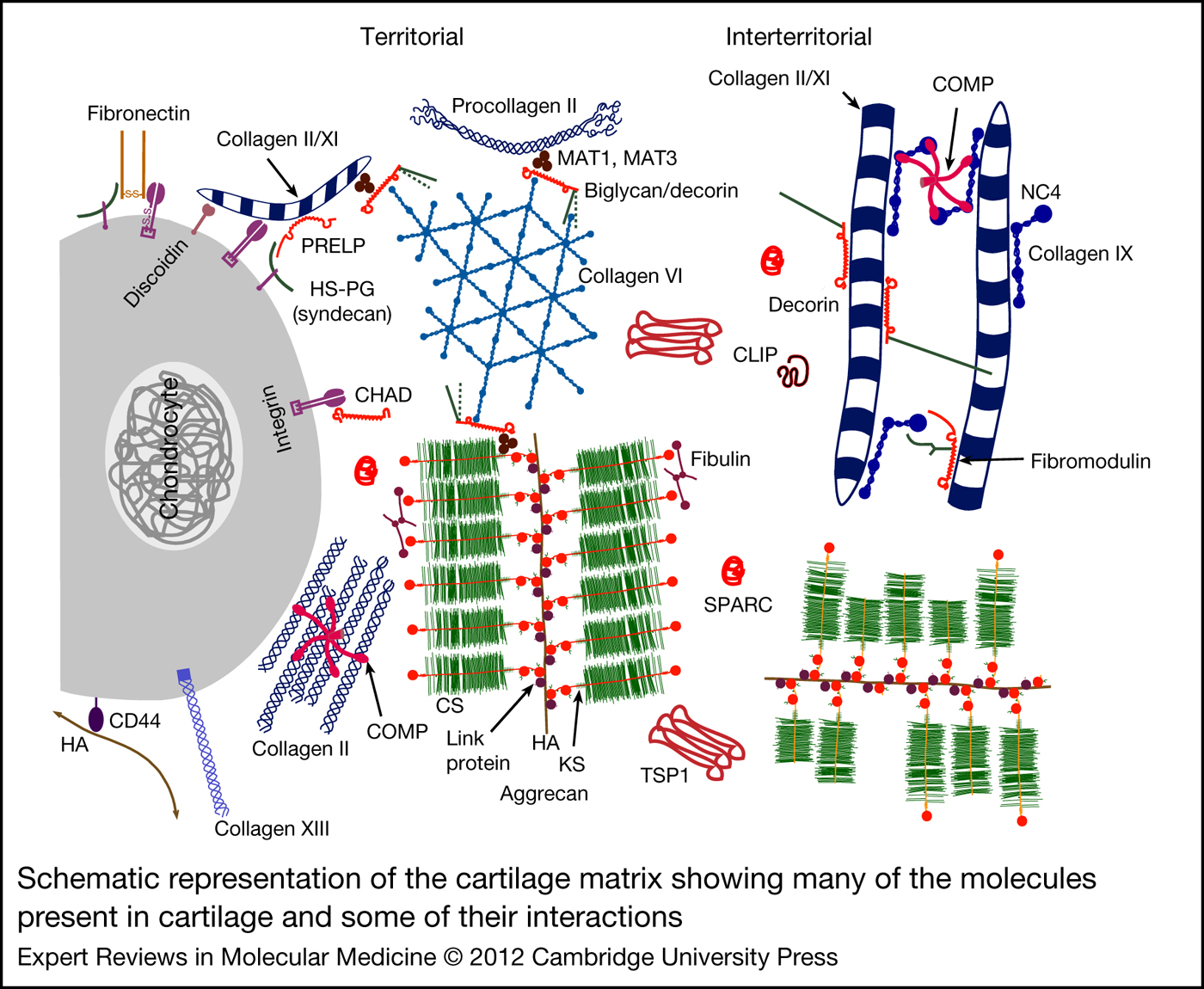
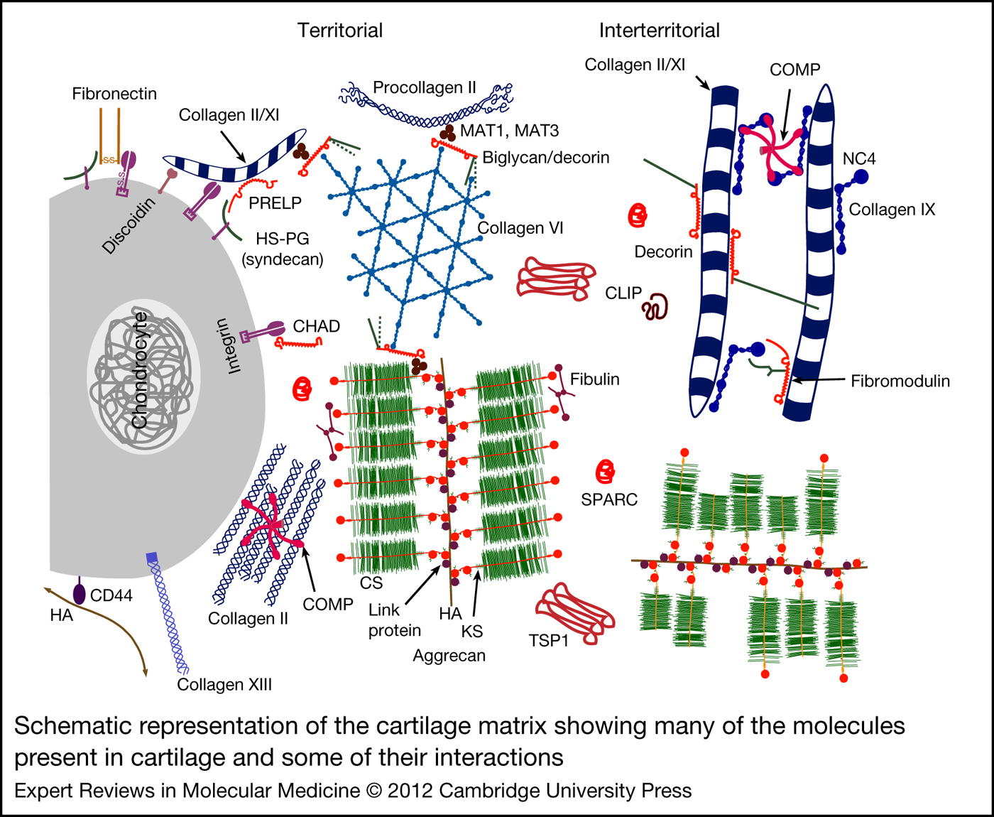
The characteristic that sets cartilage apart is that it is avascular, and this has piqued the interest of investigators who have sought to understand its avascular nature and to apply its principles to therapeutic applications. It stands to reason that the avascular nature of cartilage would be due to its biochemical composition that antagonises vascular invasion and there has been considerable interest in identifying these antiangiogenic components in cartilage. The primary components of cartilage are the two major macromolecules, type II collagen (Col II) and aggrecan (Fig. 1). Minor components include collagens types IX, XI, III, V, VI, X, XII, XIV as well as versican, perlecan, lubricin, biglycan, fibromodulin, thrombospondins (TSPs), chondromodulin-1 (ChM-1), endostatin and secreted protein acidic and rich in cysteine (SPARC), among others (reviewed in Ref. Reference Sandell, Heinegard and Hering2). Although in adults cartilage serves primarily to lubricate the joints, during embryonic development its chief function is to form the template for future skeletal development in mammals.
Schematic representation of the cartilage matrix showing many of the molecules present in cartilage and some of their interactions. The matrix closest to the chondrocytes is indicated as territorial and between the chondrocytes is interterritorial. The cartilage extracellular matrix (ECM) is made of molecules that are synthesised by the chondrocyte. It has two major macromolecules, collagen type II and the large proteoglycan, aggrecan. The aggrecan protein consists of a globular core protein to which are attached chains of chondroitin sulfate (CS) and keratan sulfate (KS). The figure shows aggrecan monomers attached to hyaluronic acid (HA), which is stabilised by the link protein, to form a larger proteoglycan aggregate in interaction with the smaller proteoglycan, fibulin. In addition to these, the cartilage ECM is also composed of minor amounts of other collagens such as collagen types IX, XI, III, V, VI, X, XII, XIV and XVIII (from which endostatin is derived). Collagen type IX has four noncollagenous (NC) domains and has the ability to form heterotypic fibrils with collagen II/XI (shown in the interterritorial matrix) that is coated with smaller proteoglycans such as decorin and fibromodulin. The cartilage intermediate layer protein (CILP) is found primarily in the interterritorial matrix. Other smaller proteoglycans include biglycan, decorin and chondromodulin-1. The other major cartilage proteins are the thrombospondins (TSPs), which consists of TSP1, TSP2 and COMP (cartilage oligomeric matrix protein also known as TSP5). Matrilins 1 and 3 (MAT1, MAT3) are the primary members of the multimeric matrilin family of proteins found in the cartilage. PRELP (proline arginine-rich end leucine-rich repeat protein) and CHAD (chondroadherin) are small leucine-rich repeat proteins (LRRs) that bind to fibre forming collagens with high affinity due to the LRR domain. Syndecan is a heparan sulfate proteoglycan (HSPG) found on the chondrocyte cell surface where it acts as receptors for matrix components such as fibronectin. A number of matricellular proteins such as SPARC (secreted protein acidic and rich in cysteine) are also seen in the cartilage ECM. The cartilage matrix and its composition are reviewed in depth elsewhere (Ref. Reference Sandell, Heinegard and Hering2). This figure is modified from Ref. Reference Sandell, Heinegard and Hering2 with permission from Lippincott, Williams & Wilkins (Philadelphia).
The process by which cartilage template is replaced by bone is called endochondral ossification, which occurs when the avascular privilege of cartilage is broken down, allowing for vascular invasion and endochondral bone formation. This topic has been well reviewed (Refs Reference Kronenberg3, Reference Ortega, Behonick and Werb4), but to briefly summarise, the process begins with mesenchymal cell condensations at sites where the future skeletal elements will form; cells in these condensates differentiate to form chondrocytes that secrete a Col-II-rich avascular cartilage, with cells surrounding the chondrocytes forming the perichondrium. To allow for bone formation, the innermost chondrocytes differentiate to form hypertrophic chondrocytes that secrete a type-X-collagen-rich matrix that gets calcified, with the perichondral cells surrounding the hypertrophic chondrocytes differentiating into osteoblasts to form the bone collar. This allows for the vascular endothelial growth factor (VEGF)-dependent vascular invasion of the calcified cartilage matrix from the bone collar, which brings along with it chondroclasts and osteoblasts that degrade and replace the calcified matrix with bone and results in removal of the hypertrophic chondrocytes by apoptosis. This process thus allows for vascular invasion only of the calcified matrix, leaving the rest of cartilage matrix avascular. This is crucial to bone growth because the avascular cartilage continuously provides chondrocytes that differentiate to form hypertrophic chondrocytes that are removed during endochondral bone growth, thus allowing for continuous bone growth. With maturity, the centre of the avascular cartilage undergoes hypertrophic differentiation again to allow vascular invasion and the development of a secondary ossification centre. In adults, most of the cartilage is thus replaced by bone, with the exception of the articular cartilage at the bone ends, which remains avascular to maintain cartilage phenotype and functions to lubricate the joint. Breakdown of the antiangiogenic barrier during diseases such as osteoarthritis and rheumatoid arthritis results in undesirable vascular invasion of the articular cartilage and irreversible cartilage degeneration.
Tumours that exceed 1–2 mm in diameter require new capillary formation or angiogenesis that feeds their growth (Ref. Reference Brem and Folkman5). The recognition of this fundamental principle behind tumour growth has triggered the search for antiangiogenic molecules that would prevent tumour growth. Blessed with its avascular state, cartilage has since then become a prime target for investigation of its antiangiogenic properties (Refs Reference Brem and Folkman5, Reference Langer6). Although primarily articular cartilage was tested, growth plate chondrocytes also demonstrated antiangiogenic effects (Ref. Reference Cheung7). These discoveries propelled an interest in the identification of cartilage components that produce these antiangiogenic and antitumour effect with the expectation that these cartilage-derived molecules could be used in therapeutic applications to treat tumours and related malignancies. This review summarises the status quo of some antiangiogenic molecules that are present in cartilage.
Neovastat and shark cartilage extract
Neovastat and shark cartilage extract (SCE) are preparations with antiangiogenesis activity derived from shark cartilage. Interest in shark cartilage as a source of anticancer agents grew because of its avascular state coupled with the misconception that sharks do not get cancer. The cartilaginous shark endoskeleton was thus thought to be an ideal source of large quantities of anticancer agents. Current literature finds researchers at extreme ends of the spectrum in their faith in shark-cartilage-derived products. The discovery of cancer in sharks (Ref. Reference Ostrander8) and the lack of promising results from the most recent clinical trial with Neovastat (see below) were serious setbacks to its use in cancer treatment.
Neovastat (or AE-941) is not a single molecule but a mix of water-soluble components derived from shark cartilage prepared by a proprietary manufacturing process developed by Aeterna Laboratories (Quebec, Canada) (Ref. Reference Dupont E. and Juneau9). Homogenisation of the shark cartilage in water followed by sequential extraction to remove water-insoluble and inactive molecules results in a concentrated biological derivative with a mixture of components less than approximately 500 kDa. To date, the identity of all the individual components of Neovastat is not known, but extensive characterisation has attributed it with antiangiogenic properties. Neovastat induced a concentration-dependent inhibition of cell proliferation in human umbilical vein endothelial cells (HUVECs) and bovine endothelial cells; inhibited the formation of blood vessels induced by basic fibroblast growth factor (FGF) in the chicken chorioallantoic membrane model; severely inhibited in vivo the vascular invasion of bFGF-containing Matrigel implanted in C57BL6 mice fed orally with Neovastat; and inhibited lung metastases in the murine Lewis lung carcinoma model (Ref. Reference Dupont10). Neovastat combined with cisplatin, a conventional anticancer agent, exhibited greater anticancer activity than cisplatin alone. These data indicated the successful extraction of the antiangiogenic and antitumour agents from shark cartilage into Neovastat, and surprisingly, its success as an orally active biological compound.
Antiangiogenic features of Neovastat
Mechanistically, Neovastat induced apoptosis specifically in endothelial cells that involved the activation of caspase-3, caspase-8 and caspase-9 (Ref. Reference Boivin11). Neovastat interferes with VEGF binding to VEGFR-2 (Flk-1/KDR), which is required to make new blood vessels (Ref. Reference Olsson12), thereby inhibiting several mitogenic, chemotactic and antiapoptotic responses in endothelial cells (Ref. Reference Beliveau13). Neovastat also inhibited the activity of matrix metalloproteinases (MMPs), primarily MMP2, but also of MMP1, MMP7, MMP9, MMP12 and MMP13 (Ref. Reference Gingras14), limiting the metastatic potential of tumour cells (Ref. Reference Weber, Lee and Orr15). Neovastat-treated ovalbumin-sensitised mice exhibited reduced airway hyper-responsiveness and inflammation when challenged (Ref. Reference Lee, Paik and Chung16); these mice demonstrated significantly reduced levels of MMP9 activity in their bronchoalveolar lavage fluid and reduced VEGF and hypoxia-inducible factor (HIF)-2α expression in their lung tissue, suggesting that the anti-inflammatory effects of Neovastat are linked to inhibition of VEGF and HIF-2α (Ref. Reference Lee and Chung17).
Neovastat can activate the Jun amino terminal kinase (JNK) and nuclear factor-kappaB (NF-κB) signalling pathways as demonstrated by its ability to modulate the plasmin–plasminogen-activator (PA) systems. Plasmin–PA systems are vital components of the proteolytic machinery that degrade the ECM along with MMPs during angiogenesis (Refs Reference Nisato18, Reference Pepper19). Tissue PA (tPA) and urokinase PA (uPA) are the two major types of PA that convert plasminogen into plasmin (Ref. Reference Lijnen and Collen20). Binding of uPA to its cell surface receptor is thought to have a role in neovascularisation and therefore tumour angiogenesis (Refs Reference Tressler21, Reference Min22). However, tPA is synthesised primarily by endothelial cells and has a role in cleavage of fibrin-bound PA to plasmin, leading to fibrin degradation (Ref. Reference Lijnen and Collen20). Neovastat stimulates tPA-mediated plasmin generation (but only marginally inhibits uPA); BIAcore surface plasmon resonance spectroscopy demonstrated direct physical interaction of Neovastat with plasminogen that increased its catalytic efficiency (Ref. Reference Gingras23). Neovastat increased tPA production by endothelial cells, increased the phosphorylation and activity of JNK1 and JNK2, and the phosphorylation of IκB (an inhibitor of NF-κB) thereby inducing NF-κB signalling (Ref. Reference Gingras24). Administration of SCE (a generic preparation of Neovastat) to mouse glioma models resulted in tPA activity stimulation that is thought to direct the antiangiogenic property of SCE because tumours derived from glioma cells that overexpressed PAI-1, an inhibitor of tPA, did not exhibit decreased blood vessel formation (Ref. Reference Simard25). Mice orally treated with SCE demonstrated a reduction in glioblastoma infiltration into the brain, with very small tumours, in contrast to mice that did not get SCE treatment. These studies indicated beneficial aspects of orally administered Neovastat across the blood–brain barrier and beneficial aspects of SCE in immune-compromised athymic mice with glioma.
Clinical trials with Neovastat
Multi-targeting properties of Neovastat propelled its use as a multimodal, anticancer drug in humans. Phase I/II clinical trials for Neovastat in patients with renal cell carcinoma (RCC) (Ref. Reference Batist26) and non-small cell lung cancer (NSCLC) (Ref. Reference Latreille27) were conducted. In both RCC and NSCLC patients, Neovastat was well tolerated, with no dose-limiting toxicity, with oral administrations of up to 240 ml/day showing significantly longer median survival than those receiving 60 ml/day. However, neither of these studies reported any response within the tumour [in contrast to the tumour necrosis observed in mouse models on Neovastat administration (Ref. Reference Simard25)]. In the NSCLC study, about half the number of patients did not complete standard imaging assessments because of progressive disease as per clinical symptoms. A randomised, double-blinded, placebo-controlled Phase III clinical trial was conducted to determine Neovastat efficacy in treating Stage III NSCLC patients (Ref. Reference Lu28). NSCLC patients who had previously received chemoradiotherapy were treated further with chemoradiotherapy plus Neovastat (or placebo). Although no tumour responses were observed with Neovastat administration, no statistically significant difference in patient survival was observed between those receiving Neovastat plus chemoradiotherapy and those receiving chemoradiotherapy alone. To rationalise the failure of Neovastat, the authors noted that as Neovastat is a complex, natural product as opposed to a well-defined chemical molecule, it lacks pharmacokinetic/pharmacodynamic (PK/PC) assays, which makes it difficult to control the product for quality (Ref. Reference Lu28). Furthermore, no validated biomarker is available to follow the antiangiogenic effect in humans. Although the authors conclude that their study does not support the use of shark-cartilage-derived products as a therapy, the study design limits the conclusion and leaves behind an open question regarding their value in clinical applications (Ref. Reference White29).
Chondromodulin-1
ChM-1 is an extensively studied molecule identified in cartilage with demonstrated antiangiogenic properties in vitro. ChM-1 (also known as the leukocyte cell-derived chemotaxin 1 encoded by the LECT1 gene in humans) is a 25 kDa glycoprotein first identified in fetal bovine epiphyseal cartilage extracts (Ref. Reference Hiraki30) with ability to stimulate DNA synthesis in cultured rabbit growth plate chondrocytes in the presence of FGF. The mature ChM-1 protein is the C-terminal portion of a larger precursor protein with one N-linked and two O-linked potential glycosylation sites. Interest in ChM-1 grew when it was discovered that it could inhibit bovine carotid artery endothelial cell growth and prevent a tube-like cellular network in vitro (Ref. Reference Hiraki31). This was considered a prognostication of its ability to inhibit proliferation and tube morphogenesis of invading endothelial cells during angiogenesis in vivo. In situ hybridisation analyses in bovine embryonic growth plate (Ref. Reference Hiraki31) and developing mice (Ref. Reference Shukunami32) detected ChM-1 mRNA in the proliferating and pre-hypertrophic cartilage zones (which are avascular) with expression missing from the mature hypertrophic zone (where vascular invasion during endochondral bone growth takes place) and vascular bone. The detection of ChM-1 protein in the cartilage inter-territorial matrix in the same avascular zones as its mRNA strengthened the notion of ChM-1 as an antiangiogenesis molecule in cartilage. Once considered specific to cartilage, ChM-1 has since then been identified in other avascular tissues, such as the eye, thymus (Ref. Reference Brandau33) and cardiac valves (Ref. Reference Yoshioka34).
Antiangiogenic features of ChM-1 and skeletal homeostasis
In humans, ChM-1 protein was detected in the ECM surrounding mature hypertrophic chondrocytes in neonatal vertebrae, at the junction with bone (Ref. Reference Kusafuka35). This is not consistent with results in mice or bovine growth plate, nor can this be deemed favourable for vascular invasion during endochondral bone growth if it is antiangiogenic in vivo. The results from mouse models cast doubt on the ability of ChM-1 to regulate angiogenesis in vivo. Based on its distribution in the bovine and murine growth plate, it was predicted that ChM-1 allows the cartilage to remain avascular and its reduced expression in the hypertrophic cartilage allows for vascular invasion during endochondral bone development. However, ChM-1-null mice demonstrated normal cartilage and bone development, with no reported abnormal cartilage vascularisation (Ref. Reference Brandau36). Even though recombinant human ChM-1 demonstrated inhibition of human retinal endothelial cell tube morphogenesis in vitro (Ref. Reference Funaki37), suggesting that it might be required to maintain normal avascular state of the retina, mice lacking ChM-1 did not show any abnormal vascularisation of the retina or any other abnormalities in eye development. It was thought that loss of ChM-1 in mice was compensated by its close homologue, tenomodulin (also called tendin) (Ref. Reference Brandau33). Like ChM-1, tenomodulin inhibited the growth of HUVECs and tube morphogenesis in vitro (Ref. Reference Oshima38). However, mice that lack both ChM-1 and tenomodulin also did not show any abnormalities in eye development, nor were any abnormalities in cartilage and endochondral bone development reported (Ref. Reference Docheva39).
Despite these contradictions, the notion of ChM-1 being an antiangiogenic factor in vivo cannot be overruled. During endochondral ossification in mice, chondrocyte hypertrophic differentiation results in loss of ChM-1 expression prior to matrix calcification in the cartilage anlagen (Ref. Reference Shukunami32). Hypertrophic chondrocyte differentiation and osteoblast differentiation is governed among other factors, by the transcription factor Runx2 (Cbfa1) (Refs Reference Ducy40, Reference Komori41, Reference Otto42, Reference Takeda43). Runx2-null mice lack hypertrophic differentiation and osteoblast development and show no endochondral bone formation. Interestingly, this was accompanied by an increase in ChM-1 in the cartilage of these mice (Ref. Reference Takeda43). However, rescue of Runx2-null mice by a transgene that expressed Runx2 in chondrocytes reduced ChM-1 expression in the cartilage and allowed for vascular invasion. This indicated that Runx2 regulates ChM-1 expression in chondrocytes and that ChM-1 is required to maintain cartilage avascular state. Likewise, in experimental osteoarthritis in rats, ChM-1 levels decreased in all cartilage zones with advancing osteoarthritis with a concomitant increase in VEGF expression (Ref. Reference Hayami44). However, inhibition of VEGF by administration of antibodies against VEGF improved articular cartilage repair in an osteochondral defect model with parallel increases in ChM-1 (Ref. Reference Nagai45).
ChM-1-null mice exhibited delayed union of the fractured bone in a tibial fracture repair model (Ref. Reference Yukata46). Fracture repair usually mimics endochondral bone development where a cartilaginous callus is initially formed enveloping the fracture site that is replaced by bone. ChM-1-null mice exhibited severely reduced external cartilaginous callus formation restricted only to the fracture site, with reduced gene expression for the cartilage-promoting factor Sox9 and for type X collagen. ChM-1-null mice exhibited a bony callus instead in the periosteal region and an enhanced internal fracture callus. ChM-1 might also be responsible for the good-quality cartilaginous repair tissue formed when femoral chondral lesions in miniature pig knee joint were treated with matrix-associated autologous chondrocyte transplantation (MACT) (Ref. Reference Blanke47). In the absence of MACT, these chondral lesions were filled with excessive bony tissue and vascular invasion from the subchondral bone plate with overlying fibrocartilage repair tissue of poor quality that lacked ChM-1 expression. With MACT treatment, the cartilage repair tissue formed was rich in proteoglycan and Col II, and positive for ChM-1, with calcification of repair tissue and subchondral bone overgrowth inhibited. ChM-1 was also detected in normal healthy noncalcified articular pig cartilage.
In the same porcine model, chondroprogenitor cells overexpressing recombinant human ChM-1 when transplanted into the chondral lesions stimulated chondrogenic differentiation that produced a superior hyaline-like matrix than those in the absence of recombinant human ChM-1, by inhibiting chondrocyte hypertrophy, preventing vascularity and therefore endochondral ossification, thus stabilising the chondrocyte phenotype (Ref. Reference Klinger48). In an immobilisation-induced cartilage degeneration model (Ref. Reference Sakamoto49), rats with plaster-cast-immobilised ankle joints demonstrated thinning of the ankle joint cartilage with reduced ChM-1, in contrast to rats with continuous passive motion. Immobilised ankle joints also exhibited increased VEGF and HIF-1α expression, with vascular invasion of the calcified cartilage zone from the subchondral bone plates. ChM-1 is also abundantly expressed in other avascular tissues such as cardiac valves in mice (Ref. Reference Yoshioka34). Young ChM-1-null mice exhibited normal cardiac valves; but as these mice aged, in strong contrast to wild-type mice, they exhibited bulky aortic valves that showed VEGF expression, enhanced angiogenesis of the aortic valves, increased calcium deposits and consequent aortic stenosis characterised by turbulent blood flow. In human heart diseases such as in congenital bicuspid aortic valves stenosis, cardiac valves exhibited ChM-1 downregulation in regions of neovascularisation, calcification and VEGF-A expression, with aggressive macrophage infiltration and inflammation, indicating that ChM-1 is important for normal valvular function (Ref. Reference Moreno50).
Modes of ChM-1 antiangiogenic inhibition
Application of ChM-1 in clinical trials as an antitumour agent has not been reported. However, ChM-1 is able to suppress tumourigenesis in mouse tumour models. The application of recombinant human ChM-1 to OUMS-27 (that do not express ChM-1) xenograft tumour model in mice (Ref. Reference Hayami51) or to HepG2-induced tumours in athymic mice (Ref. Reference Mera52) suppressed tumour growth and prevented blood vessel invasion into the tumour. Interestingly, ChM-1 can act directly on tumour cells and not just endothelial cells. ChM-1 inhibited DNA synthesis and reduced growth of human tumour cells such as HepG2, PC-3 and NOS-1, presumably by suppressing the STAT signalling pathway (Ref. Reference Mera52). ChM-1 also downregulated cell cycle proteins such as cyclin D1, cyclin D3 and cdk6 and upregulated the cell cycle inhibitor protein p21cip1, although no apoptosis was observed. These data suggested that ChM-1 mediates its cytotoxic effect on human tumour cells primarily by causing cell cycle arrest. However, in HUVECs, ChM-1 inhibited their migration through disruption of actin reorganisation and suppression of Rac1/Cdc42 GTPase activity (Ref. Reference Miura53). In a mouse model for rheumatoid arthritis, recombinant human ChM-1 suppressed proliferation of mouse splenic T cells and production of interleukin (IL)-2 from CD4+ T cells (Ref. Reference Setoguchi54). It also suppressed the development of antigen-induced arthritis and the proliferation of synovial cells from joints of rheumatoid arthritis patients. How ChM-1 manifests these diverse inhibitory physiological responses in different cell types remains to be investigated. In addition to its antiangiogenic activity, ChM-1 might have additional attributes (Ref. Reference Klinger48) that remain to be identified. These data suggest that ChM-1 has a strong potential to be used as a therapeutic agent for treatment of localised tumours as well as for suppressing the severity of symptoms in rheumatoid arthritis patients.
Thrombospondins
TSPs are a family of five cartilage matrix proteins of which only TSP1 and TSP2 have antiangiogenic properties and are of interest in clinical applications as anticancer agents. Although TSP1 and TSP2 are detected in cartilage and have important roles in maintaining growth plate homeostasis (Refs Reference Kyriakides55, Reference Iruela-Arispe56, Reference Posey57), their distribution is not limited to cartilage and their antiangiogenic properties discovered in other systems have been well reviewed (Refs Reference Chen, Herndon and Lawler58, Reference Lawler59). In addition to their antiangiogenic properties, TSP1 and TSP2 also have angiogenic functions (reviewed in Ref. Reference Bornstein60).
Antiangiogenic features of TSP1 and TSP2
The antiangiogenic properties of TSP1 and TSP2 are complex and may arise from several mechanisms. In the presence of the CD36 receptor, TSP1 mediates its antiangiogenic effect on endothelial cells in vitro, and inhibition of neovascularisation in vivo, through apoptosis by sequential upregulation of the Src family kinase p59fyn, caspase-3 like proteases and the p38 mitogen-activated protein kinase (MAPK)-mediated signalling and activation of Fas ligand (FasL) (Refs Reference Jimenez61, Reference Volpert62), or by tumour necrosis factor (TNF)-receptor 1 and TNFα in the absence of FasL (Ref. Reference Rege63). TSP1 also inhibits lymphangiogenesis (formation of lymphatic vessels) through CD36 ligation on monocytic cells in a mechanism that inhibits transforming growth factor β (TGFβ)-mediated expression of VEGF-C (Ref. Reference Cursiefen64), providing an added anticancer benefit because lymphangiogenesis facilitates cancer metastases to distant lymph nodes and organs (Refs Reference Cao65, Reference Gao66). TSP2 also mediates apoptosis through CD36 interaction (Refs Reference Simantov, Febbraio and Silverstein67, Reference Mirochnik, Kwiatek and Volpert68). However in HUVECs, in the absence of CD36, TSP1 induced upregulation of p21cip/waf, p53 phosphorylation and Rb dephosphorylation resulting in cell cycle arrest (Ref. Reference Yamauchi, Imajoh-Ohmi and Shibuya69). Ligation of the VLDL receptor by TSP1 or TSP2 also inhibited cell division in endothelial cells by a similar nonapoptotic pathway with cell cycle arrest (Ref. Reference Oganesian70). A third mechanism is the binding of TSP1 and TSP2 to heparan sulfate proteoglycans (HSPGs) through its N-terminal heparin-binding domain (Ref. Reference Chen71). HSPGs are co-receptors for the low-density lipoprotein receptor-related protein (LRP1). LRP1 is a scavenger receptor with endocytic and signal transmission behaviour that is required for chondrocyte differentiation (Ref. Reference Kawata72). LRP1 functions to clear complexes of TSP1 and TSP2 with MMP2, MMP9 and VEGF (Refs Reference Greenaway73, Reference Yang, Strickland and Bornstein74, Reference Hahn-Dantona75) resulting in decreased activity of these enzymes to inhibit vascular invasion.
TSP1- and TSP2-deficient mouse models and skeletal homeostasis
It is not known as to whether the mechanisms enumerated above are used in cartilage, but antiangiogenic benefits from TSP1 and TSP2 can be surmised from the following. Mice deficient in TSP1 or TSP2 show only mild chondrocyte growth plate disorganisation, but no abnormal vascularisation of the cartilage was reported (Ref. Reference Posey57), although TSP2-deficient mice showed enhanced cortical bone formation (Ref. Reference Hankenson76). However, loss of TSP1 in mice exhibited increased retinal vascular density, a reduced rate of endothelial cell apoptosis, and excessive capillary formation of heart and skeletal muscles (Refs Reference Wang77, Reference Malek and Olfert78). In a collagen-induced arthritis model in rats, recombinant murine TSP1 delivered intraarticularly by adenoviral vectors (AdTSP1) into the ankle joints of collagen-sensitised rats, reduced the pathogenesis of arthritis with lower levels of VEGF, IL-1β, blood vessels and synovial hypertrophy (Ref. Reference Jou79). Absence of TSP2 altered the dynamics of osteogenic and chondrogenic differentiation in the callus in a tibial fracture model such that the invading mesenchymal cells showed decreased chondrogenic differentiation with less cartilage formation and increased bone formation in the callus characterised by increased blood vessel density (Ref. Reference Taylor80).
In cartilage repair models, as seen with ChM-1, femoral chondral lesions in miniature pig knee joint treated with MACT developed superior cartilage that stained positively for TSP1 (Ref. Reference Blanke47). Application of recombinant human TSP1 to microfracture lesions discouraged ingrowing mesenchymal bone marrow stromal cells from terminal differentiation thus preventing chondrocyte hypertrophy and bone outgrowths from the subchondral plate, though this did not induce chondrogenesis and the lesions did not repair (Ref. Reference Gelse81). But application of recombinant human TSP1 along with osteogenic protein-1, which can induce chondrogenesis, but cannot prevent chondrocyte differentiation and endochondral ossification, resulted in the lesions repairing favourably with production of cartilage that resisted hypertrophic differentiation and bone formation. RT-PCR analysis of human chondrocytes treated with TSP1 showed downregulation of GADD45β, which is required for chondrocyte hypertrophy, although no increase in p21cip/waf was observed (Ref. Reference Gelse81). In a rat model of osteoarthritis with anterior cruciate ligament transection (ACLT), intraarticular injection of AdTSP1 in the ACLT-treated knee joint resulted in angiogenesis inhibition, with reduced microvessel formation, macrophage infiltration, IL-1β and MMP13 levels, and reduced synovial tissue hyperplasia that suppressed osteoarthritis progression (Ref. Reference Hsieh82). How TSP1 modulates these effects is not completely understood but TSP1 induced TGFβ in this study, which is known to aid cartilage repair and chondrogenesis (Refs Reference Guo83, Reference Yamane and Reddi84).
Clinical applications with TSP1 and TSP2
The benefits from TSP1 and TSP2 in preclinical applications in treating cancer are considerable and have been well reviewed (Refs Reference Mirochnik, Kwiatek and Volpert68, Reference Carlson, Lawler and Mosher85, Reference Zhang and Lawler86, Reference Ren87, Reference Kazerounian, Yee and Lawler88). TSP1 applications have gained further credibility as a result of some recent noteworthy observations. Treatment of prostate tumour cells with quinoline-3-carboxamide tasquinimod (chemotherapy for metastatic prostate cancer) induced upregulation of TSP1 that was paralleled by reduced VEGF expression in the tumour tissue (Ref. Reference Olsson89). TSP1 was tested for its ability to treat cerebral arteriovenous malformation (AVM) as a noninvasive therapy. Cerebral AVM endothelial cells have different angiogenic characteristics than endothelial cells from normal control brain; TSP1 was able to normalise the rate of proliferation and migration of AVM endothelial cells (Ref. Reference Stapleton90). The contradictory absence of angiogenesis in hypoxic tissues of patients suffering from systemic sclerosis could be explained by the aberrant expression of TSP1 in these tissues (Ref. Reference Morgan-Rowe91). A fusion protein of TSP2 with IgG-Fc1 could inhibit both primary tumour growth of MDA-MB-435 tumour cells in vivo and lymph node and lung metastases from the mammary gland in athymic nude mice (Ref. Reference Koch92).
Clinical applications with TSP1-derived ABT-510 and ABT-526
Several logistical considerations precluded the use of the whole molecule for clinical development and application in humans. These include the large size of TSP1, cost-prohibitive large-scale production and the possibility of unwanted side effects that could arise from multiple biological complications because of the multiple TSP1 receptors and target cells, including increased angiogenesis with TSP1 overexpression (reviewed in Ref. Reference Mirochnik, Kwiatek and Volpert68). A possible solution has been the use of TSP1-derived peptide mimetics. Successful peptides were based on the antiangiogenic sequence GVITRIR derived from the second type I repeat of TSP1 (Ref. Reference Reiher93), later modified for better half-life and PK/PC profiles, through the incorporation of non-natural amino acids, resulting in ABT-510 and ABT-526 (Ref. Reference Haviv94). ABT-510 and ABT-526 demonstrated both safety and efficacy in treatment of dogs with cancer (Refs Reference Rusk95, Reference Rusk96), in mice with malignant glioma and in inducing apoptosis of human brain endothelial cells in vitro (Ref. Reference Anderson97), and in combination with valproic acid to inhibit neuroblastoma in vivo (Ref. Reference Yang98). In an orthotopic syngeneic mouse model of epithelial ovarian cancer, treatment of mice with cisplatin or paclitaxel was more effective in combination with ABT-510, primarily because ABT-510 normalised the tumour vasculature by direct apoptotic effect on immature blood vessel endothelial cells, allowing for better uptake of cisplatin or paclitaxel, thereby significantly reducing tumour size (Ref. Reference Campbell99). ABT-510 was tested successfully for its ability to reduce tumours in mouse models of head and neck squamous cell cancer (Ref. Reference Hasina100) and epithelial ovarian cancer (Ref. Reference Greenaway101) that merited clinical trials in humans.
Although ABT-526 has remained largely preclinical, ABT-510 has been tested in clinical trials. The impact of ABT-510 in human clinical trials, however, is not impressive. Phase I/II clinical trials to determine safety, toxicity and PK/PC responses with ABT-510 in patients with a variety of advanced cancers (Refs Reference Hoekstra102, Reference Gordon103), metastatic melanoma (Ref. Reference Markovic104), advanced RCC (Ref. Reference Ebbinghaus105) and advanced soft tissue sarcoma (Ref. Reference Baker106) demonstrated that ABT-510 was well tolerated with negligible adverse effects at the doses studied (although the maximum tolerated dose was never defined). These studies, however, failed to demonstrate any significant favourable clinical benefit to patients on ABT-510 treatment. This led to Phase I/II trials for ABT-510 in combination with gemcitabine and cisplatin for treatment of solid tumours (Ref. Reference Gietema107), or 5-fluorouracil and leucovorin (Ref. Reference Hoekstra108), or in combination with chemoradiation for treatment of glioblastoma (Ref. Reference Nabors109). Although these combinations were deemed safe, the efficacy of ABT-510 in treatment of human cancers remains inconclusive because the number of patients showing improvement due to ABT-510 treatment was not impressive. Randomised Phase III trials with ABT-510 remain to be reported.
Endostatin
Endostatin is a 20 kDa fragment derived from the C-terminus of collagen XVIII by the activity of several proteinases, chiefly MMP9 (Refs Reference Ferreras110, Reference Nilsson and Dabrosin111). Endostatin, similar to other inhibitors of angiogenesis, inhibits endothelial cell proliferation and migration and induces apoptosis. It is expressed in cartilage, but is not specific to cartilage and has been detected in both fetal epiphyseal cartilage and adult articular cartilage in humans, rats and mice, where its spatio-temporal distribution might provide an antiangiogenic benefit against cartilage vascularisation (Refs Reference Pufe112, Reference Stempel113). In the meniscal fibrocartilage, its concentration is highest in the completely avascular central portion as compared with the outer region that has some vascular elements.
Antiangiogenic features of endostatin
The mechanism by which endostatin mediates its antiangiogenic effect is complex and is not completely understood because of the myriad pathways that it affects. To highlight some major pathways and recent advances, endostatin can suppress VEGF expression and induce expression of the antiangiogenic pigment epithelium-derived factor (Refs Reference Tan114, Reference Xu115, Reference Yang116); it can also upregulate the antiangiogenic VEGF165b isoform through inhibition of specificity protein 1 (Ref. Reference Li117). Endostatin can directly bind the VEGF receptor KDR/Flk-1 (VEGFR-2) on endothelial cells and therefore inhibit VEGF-induced activation of p38 MAPK (Ref. Reference Kim118). It can downregulate TNFα and vascular cell adhesion molecule-1 (VCAM-1) (Ref. Reference Xu115), which induces chemotaxis and angiogenesis in quiescent endothelial cells. Endostatin also binds to other receptors on endothelial cells such as glypicans (Ref. Reference Karumanchi119), but most of its effect is manifested by its binding to its high-affinity receptor, nucleolin. Nucleolin is tissue specific and present only on angiogenic, but not mature, blood vessels; antinucleolin antibodies can inhibit the antiangiogenic and antitumour activities of endostatin in vivo (Ref. Reference Shi120). Endostatin binding to nucleolin results in the internalisation of endostatin and transportation to the nucleus, an event that requires integrin α5. Endostatin then inhibits the phosphorylation of nucleolin in the nucleus that is inhibitory to nucleolin activity and retards endothelial cell proliferation. Mechanistically, how endostatin is internalised and translocated to the nucleus is not known, but the heparin-binding motif in its N-terminus is a key structural motif required for nucleolin interaction (Ref. Reference Fu121); mutation of six arginine residues (individually or in combinations) to alanine in this motif eliminates endostatin–nucleolin interactions and its antiangiogenic and antitumour activity. Endostatin–nucleolin interaction also inhibits lymphangiogenesis by inhibiting lymph endothelial cells in active lymphangiogenic vessels, and prevents liver metastasis in a mouse breast cancer model (Ref. Reference Zhuo122).
Endostatin can negatively impact on endothelial cell migration and adhesion in several ways. It binds to α5β1 integrins on endothelial cells, resulting in the activation of Src kinase and downregulation of RhoA GTPase in a caveolin-1- and heparin-sulfate-dependent manner resulting in actin cytoskeleton disorganisation (Refs Reference Wickstrom, Alitalo and Keski-Oja123, Reference Wickstrom, Alitalo and Keski-Oja124). It inhibits MT1-MMP-mediated activation of proMMP2 protease (Ref. Reference Kim125) and directly inhibits MMP2 activity by binding to its catalytic site (Ref. Reference Lee126), thereby eliminating their activities vital to endothelial cell migration. Endostatin downregulates uPA and PAI-1 and removes uPA from focal adhesion molecules to effect actin cytoskeletal disorganisation (Ref. Reference Wickstrom127). Endostatin downregulates antiapoptotic members Bcl-2 and Bcl-xL (with no effect on the pro-apoptotic Bax) to facilitate apoptosis (Ref. Reference Dhanabal128). It inhibits the Wnt-signalling pathway through the glypican receptors to target β-catenin for degradation (Ref. Reference Hanai129), which eventually suppresses Myc and cyclin D1 (Ref. Reference Hanai130). Endostatin can also upregulate levels of Beclin-1, a physiological partner of Bcl-2 and Bcl-xL; this upregulation distorts the Bcl-2 (or Bcl-xL) to Beclin 1 ratio, which can modulate endothelial cell apoptosis (Ref. Reference Nguyen131). It can also induce apoptosis in endothelial cells by inducing tyrosine phosphorylation of the Shb adaptor protein by binding to HSPG (Ref. Reference Dixelius132).
In animal models of ulcerative colitis, increase in VEGF in the colonic tissue was paralleled by a concomitant increase in endostatin as a defense mechanism; the larger the colonic lesion, the greater the increase in VEGF and therefore endostatin (Ref. Reference Tolstanova133). Induction of ulcerative colitis in MMP9-deficient mice resulted in less endostatin than in wild-type mice pointing to the in vivo role of MMP9 in generating endostatin from collagen XVIII in lesions. The levels of VEGF and endostatin are often linked, and although the molecular mechanism is not completely understood, VEGF might positively influence endostatin levels through the activation of MMP9 (Ref. Reference Belotti134). Finally, the large number of genes that are up- or downregulated by endostatin in a genome-wide microarray analysis (Ref. Reference Abdollahi135) suggests that a lot remains to be understood about endostatin's role in antiangiogenesis.
Derivatives of endostatin with antiangiogenic properties
Several studies have explored structural modifications of endostatin to improve function, routes of endostatin administration and combinatorial therapeutic applications as an antitumour agent in preclinical and clinical studies. Structural modifications include a single amino acid substitution (P125A endostatin) that was more antiangiogenic than wild-type endostatin (Ref. Reference Yokoyama and Ramakrishnan136). Zinc has an important role in both structure and biological functions of endostatin with the amino acid residues H1, H3, H11 and D76 coordinating with a single zinc ion (Refs Reference Ding137, Reference Hohenester138, Reference Han139). Impaired zinc binding in recombinant endostatin expressed in Pichia pastoris showing N-terminal truncations demonstrated different biological activity depending on the extent of the truncations. However, addition of an extra zinc binding motif (ZBP) to its N-terminal region (ZBP-endostatin) showed more potent antiendothelial and antitumour activity than wild-type endostatin (Ref. Reference Fu and Luo140). Addition of the integrin-targeting Arg-Gly-Asp (RGD) moiety to its N-terminus improved targeted localisation of endostatin to the tumour vasculature, which overexpresses αVβ3, αVβ5 and αVβ1 integrins compared with normal tissues, and improved its antiangiogenic activity (Ref. Reference Yokoyama and Ramakrishnan141). Addition of the IgG Fc fragment increased endostatin half-life in systemic circulation (Ref. Reference Jing142); the addition of both RGD and Fc effectively inhibited tumour-cell-induced angiogenesis, and in combination with Bevacizumab (monoclonal antibody to VEGF-A), additively inhibited ovarian cancer growth in vivo (Ref. Reference Jing142). A novel therapeutic fusion protein, EndoCD, which links endostatin to cytosine deaminase and uracil phosphoribosyl transferase (CD) (the latter converts 5-fluorocytosine to the antitumour drug 5-fluorouracil) suppressed tumour growth and metastasis in human breast and colorectal animal models (Ref. Reference Chen143). The P125A endostatin fusion protein with anti-Her2 antibody (αHer2-IgG3-huEndo-P125A) showed better half-life in serum and improved activity in breast cancer xenograft models (Ref. Reference Shin144). Combining endostatin with another angiostatic protein, the angiostatin–endostatin hybrid, provided a potent synergistic effect (Ref. Reference Paek, Kim and Choi145).
Clinical applications of endostatin
Recent years have seen an increased interest in applications of endostatin through different vectors, and combinatorial therapies, for various tumour models. Application routes tested include adenoviral vectors (Ref. Reference Zhao146), adeno-associated vectors (Ref. Reference Pan147), the oncolytic herpes simplex virus (Ref. Reference Zhu148) and mesenchymal stem cells (Ref. Reference Jiang149), all of which deliver endostatin; the Lister vaccine vaccinia viral strain, which delivers an endostatin–angiostatin fusion protein (Ref. Reference Tysome150) or endostatin plus carboxylesterases (which converts a latent drug into an active drug) (Ref. Reference Yin151); and NIH/3T3 cells expressing murine endostatin from retroviral vectors to provide sustained levels of endostatin and a long-term antiangiogenic effect (Ref. Reference Coutinho152). Recent attempts at combinatorial therapies in preclinical models included endostatin plus oxaliplatin (Ref. Reference Zhang153) or paclitaxel (Ref. Reference Huang and Chen154), which decreased tumour blood vessel density and VEGF expression, and normalised the tumour blood vasculature, allowing for increased drug uptake. Combined with radiotherapy, endostatin inhibited tumour growth and induced tumour regression (Ref. Reference Jiang155), or reduced VEGF levels in combination with gemcitabine (Ref. Reference Peng156); ZBP-endostatin potentiated the effects of chemo-radiotherapy (Ref. Reference Jia157) or the antitumour effect of dexamethasone (Ref. Reference Li158); it disrupted the actin cytoskeleton in lymph endothelial cells by inhibiting the fibronectin alternative extra domain A and integrin α9 (which facilitates lymphangiogenesis of colorectal tumours) expression and interactions (Ref. Reference Ou159). In cartilage repair studies, collagen scaffolds supplemented with chondrocytes and endostatin-expressing mesenchymal stem cells allowed the formation of a cartilaginous implant suffused with endostatin, which promoted the anabolic activity of the chondrocytes to produce more sulfated glycosaminoglycans indicating better cartilage production (Refs Reference Jeng, Olsen and Spector160, Reference Feng161). It is hoped that endostatin-infused scaffolds when implanted into a cartilage fissure in vivo, such as during osteoarthritis, would allow for better cartilage repair.
Clinical trials with endostatin and its derivatives
Phase I/II trials with endostatin that showed any benefit were largely conducted with ZBP-endostatin referred to as Endostar (trade name) or YH-16 (in Chinese publications that have been reviewed (Ref. Reference Fu162)); however, those conducted with recombinant human endostatin showed no benefit (Refs Reference Kulke163, Reference Eder164, Reference Herbst165, Reference Thomas166, Reference Hansma167). The better response to Endostar was due to the ZBP domain that supported the vital nature of the N-terminus in endostatin functions. More recently, Phase I/II trials were conducted with Endostar in patients with solid cancers (Ref. Reference Li168), in patients with extensive-stage small-cell lung cancer combined with cisplatin and etoposide (Ref. Reference Zhou169), or recombinant human endostatin in combination with gemcitabine/cisplatin for NSCLC (Ref. Reference Zhao170), demonstrated that endostatin was safe for human administration with minimal adverse reactions and that patient responses were better in combination therapies compared with the use of endostatin alone. However, a recent, Phase II, multicentre randomised double-blind placebo-controlled study in NSCLC patients receiving Endostar plus paclitaxel-carboplatin showed that although this combination had a good safety profile, overall increased patient survival was not significant (Ref. Reference Han171). No Phase III trial has been reported in an English language publication.
Secreted protein acidic and rich in cysteine
SPARC is a matricellular glycoprotein that is not only multifunctional, but is perhaps the only molecule here that has extensive reviews devoted to both its angiogenic (or tumour-promoting) and antiangiogenic (or tumour-inhibiting) roles (Refs Reference Podhajcer172, Reference Nagaraju and Sharma173, Reference Arnold and Brekken174). Also known as osteonectin/BM40 and known initially as a bone protein (Ref. Reference Termine175), SPARC is present in cartilage (Refs Reference Chandrasekhar176, Reference Copray177), where it is thought to have a function in secretion of the ECM components (Ref. Reference Hecht and Sage178). SPARC has been tested extensively in vitro and in preclinical tumour models that demonstrated its antiangiogenic ability to induce endothelial cell apoptosis, inhibit endothelial cell migration (Ref. Reference Chlenski179), directly induce apoptosis of ovarian cancer cells (Ref. Reference Yiu180), and inhibit breast cancer cells (Ref. Reference Dhanesuan181) and neuroblastoma (Ref. Reference Chlenski182). As with other molecules discussed above, the exact mechanism of SPARC antiangiogenesis behaviour remains unclear, but it can inhibit the mitogenic activity of bFGF (Ref. Reference Hasselaar and Sage183), and by binding directly to platelet-derived growth factor (Ref. Reference Raines184) and VEGF (Ref. Reference Kupprion, Motamed and Sage185) can suppress their binding to their respective receptors. No abnormal cartilage vascularisation in SPARC-null mice has been reported, but these mice exhibit cataract formation in the eye lens (Ref. Reference Norose186) that is not associated with angiogenesis. There are no reported clinical trials of SPARC. Of late, SPARC has been used primarily in disease prognosis, where its expression is associated with better prognosis for colon cancer (Refs Reference Liang187, Reference Chew188), although high SPARC expression is associated with enhanced tumour and poor prognosis of gastric carcinoma (Ref. Reference Wang189) or oesophageal carcinoma (Ref. Reference Yamashita190).
The N-terminal propeptide of the type IIB collagen protein
The trimeric fibrillar type II collagen protein (Col II) has only recently been attributed as a source of antiangiogenic activity in cartilage. It is synthesised as a procollagen with N- and C-terminal propeptides that are removed by proteinases from the collagen trimers prior to its incorporation into the ECM (Ref. Reference Prockop, Sieron and Li191). The Col II protein exists in two splice forms, type IIA and type IIB (Ref. Reference Sandell192). The type IIB form (Col IIB) is distinguished from type IIA (Col IIA) by the absence of exon-2-coded sequences and is chondrocyte specific, as opposed to the chondroprogenitor-derived Col IIA. The N-terminal propeptide is not removed from the Col IIA procollagen and is deposited intact into the ECM, where it binds growth factors such as BMP-2 and TGFβ through the exon-2-coded sequences (Ref. Reference Zhu193). The removal of the N-terminal propeptide (PIIBNP) from the Col IIB procollagen results in a free peptide whose molecular function is not understood. The presence of two RGD sequences in tandem (RGDRGD) that is well conserved across mammals in PIIBNP suggests its interaction with integrin family members.
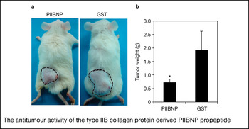
Recent work indicated that PIIBNP has strong antiangiogenic and antitumour activities (Ref. Reference Wang194) that might have a role in keeping the cartilage avascular and could be exploited to kill tumours in vivo. The interaction of recombinant human PIIBNP (GST-PIIBNP) with hCh-1 cells (human chondrosarcoma cell line) was dependent on αVβ5 and αVβ3 integrins and took place in an RGDRGD-dependent manner in vitro. Notably, PIIBNP was able to directly induce cell death in hCh-1 cells, in HeLa cells (cervical carcinoma cell line) and in MDA-MB231 (breast cancer cell line) in a dose-dependent manner. Disruption of the RGD-integrin interaction by mutation of the RGDRGD motif in PIIBNP or downregulation of integrin αV in the cell line using siRNA technology significantly reduced cell death. The significance of these in vitro observations hit home when it was realised that PIIBNP exists in cartilage and that normal chondrocytes do not express, or have very low levels of αV, β5 and β3 integrins, and presumably therefore are not killed by PIIBNP in vivo. GST-PIIBNP induced necrosis as opposed to apoptotic cell death and was also inhibitory to tumour cell migration. When tested in vivo, GST-PIIBNP suppressed MDA-MB231 induced tumours in nude mice (Fig. 2) demonstrating a 75% reduction in tumour growth over untreated tumours. Several cell types such as endothelial cells, osteoclasts and tumour cells express αVβ5 and αVβ3 integrins, suggesting that PIIBNP might function in vivo to keep the cartilage avascular and free from bone invasion. Indeed, PIIBNP was able to induce apoptotic death in osteoclasts (but not osteoblasts and macrophages) in an RGD- and integrin-dependent manner both in vitro and in vivo (Ref. Reference Hayashi195). These data indicate that PIIBNP might have antiangiogenic role in cartilage and could be used as an anticancer agent.
The antitumour activity of the type IIB collagen protein derived PIIBNP propeptide. (a) An in vivo tumour assay showing the antitumour activity of PIIBNP peptide. NOD/SCID mice were injected into the dorsal surface with MDA-MB231 breast cancer cells to form solid tumours. When the tumours grew to 0.5 cm in diameter, the mice were divided into two groups with three mice in each. Mice received either 6 nmol of PIIBNP or GST per day by subcutaneous injection into the tumour for 2 weeks. The dashed line shows the boundary of the tumour. (b) When the largest tumour size in the control (GST) group reached 2 cm in diameter, the mice were sacrificed and tumours removed and weighed. The graph shows tumour weights in grams (g) represented as mean ± s.d. (*P < 0.01 compared with GST control, n = 6) derived from these mice after treatment with PIIBNP or GST. PIIBNP suppressed tumour growth in mice. Reproduced from Ref. Reference Wang194, with permission. © The American Society for Biochemistry and Molecular Biology, 2010.

Future prospects for antiangiogenic research for cartilage-derived molecules
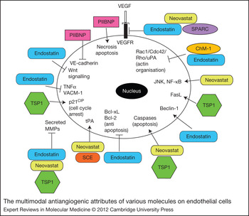
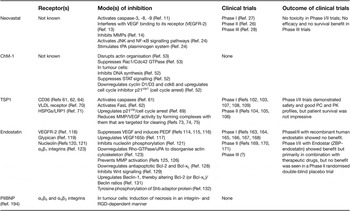
Interest in antiangiogenic molecules has also triggered an interest in their application as anticancer agents in the clinic. The current antiangiogenic modality approved to treat different forms of cancer in the clinic uses monoclonal antibodies against VEGF or to its receptor. Bevacizumab, an anti-VEGF humanised antibody is used as a treatment for pancreatic cancer, colorectal cancer, nonsmall lung cancer, metastatic renal cancer, carcinoma, glioblastoma and metastatic breast cancer, often in combination with chemotherapy. It is also under investigation for treatment of solid tumours. Despite the tremendous interest generated by the applications of anti-VEGF therapy, it is beleaguered with several drawbacks, primarily its sporadic success in clinical treatments and an inability to provide significant survival benefit (Refs Reference Allegra196, Reference Van Cutsem197). In fact Avastin, a commercially marketed bevacizumab preparation, was banned recently by the FDA for breast cancer treatment, although it continues to be used for lung and kidney cancers. Furthermore, some tumours demonstrate initial sensitivity to anti-VEGF therapy, but acquire resistance later as a result of the upregulation of bFGF within the tumour after treatment with an anti-VEGFR2 antibody (Ref. Reference Casanovas198). Some tumours also have a natural resistance to anti-VEGF therapy because of CD11b+Gr1+ myeloid cells (Ref. Reference Shojaei199). The high cost of these therapies and their lack of efficacy, coupled with significant toxicity in the form of hypertension (Ref. Reference Syrigos200), suggest a need for designing additional antiangiogenic agents. Cartilage as a naturally occurring avascular tissue was considered a good source of antiangiogenic molecules that might fill the gaps from anticancer therapies resulting from targeting VEGF alone. Indeed, studies on these cartilage-derived antiangiogenic molecules have demonstrated the versatile and complex nature of their antiangiogenic, and in some cases, direct anticancer (ChM-1 and PIIBNP) effect, that is not restricted to targeting VEGF (see Table 1 and Fig. 3).
The multimodal antiangiogenic attributes of various molecules on endothelial cells. Representation of the effects of Neovastat, chondromodulin-1 (ChM-1), thrombospondin-1 (TSP1), endostatin, SPARC (secreted protein acidic and rich in cysteine) and the type II collagen-derived N-terminal propeptide (PIIBNP) on endothelial cells (see also Table 1 and text for further details).

Summary of cartilage-derived antiangiogenic molecules, their modes of inhibition in endothelial cells and outcomes in clinical trials

Question mark indicates lack of publication in an English language publication and therefore inability to review (see Further Reading section).
However, none of these cartilage-derived molecules have so far had much success in clinical trials. Their in vitro assessments and highly successful preclinical trials have had little bearing on their success in humans. Although their in vivo antiangiogenic benefit was largely visible only under certain experimental conditions, such as in a fracture model or cartilage lesion model, deficiency of these molecules in mouse models did not create any abnormal pathology. Perhaps redundancies in functions are responsible for this effect. Discovery of these molecules has not helped to explain how cartilage remains avascular either. Furthermore, Col IIB, ChM-1 and endostatin levels in cartilage drop drastically with maturity and yet the cartilage remains avascular in a healthy individual. Thus, cartilage might derive its antiangiogenic benefit, not from a single molecule, but from the sum total from several individual antiangiogenic molecules, or all cartilage components might need to be present together for it to be antiangiogenic. This suggests that it would be improbable to expect total comprehensive benefit based on administration of a single antiangiogenic molecule in clinical trials. Combinations of molecules, such as TSP1 plus endostatin, in trials might provide better benefit. In preparations of Neovastat, the individual molecules remain largely unidentified. It needs to be ascertained whether Neovastat extract by any chance contains the water-soluble fractions of all the single molecules discussed above. The only identified molecule in Neovastat is the kappa light chain (which had the ability to stimulate tPA activity; see Ref. Reference Boivin201), which cannot be considered cartilage specific.
Attempts to identify individual shark cartilage components that demonstrate antiangiogenic properties showed sequence similarity to alpha parvalbumin family (SCP1 protein) (Ref. Reference Rabbani-Chadegani202) or novel proteins (Refs Reference Zheng203, Reference Bargahi and Rabbani-Chadegani204), but not to the molecules discussed above. Although Col IIB has recently been recognised, molecules in cartilage identified as antiangiogenic so far are largely the minor cartilage components. Not many reports on articular cartilage aggrecan, the other major macromolecule, as an antiangiogenic compound exist, although intervertebral disc aggrecan has been demonstrated to be inhibitory to endothelial cell adhesion and migration (Ref. Reference Johnson205), and a polysaccharide isolated from porcine cartilage, an aggrecan-derived component, has demonstrated apoptosis of tumour cells by activation of caspase-3 and caspase-9 (Ref. Reference Liu206). Recently, articular cartilage glycosaminoglycan has been demonstrated to inhibit endothelial cell adhesion (Ref. Reference Bara207). Other cartilage components such as Troponin 1 (Ref. Reference Moses208), which interferes with bFGF function by binding to bFGF receptor (Ref. Reference Feldman and Rouleau209), metastatin complex, which disrupts endothelial cell proliferation by binding to hyaluronan (Ref. Reference Liu210), the 16 kDa N-terminal fragment of prolactin with ability to inhibit bFGF (Ref. Reference Macotela211), or the tissue inhibitors of metalloproteinases that are often upregulated by drugs that inhibit angiogenesis (Ref. Reference Donnini212), have not been pursued much beyond their initial recognition of their antiangiogenic properties.
There is no simple explanation for the overall disappointment with these molecules. In the case of Neovastat, lack of PK/PC data was considered a hindrance to analyse its failure. But PK/PC values are available for TSP1 and endostatin, but have not helped in alleviating their failures. Peptide mimetics of TSP2 have not been reported and considering the lack of significant benefit from TSP1, antiangiogenic properties of TSP2 might need further evaluation. The mechanisms of inhibitory processes for most of these molecules are only marginally understood. Understanding the complete antiangiogenic mechanism might better prepare us for clinical trials. This can be exemplified for endostatin where a lack of understanding of the ZBP domain in the N-terminus led to the use of P. pastoris expressed, N-terminally truncated, recombinant human endostatin in clinical trials, which did not demonstrate any therapeutic efficacy. The genetic heterogeneity of the human population could also be a hindrance to translating the success from the preclinical, syngeneic mouse models. Formulating a new strategy or criteria for testing antiangiogenic molecules might be necessary before any further investments in clinical trials.
Designing effective administration, improving half-life, or identification and modification of peptide derivatives are being tested to tackle these shortcomings. For example, attachment of polyethylene glycol to the N-terminus of Endostar to reduce proteolytic susceptibility (Ref. Reference Tong213), peptide derivatives of endostatin further modified by the addition of RGD sequence for improved activity (Refs Reference Xu214, Reference Yin215), addition of the tumour-penetrating peptide internalisation sequence (iRGD) to endostatin to facilitate penetration into extravascular tumour tissue that demonstrated stronger antiangiogenic/antitumour activity (Ref. Reference Hai-Tao216), and finally, endostatin-expressing Ad vectors encapsulated in cationic liposome to allow delivery to cancer cells lacking Ad receptors (Refs Reference Wang217, Reference Yang218), have all found success in preclinical trials, although not yet in humans. The criteria of what is a better biomarker of a successful clinical trial might also need to be revisited. Antiangiogenesis prevents blood vessel formation that shrinks tumours, rather than by being cytotoxic; this is a slow process that might need time for characterisation and recognition of the clinical end point. Antiangiogenesis remains an attractive therapeutic application, but it would appear that its sources need to be scrupulously and completely understood to translate them into beneficial clinical applications.
Acknowledgements and funding
We thank the reviewers and the editor of ERMM for reading the article and for their expert comments that helped to better the article. We thank Zhepeng Wang for his assistance with Figure 2. This work was supported by National Institutes of Health grants RO1 AR050847, and RO1 AR045550, to L.J. Sandell.