Introduction
One of the main factors for successful pregnancies in assisted reproduction (AR) cycles is oocyte quality, which has a direct impact on embryo development and implantation (Cha & Chian, Reference Cha and Chian1998). Only mature oocytes (gametes arrested in metaphase of the second meiotic division – the MII stage) can be fertilized and used for AR. In the menstrual cycle, the maturation process occurs after recruitment, selection and dominance of the ovulatory follicle. Physiologically, the peak of luteinizing hormone (LH) levels triggers oocyte maturation. This event comprises germinal vesicle breakdown (GVBD), first metaphase plate formation and polar body (PB) extrusion (Dekel, Reference Dekel1995; Cha & Chian, Reference Cha and Chian1998; Sathananthan et al., Reference Sathananthan, Selvaraj and Trounson2000; Adona et al., Reference Adona, Pires, Quetglas, Schwarz and Leal2008).
Since the first description of human oocyte in vitro maturation (IVM) (Edwards, Reference Edwards1965), there has been increasing interest in this procedure. The main benefit of IVM is the possibility of increasing the number of gametes that can be fertilized, improving the source of embryos available for transfer or cryopreservation, thus enhancing the chance of pregnancy (De Vos et al., Reference De Vos, Van de Velde, Joris and Van Steirteghem1999; Kim et al., Reference Kim, Lee, Kim, Han and Kim2000; Son et al., Reference Son, Lee and Lim2005). Furthermore, delivery of healthy children has been described after IVM and ICSI (Nagy et al., Reference Nagy, Cecile, Liu, Loccufier, Devroey and Van Steirteghem1996; Edirisinghe et al., Reference Edirisinghe, Junk, Matson and Yovich1997; Jaroudi et al., Reference Jaroudi, Hollanders, Sieck, Roca, Nour and Coskun1997; De Vos et al., Reference De Vos, Van de Velde, Joris and Van Steirteghem1999; Liu et al., Reference Liu, Lu, Qian, Mao and Ding2003; Son et al., Reference Son, Lee and Lim2005).
IVM can be used in stimulated in vitro fertilization (IVF) cycles with recovery of oocytes at different stages of meiotic maturity, either at metaphase I (MI) or prophase I (PI) (germinal vesicle) (Edirisinghe et al., Reference Edirisinghe, Junk, Matson and Yovich1997; Jaroudi et al., Reference Jaroudi, Hollanders, Sieck, Roca, Nour and Coskun1997; Liu et al., Reference Liu, Katz, Garcia, Compton and Baramki1997; De Vos et al., Reference De Vos, Van de Velde, Joris and Van Steirteghem1999). The retrieval of immature oocytes in an IVF cycle is the result of asynchrony of the follicle cohort at the time of human chorionic gonadotrophin (hCG) administration (Edirisinghe et al., Reference Edirisinghe, Junk, Matson and Yovich1997; Kim et al., Reference Kim, Lee, Kim, Han and Kim2000). The rate of immature oocyte retrieval in stimulated IVF cycles ranges from 0 to 50% of all oocytes recovered (Edirisinghe et al., Reference Edirisinghe, Junk, Matson and Yovich1997; Cha & Chian, Reference Cha and Chian1998), and can occur in the same woman in more than one cycle (Lee et al., Reference Lee, Kim, Jee, Suh and Kim2011). Immature oocytes retrieved from stimulated ovaries can result in pregnancy, even after oocyte cryopreservation (Tucker et al., Reference Tucker, Wright, Morton and Massey1998) or after cryopreservation of embryos derived from IVM oocytes (Edirisinghe et al., Reference Edirisinghe, Junk, Matson and Yovich1997). Even though oocyte IVM can improve the success rate of AR procedures, data in the literature are contradictory with regard to the use of embryos derived from immature gametes; doubts remain as to whether these oocytes are intrinsically compromised or whether culture conditions remain suboptimal. Therefore, immature gametes are sometimes discarded in stimulated IVF cycles. It should be noted that the apparent low quality of embryos derived from in vitro matured oocytes recovered from stimulated ovaries may reflect unsuitable culture conditions when the majority of studies were carried out (Jaroudi et al., Reference Jaroudi, Hollanders, Sieck, Roca, Nour and Coskun1997; Kim et al., Reference Kim, Lee, Kim, Han and Kim2000). It is widely known that culture conditions have improved in recent years. In this study, the performance of immature oocytes (PI and MI) and mature oocytes (MII) was evaluated, and IVM and oocyte fertilization rates, cleavage, transfer, cryopreservation and utilization rates of the embryos derived from immature and mature retrieved oocytes were compared.
Materials and methods
Patients
This study was approved by the Hospital Moinhos de Vento (HMV) Research Ethics Committee (protocol #2011/76). All patients who underwent AR and had immature oocytes retrieved during ovarian puncture at Núcleo de Reprodução Humana Gerar between March 2006 and June 2011 were included in the study.
Controlled ovarian stimulation and oocyte recovery
Patients included in the study underwent controlled ovarian stimulation using a GnRH agonist or antagonist and purified or recombinant gonadotropins as prescribed, independent of this study. According to ultrasound control, recombinant or urinary hCG was administered 36 h before oocyte retrieval, which was carried out by guided transvaginal ultrasound. Retrieved cumulus–oocyte complexes (COCs) were transferred to plastic dishes containing ECM (Early Cleavage Medium, –Irvine Scientific) supplemented with 10% (v/v) Serum Substitute Supplement (Irvine Scientific), where they remained for 4 h at 37°C at 100% relative humidity and a 5% CO2 in air atmosphere. In order to evaluate the stage of maturation, oocytes were denuded of the cumulus oophorus cells by enzymatic treatment (exposure to hyaluronidase solution [Irvine Scientific] for 25 s). Denuded oocytes were observed under an inverted microscope at ×2000 magnification, and their maturity was classified according to nuclear status: PI in the presence of a germinal vesicle, MII in the presence of PB extrusion or MI in the absence of both structures (GVBD with no visible PB) (Kim et al., Reference Kim, Lee, Kim, Han and Kim2000). Immature oocytes defined as PI or MI were assigned to the IVM group, while gametes MII were used for IVF.
In vitro maturation
Immature oocytes were transferred, individually, to plastic dishes that contained droplets of ECM supplemented with 10% Serum Substitute Supplement covered with mineral oil (Irvine Scientific), where they remained for 24 h (IVM period) at 37°C at 100% relative humidity and a 5% CO2 in air atmosphere. After the IVM period, matured oocytes (confirmed by the presence of PB) were fertilized. Gametes that remained immature after 24 h of IVM were disposed of in the proper manner.
In vitro fertilization, cleavage, transfer and cryopreservation of the embryos
Semen samples were collected by masturbation and prepared using a discontinuous gradient – 50% and 90% (Isolate Concentrate and human tubal fluid (HTF) modified). Only oocytes that achieved stage MII after IVM or in vivo maturation were fertilized by the ICSI technique. We evaluated cycles that used only ICSI in order to eliminate the male factor bias. After 18 h of fertilization, using the same conditions of IVM, gametes were evaluated for the presence of two pronuclei and two PB, as an indicator of proper fertilization. Embryo development/cleavage was evaluated 48 and 72 h after fertilization. Embryos were classified as suitable in the presence of 2–4 cells at 48 h and 6–8 cells at 72 h and less than 20% fragmentation. Cryopreservation was performed using the vitrification technique (Irvine Scientific–Embryo Freeze Media), according to manufacturer instructions, and embryos were stored in CryoTips® (Irvine Scientific). In some cycles, embryos derived from oocytes matured in vitro and in vivo were transferred or cryopreserved together; hence, we were unable to evaluate implantation and pregnancy rates.
Statistical analysis
The chi-squared test, supplemented by adjusted residuals, was used for between-group comparisons. The Spearman correlation coefficient was used to analyze the relationship between the rate of immature oocytes retrieved per cycle and the potential of MII oocytes recovered (assessed by fertilization, cleavage and utilization rates). Data were analyzed in the Statistical Package for the Social Sciences (SPSS) 18.0 software environment. For all tests, differences were considered statistically significant if the P-value was ≤0.05.
Results
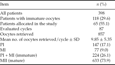
In total, 398 patients underwent oocyte retrieval and IVF at Núcleo de Reprodução Humana Gerar during the study period. Of these patients, 118 (29.6%) had at least one immature oocyte recovered, and their medical records were reviewed. Sixty-five patients (55.1%) – mean age 35.4 ± 5.1 years for women and 38.9 ± 7.2 years for men – were included in the study, and 53 (44.9%) were excluded due to use of a non-ICSI fertilization technique. The 65 patients had 87 cycles analyzed; 857 oocytes were recovered; the mean and standard deviation (SD) of gametes recovered per cycle were 9.85 ± 5.35 respectively. Of these, 147 (17.1%) were PI, 77 (9.0%) were MI and 633 (73.9%) were MII, as described in Table 1.
Sample profile

MI, metaphase I; MII, metaphase II, PI, prophase I.
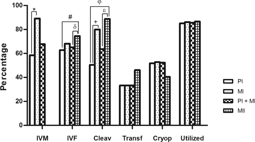
Oocyte maturation and fertilization rates, and embryo cleavage, transfer, cryopreservation and utilization rates are shown in Fig. 1. We demonstrate the results for immature gamete (PI and MI) both separately and jointly in order to demonstrate if one of them was influencing in the result of the other. The embryo utilization rate (cryopreserved plus transferred embryos) was used as an indicator of good embryo quality. The IVM rate was higher in MI gametes (66/74, 89.2%) as compared with PI (86/147, 58.5%). The fertilization rate was higher in oocytes matured in vivo (430/577, 74.5%) as compared with PI oocytes (54/86, 62.8%) and with all immature oocytes (PI + MI; 99/152, 65.1%). However, there were no significant differences in fertilization rate between MI oocytes (45/66, 68.2%) and MII oocytes (P = 0.336). It should be noted that the number of MII oocytes fertilized (577) was lower than the total number of MII oocytes retrieved (633), because some patients decided to vitrify their oocytes.
Embryonic features of in vitro or in vivo matured oocytes recovered from patients who underwent hormonal stimulation. *Values are significantly different between prophase I (PI) and metaphase I (MI) oocytes regarding in vitro maturation (IVM) rate (P < 0.001); #Values are significantly different between PI and MII oocytes regarding fertilization rate (P = 0.031). δValues are significantly different between PI + MI and MII oocytes regarding fertilization rate (P = 0.027). φValues are significantly different between PI and MII oocytes regarding embryo cleavage rate (P < 0.001). ɛValues are significantly different between PI + MI and MII oocytes regarding embryo cleavage rate (P < 0.001). +Values are significantly different between PI and MI oocytes regarding embryo cleavage rate (P = 0.004).
The embryo cleavage rate showed a similar pattern to fertilization rate, i.e. it was higher for embryos derived from gametes matured in vivo (382/430, 88.8%) than in IVM PI oocytes (27/54, 50.0%) and embryos from all immature oocytes (63/99, 63.6%). However, as observed for fertilization rate, the embryo cleavage rate was similar between embryos derived from IVM MI oocytes (36/45, 80.0%) and from in vivo matured oocytes (P = 0.135). Additionally, the embryo cleavage rate was significantly higher in the MI oocytes group than among their PI counterparts (P = 0.004). The embryo transfer rate was equal among immature oocytes (PI group, 9/27; MI group, 12/36, both representing 33.33%) and was not significantly different from that observed in the in vivo matured oocytes group (176/382, 46.1%) (P = 0.80). The cryopreservation rate was similar between embryos derived from oocytes matured in vitro (PI group, 14/27, 51.9%; MI group, 19/36, 52.8%) and in vivo (155/328, 40.5%) (P = 0.138). The utilization rate (transfer and cryopreservation) is similar among the three groups.
During one stimulated IVF cycle embryos derived only from an IVM MI oocyte were transferred, but pregnancy was not achieved.
The rate of immature oocytes recovered did not correlate with the fertilization (P = 0.077), cleavage (P = 0.155), or utilization (P = 0.160) rates of oocytes retrieved as MII in the same cycle.
Discussion
Preservation of fertility in cancer patients, avoidance of the need for ovulation-inducing drugs, use in poor responders, and reduction of the cost of AR procedures are some of the benefits of the IVM technique (Jaroudi et al., Reference Jaroudi, Hollanders, Sieck, Roca, Nour and Coskun1997; Liu et al., Reference Liu, Lu, Qian, Mao and Ding2003; Le Du et al., Reference Le Du, Kadoch, Bourcigaux, Doumerc, Bourrier, Chevalier, Fanchin, Chian, Tachdjian, Frydman and Frydman2005; Suikkari & Söderström-Anttila, Reference Suikkari and Söderström-Anttila2007; Oktay et al., Reference Oktay, Demirtas, Son, Lostritto, Chian and Tan2008; Huang et al., Reference Huang, Chian, Gilbert, Fleiszer, Holzer, Dermitas, Elizur, Gidoni, Levin, Son and Tan2010). Furthermore, IVM can raise the number of oocytes available for IVF and, probably, the ensuing pregnancy rates. In our study, 118 patients (29.6%) had at least one immature oocyte retrieved and IVM enhanced the number of mature oocytes for fertilization. A previous study of IVM cycles showed that the presence of a dominant follicle (≥1.4 cm) affects the developmental competence of immature gametes (Russell, Reference Russell1999). We demonstrated an inverse relationship: retrieval of immature gametes did not correlate with the performance of retrieved MII oocytes. In other words, the retrieval of immature oocytes was not associated with poor quality of the oocyte cohort.
The mean number of oocytes retrieved per cycle (9.85 ± 5.35) was similar to that reported in the literature, ranging from 6.4–10.3% (Kim et al., Reference Kim, Lee, Kim, Han and Kim2000; Bodri et al., Reference Bodri, Vernaeve, Guillén, Vidal, Figueras and Coll2006; Söderström-Anttila et al., Reference Söderström-Anttila, Salokorpi, Pihlaja, Serenius-Sirve and Suikkari2006; Suikkari & Söderström-Anttila, Reference Suikkari and Söderström-Anttila2007; McAvey et al., Reference McAvey, Zapantis, Jindal, Lieman and Polotsky2011). The total number of immature oocytes retrieved (PI and MI, 26.1%) was somewhat higher than that described in the literature (10–20% range) (Barnes et al., Reference Barnes, Kausche, Tiglias, Wood, Wilton and Trounson1996; Cha & Chian, Reference Cha and Chian1998; De Russell, Reference Russell1999; Vos et al., Reference De Vos, Van de Velde, Joris and Van Steirteghem1999; Kim et al., Reference Kim, Lee, Kim, Han and Kim2000), although it remained within the physiological range (0–50%) (Edirisinghe et al., Reference Edirisinghe, Junk, Matson and Yovich1997; Cha & Chian, Reference Cha and Chian1998; Bodri et al., Reference Bodri, Vernaeve, Guillén, Vidal, Figueras and Coll2006; Lee et al., Reference Lee, Kim, Jee, Suh and Kim2011). Among all immature oocytes recovered, those at the PI stage predominated, as observed by De Vos et al. (Reference De Vos, Van de Velde, Joris and Van Steirteghem1999).
It is known that the longer oocytes remain in IVM, the lower the quality of their derived embryo (Son et al., Reference Son, Lee and Lim2005). To avoid oocyte senescence (Smith et al., Reference Smith, Mikkelsen and Lindenberg2000), the IVM period was defined as 24 h. The IVM rate among MI oocytes (89.2%) was higher than that observed in the literature (approximately 70%) (Trounson et al., Reference Trounson, Wood and Kausche1994; Barnes et al., Reference Barnes, Kausche, Tiglias, Wood, Wilton and Trounson1996; Kim et al., Reference Kim, Lee, Kim, Han and Kim2000; Ben-Ami et al., Reference Ben-Ami, Komsky, Bern, Kasterstein, Komarovsky and Ron-El2010), but the IVM rate of PI oocytes (58.5%) was lower. Additionally, there was a significant difference in IVM rate between PI and MI oocytes (P < 0.001). This result may be because PI oocytes must undergo two stages of nuclear maturation that are considered independent: GVBD and PB extrusion (Cha & Chian, Reference Cha and Chian1998; Trounson et al., Reference Trounson, Anderiesz and Jones2001).
The reduced fertilization rate in PI gametes is probably due to asynchrony between nuclear and cytoplasmic maturation, as the protein content of PI oocytes matured in vitro is lower than that of their in vivo matured counterparts. The mechanisms of protein translation and accumulation on the ooplasm are essential for oocyte cytoplasm maturation, fertilization, and initiation of embryo development (Trounson et al., Reference Trounson, Anderiesz and Jones2001), therefore abnormalities in cytoplasmic maturation of PI oocytes could explain our findings. It bears stressing that the fertilization rates of in vivo matured oocytes and in vitro matured MI oocytes (68.2%) were similar, reflecting that the quality of these gametes, even when immature at the time of retrieval, is suitable for fertilization techniques, increasing the number of embryos. Contrary to our findings, De Vos et al. (Reference De Vos, Van de Velde, Joris and Van Steirteghem1999) reported a lower fertilization rate in IVM MI oocytes as compared with MII oocytes. In our study, the fertilization rate of IVM MI oocytes (68.2%) was similar to that reported by De Vos et al. (Reference De Vos, Van de Velde, Joris and Van Steirteghem1999) for their MII group (70.8%) and higher than that found in their MI group (52.7%). The improvement of IVM and fertilization conditions in recent years could explain these findings, allowing safer use of MI gametes in AR procedures. However, we have to consider that neither morphological appearance nor the cleavage rate can fully measure the genomic quality of the embryos.
The embryo cleavage rate of oocytes matured in vivo (88.8%) remained within the range reported in the literature (Kim et al., Reference Kim, Lee, Kim, Han and Kim2000; Son et al., Reference Son, Chung, Demirtas, Holzer, Sylvestre, Buckett, Chian and Tan2008), and was higher than that of embryos derived from PI oocytes (50.0%), reflecting the lower quality of these embryos. Nevertheless, the embryo cleavage rate of oocytes matured in vivo was similar to the rate of IVM MI oocytes (80.0%), confirming the ability of these immature oocytes to generate good embryos, as shown by De Vos et al. (Reference De Vos, Van de Velde, Joris and Van Steirteghem1999), who reported that in vitro matured MI oocytes produce embryos of similar quality as mature MII oocytes after ICSI. The fact that the embryo cleavage rate was higher in the MI group than among PI oocytes confirms good performance. We did not compare cleavage after the third day, because embryos were transferred or cryopreserved at day 2 or 3, but it has been demonstrated elsewhere that embryo cleavage on day 4 (Kim et al., Reference Kim, Lee, Kim, Han and Kim2000) and number of good-quality blastocysts (Son et al., Reference Son, Lee and Lim2005) were higher in the group derived from in vivo matured oocytes than in embryos derived from PI oocytes (using 24 h of IVM).
The utilization rate (transfer and cryopreservation) did not differ among the three groups, demonstrating that although only a small portion of PI oocytes were able to generate embryos with cleavage potential, when a PI oocyte does generate an embryo, it will be of good quality. Hence, we suggest that auxiliary techniques designed to identify which PI oocytes have the potential to mature and fertilize in vitro must be applied in conjunction with IVM so as to optimize outcomes.
Most studies in this field have analyzed one type of immature gamete (PI or MI) separately, or have described these jointly as immature. Our report assessed immature oocytes both separately and jointly, so as to analyze the performance of gametes under the same culture conditions. The MI oocytes group, as previously reported, performed similar to the MII oocytes group, indicating the safety of their use for AR techniques. Conversely, the results observed with PI oocytes show that further studies are required to develop techniques that would identify which PI oocytes are able to mature and fertilize, producing good embryos.