Introduction
Lungworms of the genus Angiostrongylus are bursate nematodes of the superfamily Metastrongyloidea. The Metastrongyloidea contains up to seven families, depending on taxonomic authorities – Angiostrongylidae, Crenosomatidae, Filaroididae, Metastrongylidae, Protostrongylidae, Pseudaliidae and Skrjabingylidae – with over 180 species in 46 genera (Anderson, Reference Anderson2000). In the National Center for Biotechnology Information (NCBI) taxonomy, only six families are recognized, without Skrjabingylidae but with the genus Skrjabingylus treated as a member of the Metastrongylidae.
Of the Angiostrongylus lungworms, the rat lungworm A. cantonensis (Chen, 1935) is a food-borne zoonotic parasite of public health importance in many countries of the tropics and subtropics (Eamsobhana & Tungtrongchitr, Reference Eamsobhana, Tungtrongchitr, Arizono, Chai, Nawa and Takahashi2005; Eamsobhana, Reference Eamsobhana2006). Its occurrence has now been reported in many countries worldwide (Eamsobhana, Reference Eamsobhana2006; Cross & Chen, Reference Cross, Chen, Murrel and Fried2007; Foronda et al., Reference Foronda, López-González, Miquel, Torres, Segovia, Abreu-Acosta, Casanova, Valladares, Mas-Coma, Bargues and Feliu2010; Maldonado et al., Reference Maldonado, Simŏes, Thiengo and Lorenzo-Morales2012). Infection with this parasite causes eosinophilic meningitis or eosinophilic meningoencephalitis, with marked cerebrospinal fluid eosinophilia. Rats and snails have been implicated in the global dispersal of this zoonosis (Kliks & Palumbo, Reference Kliks and Palumbo1992). Based on the affected organ, three types of eosinophilic meningitis may be recognized, namely cerebrospinal angiostrongyliasis cantonensis, ocular angiostrongyliasis due to A. cantonensis and pulmonary angiostrongyliasis cantonensis.
A related species, A. malaysiensis Bhaibulaya & Cross, Reference Bhaibulaya and Cross1971, in South-East Asia and Japan, has not been confirmed to infect humans but remains a possibility. It has been shown to produce neurological abnormality in infected rodent hosts (Cross, Reference Cross and Cross1979). When first documented, it was referred to as A. cantonensis (Bhaibulaya & Cross, Reference Bhaibulaya and Cross1971).
Another species in the Americas (from southern USA to northern Argentina in South America), A. costaricensis Morera and Céspedes, 1971, causes abdominal or intestinal angiostrongyliasis, which mimics appendicitis with eosinophilia (Graeff-Teixeira et al., Reference Graeff-Teixeira, da Silva and Yoshimura2009; Graeff-Teixeira, Reference Graeff-Teixeira and Eamsobhna2010). This species does not occur outside the Americas.
In addition to the species of human public health importance, the heartworm A. vasorum (Baillet, 1866) is of veterinary concern. In contrast to A. cantonensis, A. costaricensis and A. malaysiensis, which infect rodents, the natural definitive hosts of A. vasorum are domestic dogs and various other carnivores. This parasite infects wild and domestic canids and causes canine angiostrongylosis (Morgan et al., Reference Morgan, Shaw, Brennan, De Waal, Jones and Mulcahy2005; Conboy, Reference Conboy2011). It is enzootic to Western Europe but has been found in many parts of the world. Infections in dogs tend to be chronic but can be fatal.
Of the Angiostrongylus species, A. cantonensis has received great attention in laboratory and clinical studies (Eamsobhana, Reference Eamsobhana2006; Cross & Chen, Reference Cross, Chen, Murrel and Fried2007; Graeff-Teixeira et al., Reference Graeff-Teixeira, da Silva and Yoshimura2009). In particular, immunological diagnosis of human angiostrongyliasis has been explored extensively (Eamsobhana & Yong, Reference Eamsobhana and Yong2009). Polymerase chain reaction–restriction fragment length polymorphism (PCR-RFLP) has been employed to differentiate A. cantonensis, A. costaricensis and A. vasorum (Caldeira et al., 2003). The DNA sequences of a 66-kDa protein gene indicate a close relationship of A. cantonensis and A. malaysiensis compared to A. costaricensis (Eamsobhana et al., Reference Eamsobhana, Lim, Zhang, Gan and Yong2010a). Additionally, the DNA nucleotide sequences of internal transcribed spacer 2 (ITS-2) (Jefferies et al, Reference Jefferies, Shaw, Viney and Morgan2009; Foronda et al., Reference Foronda, López-González, Miquel, Torres, Segovia, Abreu-Acosta, Casanova, Valladares, Mas-Coma, Bargues and Feliu2010), cytochrome c oxidase subunit I (COI) (Eamsobhana et al., Reference Eamsobhana, Lim, Solano, Zhang, Gan and Yong2010b; Tokiwa et al., Reference Tokiwa, Harunari, Tanikawa, Komatsu, Koizumi, Tung, Suzuki, Kadasaka, Takada, Kumagai, Akao and Ohta2012) and nuclear small subunit (18S) ribosomal RNA (Fontanilla & Wade, Reference Fontanilla and Wade2008; van Megan et al., 2009; Tokiwa et al., Reference Tokiwa, Harunari, Tanikawa, Komatsu, Koizumi, Tung, Suzuki, Kadasaka, Takada, Kumagai, Akao and Ohta2012) have been used to determine the relationship between various component taxa of Angiostrongylus.
At the family level, the Angiostrongylus lungworms have been included in the family Angiostrongylidae (Anderson, Reference Anderson2000; NCBI Taxonomy) or the family Metastrongylidae (Bhaibulaya & Cross, Reference Bhaibulaya and Cross1971; Maldonado et al., Reference Maldonado, Simŏes, Thiengo and Lorenzo-Morales2012; Myers et al., Reference Myers, Espinosa, Parr, Jones, Hammond and Dewey2013). To date there appears to be no consensus on their family stautus.
The present study was undertaken to determine the usefulness and suitability of the nuclear 18S (small subunit, SSU) rDNA sequences for differentiating various taxa of the genus Angiostrongylus, as well as to determine the systematics and phylogenetic relationship of Angiostrongylus species and other metastrongyloid taxa. The 18S rRNA gene is generally conserved. In a barcoding study of nematodes, it has been amplified most reliably from a range of taxa compared to the nuclear 28S rRNA as well as mitochondrial cytochrome c oxidase I (COI) and 16S rRNA genes (Bhadury et al., Reference Bhadury, Austen, Bilton, Lambshead, Rogers and Smerdon2006). In the present study, with more extensive taxa sampling, the 18S (SSU) nucleotide sequences reveal a greater haplotype diversity in A. cantonensis than previously reported. The phylogenetic analyses also resolve the family status of Angiostrongylidae and Metastrongylidae, as well as the close relationship of A. cantonensis and A. malaysiensis and that of A. costaricensis and A. dujardini.
Materials and methods
Parasite species and isolates
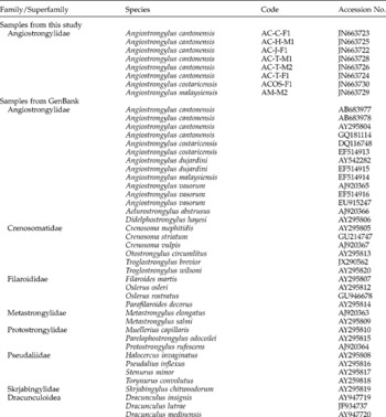
The Angiostrongylus lungworms used in this study are summarized in Table 1. Adult male and female worms of A. cantonensis from Thailand and Hawaii were obtained from experimentally infected albino rats, as described by Eamsobhana et al. (Reference Eamsobhana, Lim, Zhang, Gan and Yong2010a, Reference Eamsobhana, Lim, Solano, Zhang, Gan and Yongb). Angiostrongylus cantonensis adult worms from Japan and China were obtained from wild-caught rodents. Angiostrongylus malaysiensis adult worms were obtained from the pulmonary arteries of wild-caught Rattus tiomanicus in Malaysia, and A. costaricensis adult specimens from Costa Rica were obtained from an experimentally infected cotton rat.
Nucleotide sequences of 18S rRNA gene for 42 taxa of Metastrongyloidea used in the present study. Dracunculus spp. of Dracunculoidea were used as outgroups. Specimens of A. cantonensis in the present study were obtained from experimentally infected rats in Thailand; the specimen of A. malaysiensis was recovered from a rat in Peninsular Malaysia; the specimen of A. costaricensis was from an experimentally infected cotton rat in Costa Rica.

DNA template preparation
DNA extractions from individual adult female and male worms of A. cantonensis (Thailand, Hawaii, Japan and China isolates), A. costaricensis and A. malaysiensis, were prepared using the FTA (fast technology for analysis of nucleic acid) classic card method (Whatman BioScience, Newton Center, Massachusetts, USA) (Eamsobhana et al., Reference Eamsobhana, Lim, Zhang, Gan and Yong2010a, Reference Eamsobhana, Lim, Solano, Zhang, Gan and Yongb). Captured nucleic acids on the FTA cards were purified following the FTA manufacturer's instructions. Briefly, a 2.0 mm sample disc was removed from the dried spot of the individual worm sample on the FTA card using a Harris micro-punch. The punched disc was washed with FTA purification reagent (Whatman BioScience), rinsed with TE buffer (10 mm Tris–HCI, 0.1 mm EDTA, pH 8) and dried. Polymerase chain reaction (PCR) master mix was added directly to the DNA punch in a PCR tube, followed by amplification.
PCR amplification and nucleotide sequencing
The 18S RNA nuclear gene (SSU rRNA) was amplified using the primers SSU F07: 5′-AAAGATTAAGCCATGCATG-3′ and SSU R09: 5′-AGCTGGAATTACCGCGGCTG-3′ (Blaxter et al., Reference Blaxter, De Ley, Garey, Liu, Scheldeman, Vierstraete, Vanfleteren, Mackey, Dorris, Frisse, Vida and Thomas1998; Foronda et al., Reference Foronda, López-González, Miquel, Torres, Segovia, Abreu-Acosta, Casanova, Valladares, Mas-Coma, Bargues and Feliu2010). PCR amplification was carried out using a DNA thermal cycler (Perkin-Elmer Cetus, Waltham, Massachusetts, USA). Each 50 μl reaction mixture for PCR amplifications contained 5 μl of 10 × PCR buffer (Tris–HCl, KCl, (NH4)2SO4, 15 mm MgCl2, pH 8.7) (QIAGEN, Hilden, Germany), 1 μl of each deoxynucleoside triphosphate (dNTP) mix (10 mm), 1 μl of each primer (12.5 ng/μl), 0.2 μl of Taq DNA polymerase (5 U/μl), 42.8 μl of distilled water and a DNA sample disc (FTA card). Amplification conditions were: 94°C for 2 min, followed by 38 cycles of 94°C for 30 s, 45°C for 30 s and 65°C for 1 min. PCR products were analysed by 1.5% agarose gel electrophoresis stained by ethidium bromide. Amplification products were purified using QIAquick PCR Purification Kit (QIAGEN). The purified PCR products were sent to a commercial company for sequencing. Samples were sequenced using an ABI PrismDyeTerminator Cycle Sequencing Core kit (Thermo Fisher Scientific, Waltham, Massachusetts, USA) and analysed on an ABI PRISMH 377 Genetic Analyzer.
SSU sequences from GenBank
In order to elucidate the phylogenetic relationship among the different species of Angiostrongylus and related taxa, sequences generated from this study and some of the GenBank sequences (table 1) were used for construction of phylogenetic trees. Additonally, the nucleotide sequences of 12 protein-coding genes (cox1–3, nad1–6, nad4L, cytb and atp6) of the mitochondrial genome of A. cantonensis (NC_013065), A. costaricensis (NC_013067), A. vasorum (NC_018602), Aelurostrongylus abstrusus (NC_019571), Metastrongylus pudentotectus (NC_013813) and Metastrongylus salmi (NC_013815) were used for constructing the phylogenetic tree, with Dracunculus medinensis (NC_016069) and Syngamus trachea (NC_013821) as outgroups. The partial sequences (461 bp) of the nuclear 18S (SSU) rDNA were used to understand the phylogenetic relationship between species and genera, while the long sequences (1573 bp) were used to infer family-level relationships.
Sequence alignment and data analysis
The nucleotide sequences were initially aligned using BioEdit v. 7.0.9.0 (Hall, Reference Hall1999) with the Clustal W program (Thompson et al., Reference Thompson, Higgins and Gibson1994) and then aligned manually. Phylogenetic and molecular evolutionary analyses were conducted using MEGA version 5 (Tamura et al., Reference Tamura, Peterson, Peterson, Stecher, Nei and Kumar2011). Phylogenetic trees were generated by the maximum likelihood (ML) method based on the Tamura–Nei model (Tamura & Nei, Reference Tamura and Nei1993). Initial tree(s) for the heuristic search were obtained automatically by applying Neighbor-Join and BioNJ algorithms to a matrix of pairwise distances estimated using the maximum composite likelihood (MCL) approach, and then selecting the topology with superior log likelihood value. Analyses were conducted using the Kimura 2-parameter model (Kimura, Reference Kimura1980) to estimate the evolutionary divergence between sequences.
Results
Genetic diversity of Angiostrongylus cantonensis
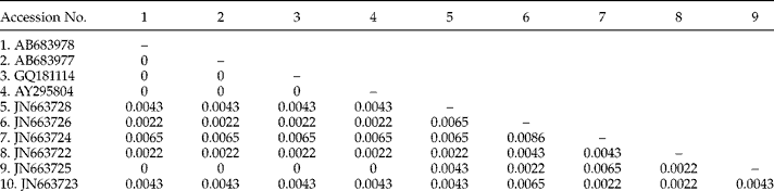
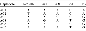
The number of base substitutions per site between sequences are summarized in Table 2. The percentage of uncorrected ‘p’ distance ranged from 0 to 0.86%. Five of the 461 sites had different bases, namely sites 315, 324, 338, 443 and 445. This resulted in six haplotypes/genotypes (table 3). Haplotype AC1 (site 443 with cytosine) was the most common, represented by isolates from China, Japan, Taiwan, Thailand and the Canary Islands. Haplotypes AC2 (site 315 with thymine), AC3 (site 338 with cytosine, site 445 with guanine) and AC4 (site 324 with guanine, site 443 with thymine and site 445 with guanine) were represented only by the Thailand isolate, while haplotype AC5 (site 445 with guanine) was represented by the Japan Okinawa isolate and haplotype AC6 (site 443 with thymine, site 445 with guanine) by the China Guangxi isolate.
Evolutionary divergence between sequences (number of base substitutions per site) of Angiostrongylus cantonensis based 18S rDNA nucleotide sequences. AB683978 (Japan: Miyagi; Japan: Aichi; China: Wenzhou); AB683977 (Japan: Tokyo, Kanagawa, Chiba, Ishikawa, Kagoshima, Okinawa; Taiwan: Taicung; China: Shenzhen, Lianjiang; Thailand: Bangkok); GQ181114 (Spain: Canary Islands); AY295804 (Japan: Akita University); JN663723 (China: Guangxi); JN663725 (Hawaii isolate); JN663722 (Japan: Okinawa); JN663724, JN663726, JN663728 (Thailand isolate).

Variable sites in 18S rDNA nucleotide sequences of Angiostrogylus cantonensis from various localities. Haplotype AC1: AB683978 (Japan), AB683977 (Japan), GQ181114 (Spain, Canary Islands), AY295804 (Japan), JN663725 (Hawaii); AC2: JN663726 (Thailand); AC3: JN663728 (Thailand); AC4: JN663724 (Thailand); AC5: JN663722 (Japan); AC6: JN663723 (China). Bold letters indicate unique bases.

Phylogeny of angiostrongylid and metastrongylid taxa
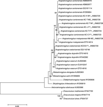
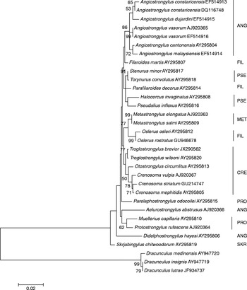
Based on the partial nucleotide sequences (461 base pairs) of the 18S rRNA gene, the phylogenetic relationships of the metastrongyloid genera (Angiostrongylus, Aelurostrongylus, Didelphostrongylus, Metastrongylus and Skrjabingylus) separated the genera Angiostrongylus and Metastrongylus into distinct clades (fig. 1). The genera Aelurostrongylus, Didelphostrongylus and Skrjabingylus formed the basal group. The Angiostrongylus clade consisted of three groups: (1) A. cantonensis clustered with A. malaysiensis; (2) A. costaricensis with A. dujardini; and (3) A. vasorum. The [A. cantonensis–A. malaysiensis] group and the [A. costaricensis–A. dujardini] group were, however, weakly supported. Likewise, the Angiostrongylus and Metastrongylus clades had weak support.
Phylogenetic tree of the metastrongyloid genera Angiostrongylus, Aelurostrongylus, Didelphostrongylus, Metastrongylus and Skrjabingylus (with Dracunculus spp. of Dracunculoidea as outgroups) generated by the maximum likelihood method based on partial 18S rDNA nucleotide sequences, conducted in MEGA5. The tree with the highest log likelihood ( − 1427.5718) is shown. The percentage of trees in which the associated taxa clustered together is shown next to the branches. The tree is drawn to scale, with branch lengths measured in the number of substitutions per site. The analysis involved 27 nucleotide sequences. Codon positions included were 1st +2nd +3rd. All positions containing gaps and missing data were eliminated. There was a total of 461 positions in the final dataset.

Phylogeny of metastrongyloid families
The phylogenetic relationships of the component families of the Metastrongyloidea inferred from long 18S rDNA nucleotide sequences (1573 bp) are depicted in fig. 2. Only the family Crenosomatidae appeared to be monophyletic. The genera Angiostrongylus and Metastrongylus were distinctly separated, while the genera Aelurostrongylus, Didelphostrongylus and Skrjabingylus did not cluster with each other or with Angiostrongylus and Metastrongylus. Metastrongylus formed a cluster with the genus Oslerus of Filaroididae. The genus Skrjabingylus appeared most basal in the superfamily.
Phylogenetic tree of the component families of Metastrongyloidea (with Dracunculus spp. of Dracunculoidea as outgroups) generated by the maximum likelihood method based on 18S rDNA nucleotide sequences, conducted in MEGA5. The tree with the highest log likelihood ( − 5098.1772) is shown. The percentage of trees in which the associated taxa clustered together is shown next to the branches. The tree is drawn to scale, with branch lengths measured in the number of substitutions per site. The analysis involved 32 nucleotide sequences. Codon positions included were 1st +2nd +3rd. All positions containing gaps and missing data were eliminated. There were a total of 1573 positions in the final dataset. ANG, Angiostrongylidae; CRE, Crenosomatidae; FIL, Filaroididae; MET, Metastrongylidae; PRO, Protostrongylidae; PSE, Pseudaliidae; SKR, Skrjabingylidae.

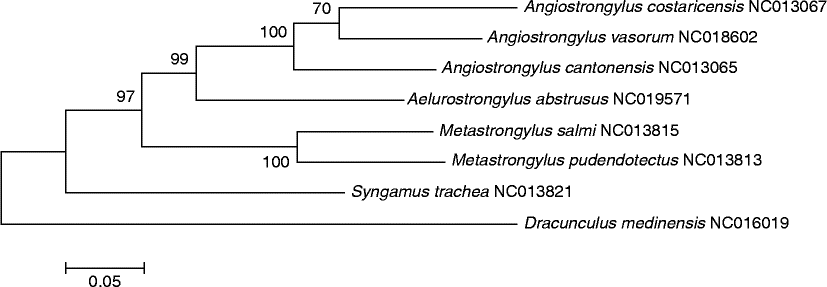
Based on the 12 protein-coding genes of the mitochondrial genome, the genera Angiostrongylus and Aelurostrongylus were more closely related compared to the genus Metastrongylus (fig. 3).
Phylogenetic tree of six taxa of Metastrongyloidea (with Syngamus trachea and Dracunculus medinensis as outgroups) generated by the maximum likelihood method based on 12 protein-coding genes (cox1–3, nad1–6, nad4L, cytb and atp6) of the mitochondrial genome, conducted in MEGA5. The tree with the highest log likelihood (–55, 523.3733) is shown. The percentage of trees in which the associated taxa clustered together is shown next to the branches. The tree is drawn to scale, with branch lengths measured in the number of substitutions per site. The analysis involved eight nucleotide sequences. Codon positions included were 1st +2nd +3rd. All positions containing gaps and missing data were eliminated. There were a total of 10 ,121 positions in the final dataset.

Discussion
The nuclear small subunit (SSU) ribosomal RNA (18S rRNA) gene has been widely used for phylogenetic studies of invertebrate organisms. It is the most conserved among the ribosomal encoding genes (Holterman et al., Reference Holterman, van der Wulff, van den Elsen, van Megan, Bongers, Holovachov, Bakker and Helder2006). It was first used to infer the relationships of 53 species of nematodes (Blaxter et al., Reference Blaxter, De Ley, Garey, Liu, Scheldeman, Vierstraete, Vanfleteren, Mackey, Dorris, Frisse, Vida and Thomas1998). The partial sequences from the 18S (SSU) rRNA gene of A. cantonensis were among those used for phylogenetic analysis of the Metastrongyloidea (Carreno & Nadler, Reference Carreno and Nadler2003). A phylo genetic tree of nematodes based on about 1200 full-length small subunit ribosomal DNA sequences indicated a considerable congruence with morphology at the family level and below (van Megan et al., 2009). The nematode 18S rDNA dataset has been proposed to be of greater use within major clades, where alignments are likely to be more robust (Smythe et al., Reference Smythe, Sanderson and Nadler2006).
The 18S (SSU) rDNA sequences have enabled the differentiation of closely related nematode species (Gasser & Newton, Reference Gasser and Newton2000). This gene has been used to identify the rat lungworm A. cantonensis in Tenerife, Canary Islands, Spain (Foronda et al., Reference Foronda, López-González, Miquel, Torres, Segovia, Abreu-Acosta, Casanova, Valladares, Mas-Coma, Bargues and Feliu2010), and as a genetic marker for identifying the infective third-stage A. cantonensis (Fontanilla & Wade, Reference Fontanilla and Wade2008). It has also been used to distinguish A. cantonensis from A. costaricensis, A. dujardini, A. malaysiensis and A. vasorum (Fontanilla & Wade, Reference Fontanilla and Wade2008; Foronda et al., Reference Foronda, López-González, Miquel, Torres, Segovia, Abreu-Acosta, Casanova, Valladares, Mas-Coma, Bargues and Feliu2010). The present study on 18S (SSU) rDNA sequences also unequivocally distinguishes the five Angiostrongylus species (fig. 1). Unlike the cytochrome c oxidase subunit I (COI) sequences which separated the isolates from Thailand, China and Hawaii (Eamsobhana et al., Reference Eamsobhana, Lim, Solano, Zhang, Gan and Yong2010b), the 18S sequences could not unequivocally separate the six geographical isolates (China, Hawaii, Japan, Spain – Canary Islands, Taiwan, Thailand) of A. cantonensis. Nonetheless, in all cases the congeneric metastrongyloid species clustered together, indicating close genetic affinity (fig. 2).
A recent study (Tokiwa et al., Reference Tokiwa, Harunari, Tanikawa, Komatsu, Koizumi, Tung, Suzuki, Kadasaka, Takada, Kumagai, Akao and Ohta2012) indicated extremely low intraspecific variation of the 18S (SSU) rRNA gene in A. cantonensis, with two genotypes in 13 localities in Japan and three localities in China; only a single specimen each from Taiwan and Thailand was included in that study. The present study uncovered four additional haplotypes (two from Thailand, one each from China and Japan), bringing the total to six (table 3). This is the result of a larger sample size. The uncorrected pairwise ‘p’ distances among A. cantonensis ranged from 0 to 0.86% (table 2).
In the present study, based on a larger number of taxa and 18S (SSU) sequences of the genus Angiostrongylus, the phylogenetic analysis indicated a close relationship between A. cantonensis and A. malaysiensis (fig. 1). This concurs with the earlier findings based on 18S rDNA sequences (van Megan et al., 2009), 66-kDa protein gene (Eamsobhana et al., Reference Eamsobhana, Lim, Zhang, Gan and Yong2010a) and COI sequences (Eamsobhana et al., Reference Eamsobhana, Lim, Solano, Zhang, Gan and Yong2010b). It differs from the finding of A. malaysiensis being more closely related to A. costaricensis than to A. cantonensis (Fontanilla & Wade, Reference Fontanilla and Wade2008; Tokiwa et al., Reference Tokiwa, Harunari, Tanikawa, Komatsu, Koizumi, Tung, Suzuki, Kadasaka, Takada, Kumagai, Akao and Ohta2012), based on fewer sequences.
The overall phylogenetic relationships of the five Angiostrongylus species in the present study are in agreement with the findings of van Megan et al. (2009). It is beyond reasonable doubt that A. costaricensis is more closely related to A. dujardini, compared to the findings of A. costricensis being more closely related to A. vasorum (Foronda et al., Reference Foronda, López-González, Miquel, Torres, Segovia, Abreu-Acosta, Casanova, Valladares, Mas-Coma, Bargues and Feliu2010) and A. dujardini being more closely related to A. vasorum (Fontanilla & Wade, Reference Fontanilla and Wade2008).
Both the long and shorter 18S (SSU) nucleotide sequences in the present study unequivocally separated the genera Angiostrongylus and Metastrongylus (figs 1 and 2), rendering support to the placement of Angiostrongylus in Angiostrongylidae and Metastrongylus in Metastrongylidae. Indeed, the genus Metastrongylus appeared to be closer to the genus Oslerus of Filaroididae (fig. 2). The genera Aelurostrongylus and Didelphostrongylus, however, were not recovered with Angiostrongylus, indicating that the Angiostrongylidae, as presently constituted, may not be monophyletic. However, based on the 12 protein-coding genes of available mitochondrial genomes, Aelurostrongylus was closer to Angiostrongylus (fig. 3). Such non-concordance has also been reported for other organisms, for example in filarial worms (Eamsobhana et al., Reference Eamsobhana, Lim and Yong2013). The need for employing multiple genes and extensive sampling of taxa is warranted to understand the phylogenetic relationships of the component taxa of the Metastrongyloidea.
The higher taxonomic position of Skrjabingylus could not be resolved by the present study based on 18S (SSU) sequences. Until further evidence based on a larger number of congeneric taxa becomes available, it may be reasonable to treat it as belonging to Skrjabingylidae (Anderson, Reference Anderson2000; Carreno et al., Reference Carreno, Reif and Nadler2005). At the family level, excepting Crenosomatidae, the other families appeared to be non-monophyletic (fig. 2).
In summary, the present study revealed six 18S (SSU) haplotypes in A. cantonensis, indicating considerable intraspecific genetic diversity compared to earlier findings. The uncorrected pairwise ‘p’ distances among A. cantonensis ranged from 0 to 0.86%. This study confirmed the close relationship of A. cantonensis and A. malaysiensis and that of A. costaricensis and A. dujardini. The 18S (SSU) rDNA sequences support the family status of Angiostrongylidae and Metastrongylidae. There is no supporting evidence to include the genus Skrjabingylus as a member of Metastrongylidae. The genera Aelurostrongylus and Didelphostrongylus were not recovered with Angiostrongylus, indicating polyphyly of the Angiostrongylidae. Of the currently recognized families of Metastrongyloidea, only Crenosomatidae appeared to be monophyletic. In view of the unsettled questions regarding the phylogenetic relationships of various taxa of the metastrongyloid worms, further analyses using more markers and more taxa are warranted.
Acknowledgements
The authors would like to thank Paibulaya Punthuprapasa and Adisak Yoolek for their excellent technical assistance. We also thank the Mahidol University and University of Malaya for support and facilities. We thank Dr Hongman Zhang (CDC, Guangxi), Dr Ichiro Miyagi and Dr T. Toma (University of the Ryukyus), Dr Akira Ishih (Hamamatsu University), Dr Elizabeth Abrahams and Dr Gabriela Solano (University of Costa Rica) for the gifts of Angiostrongylus materials. The constructive comments of the editor and two anonymous reviewers have helped to improve the manuscript.
Financial support
This study was funded in part by the Department of Disease Control, Ministry of Public Health, Thailand, and the University of Malaya (YHS: grants H-50001-00-A000025 and H-5620009).
Conflict of interest
None.