


Introduction
Cladonia norvegica Tønsberg & Holien was originally described from the vicinity of Trondheim (Norway) as an overlooked species growing on decaying wood of coniferous trees (Tønsberg & Holien Reference Tønsberg and Holien1984). Characteristics of this taxon include small, finely divided basal squamules, relatively short, farinosely sorediate and usually ascyphose podetia, and the presence of barbatic and 4-O-demethylbarbatic acids (Tønsberg & Holien Reference Tønsberg and Holien1984). Due to its ochraceous apothecia, which are rarely present, the authors assigned Cladonia norvegica to Cladonia subsection Ochroleucae, which was at that time classified in the section Coccifereae (Tønsberg & Holien Reference Tønsberg and Holien1984).
Originally considered a rare species of Norway, it was soon recorded in Sweden (Muhr Reference Muhr1986), Central Europe (Germany: Wirth Reference Wirth1987; Austria: Ruoss et al. Reference Ruoss, Mayrhofer and Pongratz1987; Czechia: Liška et al. Reference Liška, Palice and Bayerová1999), Finland, Poland and Italy (Kuusinen et al. Reference Kuusinen, Stenroos and Ahti1989), Madeira (Timdal Reference Timdal1989), Chile and Argentina (Stenroos & Ahti Reference Stenroos and Ahti1990), Canada and the USA (Tønsberg & Goward Reference Tønsberg and Goward1992), Japan (Stenroos & Ahti Reference Stenroos and Ahti1994), Estonia (Randlane & Saag Reference Randlane and Saag1999), and later in Russia (Urbanavichus & Urbanavichene Reference Urbanavichus, Urbanavichene and Korneeva2004; Kuznetsova & Dudov Reference Kuznetsova and Dudov2017) and other countries.
Cladonia norvegica typically thrives in moist, partially shaded environments, such as mossy, decayed wood and the bases or lower trunks of Betula spp., Picea abies, or Pinus spp. (Tønsberg & Holien Reference Tønsberg and Holien1984; Ahti et al. Reference Ahti, Stenroos and Moberg2013). The species prefers forests with high ecological continuity (Kuusinen et al. Reference Kuusinen, Stenroos and Ahti1989; Burgaz et al. Reference Burgaz, Ahti and Pino-Bodas2020; Pino-Bodas et al. Reference Pino-Bodas, Sanderson, Cannon, Aptroot, Coppins, Orange and Simkin2021), though it can also be found in managed forests (Lõhmus & Lõhmus Reference Lõhmus and Lõhmus2009; Malíček et al. Reference Malíček, Bouda, Palice and Peksa2011). It often has a low local abundance, and this may have led to it being frequently overlooked (Lõhmus & Lõhmus Reference Lõhmus and Lõhmus2009).
Cladonia norvegica can be easily confused with C. coniocraea (Flörke) Spreng., with which it also regularly shares its habitat. The two species can be distinguished by subtle morphological differences including finer and more deeply incised basal squamules, as well as their distinct chemical composition; C. coniocraea contains fumarprotocetraric acid (P+ red, UV−), while C. norvegica contains barbatic acid (P−, UV+ bluish) (Ahti et al. Reference Ahti, Stenroos and Moberg2013). Another similar species is C. macilenta var. bacillaris (Ach.) Schaer., which shares the same chemical pattern as C. norvegica but can be recognized by its red apothecia and pycnidia (which may not be present). Cladonia bacilliformis (Nyl.) Sarnth. also has sorediate ascyphose podetia and pale apothecia, but it additionally contains usnic acid, which gives the thallus a yellowish tint. Furthermore, C. bacilliformis also prefers well-lit, relatively dry and open habitats (Tønsberg & Holien Reference Tønsberg and Holien1984).
Interestingly, many specimens of Cladonia norvegica exhibit a striking feature not mentioned in the original description: the presence of red spots on the squamules and podetia. Timdal (Reference Timdal1989), who had collected such specimens in the Oslo area since 1981, and later also over much of southern and central Scandinavia as far north as Lycksele lappmark (Timdal Reference Timdal1989; Ahti et al. Reference Ahti, Stenroos and Moberg2013), identified this red pigment as rhodocladonic acid using TLC. Timdal (Reference Timdal1989) also reported red spots on the thallus of 14 specimens of Cladonia bacilliformis collected in Norway, Sweden and Finland. This pigment is otherwise known from the apothecia of red-fruited Cladonia species (Baker & Bullock Reference Baker and Bullock1969; Stenroos Reference Stenroos1989b) and is possibly produced by the lichen in response to mite infection (Timdal Reference Timdal1989) or other damage (Pino-Bodas et al. Reference Pino-Bodas, Sanderson, Cannon, Aptroot, Coppins, Orange and Simkin2021). In the Nordic countries, red-spotted specimens of C. norvegica are widely distributed and found most commonly in the southern part, whereas non-spotted specimens are mainly found in the boreal, oceanic spruce forests of Trøndelag County and the southern part of Nordland County in Central Norway (Timdal Reference Timdal1989). To our knowledge, specimens from European herbaria outside of Scandinavia and the British Isles consistently display these red spots. Some lichenologists rely only on this feature to identify the species (Lõhmus & Lõhmus Reference Lõhmus and Lõhmus2009).
Although considered conspecific by Timdal (Reference Timdal1989), the red-spotted material assigned to Cladonia norvegica does not always appear to correspond well with the original description of the species, even when the presence of rhodocladonic acid is ignored. In this study, we wanted to evaluate the taxonomic status of material of C. norvegica with and without red spots. We performed a thorough phylogenetic analysis of both the mycobiont and photobiont, using three and two molecular markers for fungi and algae, respectively. Since the exact placement of C. norvegica within the genus Cladonia has remained uncertain due to the lack of molecular data, we also aimed to resolve its phylogenetic position. Finally, we sought to confirm the identity of the red pigment by using sensitive HPLC and LC-HRMS analysis.
Materials and Methods
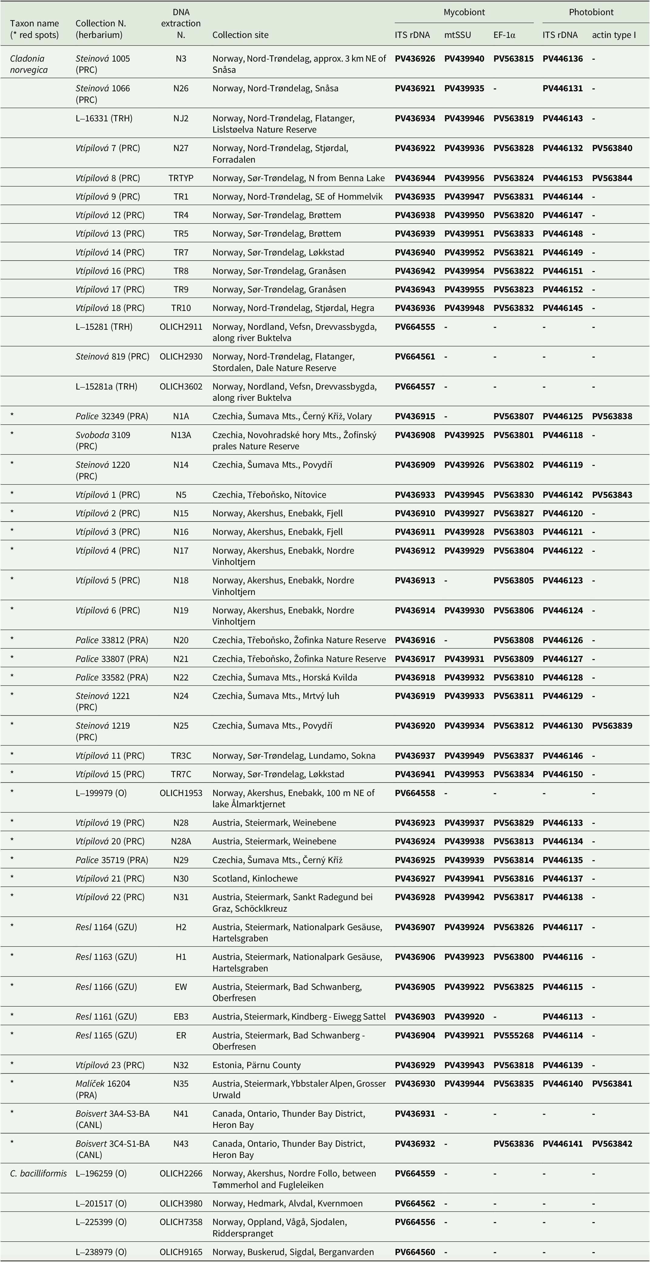
Sampling
Altogether, 46 specimens of Cladonia norvegica from Austria, Czechia, Estonia, Norway, Scotland and Canada were used for the molecular phylogenetic and chemical analyses (thin-layer chromatography, TLC). The material used included specimens both without red spots as in the original description (15 specimens collected from different sites in Norway, including the type locality) and with red spots (31 specimens collected in Austria, Canada, Czechia, Estonia, Norway and Scotland). At one locality in Trøndelag in Norway, we collected specimens both with and without red spots growing up to only 10 m apart. At all localities, C. norvegica was growing on wood (birch, pine or spruce) together with bryophytes in humid habitats. Collection data are presented in Table 1.
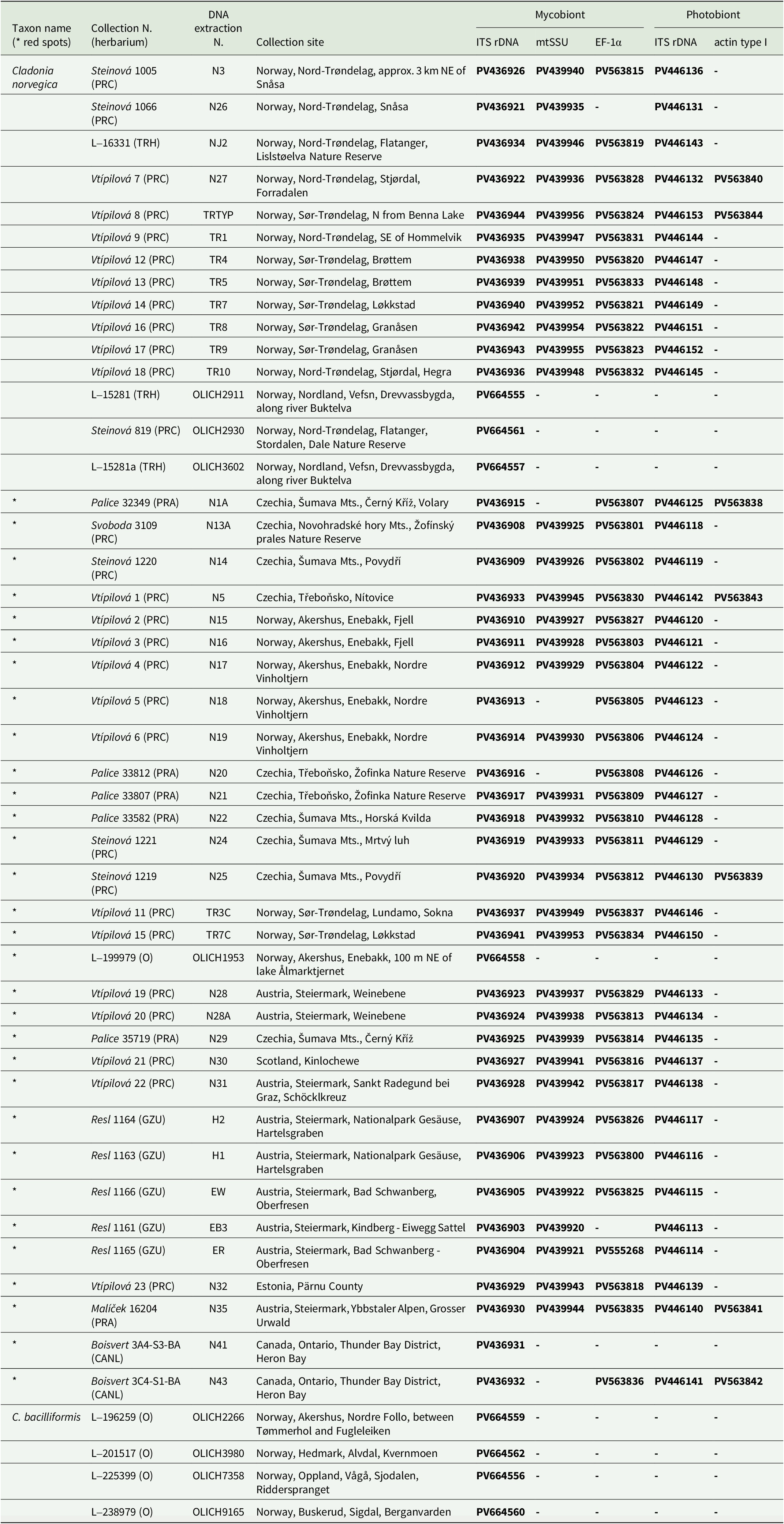
List of specimens used in the molecular analyses, including voucher information, DNA extraction codes and GenBank Accession numbers. An asterisk indicates the presence of red spots on the thallus. Newly obtained sequences (in bold) are reported for both the mycobionts and photobionts.
We confirmed the identity of the collected material using chemically-based methods, specifically negative para-phenylenediamine (P) reaction (to distinguish C. norvegica from C. coniocraea in the field; Ahti et al. Reference Ahti, Stenroos and Moberg2013), positive UV reaction (weak bluish reaction in contrast to a negative reaction in the case of C. coniocraea; Pino-Bodas et al. Reference Pino-Bodas, Sanderson, Cannon, Aptroot, Coppins, Orange and Simkin2021), and thin-layer chromatography (TLC).
TLC
Forty-six samples of Cladonia norvegica (31 with red spots and 15 without red spots) were used for TLC, which was performed according to the methodology of Culberson (Reference Culberson1972) using solvent systems A and C. Cladonia symphycarpa (Flörke) Fr. (containing norstictic acid and atranorin) was chosen as the standard. To extract secondary metabolites, one podetium from each lichen sample was used, or several squamules if the podetium was not present in the sample, (this applied to samples labelled N4, N13A, N25, N30, N31, N32 and N43).
HPLC UV-VIS and LC-HRMS analysis
The presence of rhodocladonic acid was analyzed using HPLC UV-VIS (HPLC method that measures the absorbtion of ultraviolet and visible light by molecules) and LC-HRMS (liquid chromatography high resolution mass spectrometry). Three herbarium samples of Cladonia norvegica were examined: B1 (Zdeněk Palice 32349), B2 (Jiří Malíček 2057) and B3 (Jiří Malíček 3128). For these samples, only the red parts of the lichen thalli were extracted using a razor blade. As a standard, a red apothecium from Cladonia macilenta Hoffm. (B5; Jana Steinová 1405), known to contain rhodocladonic acid as well as barbatic acid in both the thallus and apothecium, was used.
Lichen samples were extracted with 500 μl of CH2Cl2 (overnight, in dark conditions). Samples were centrifuged, and the supernatant was evaporated to dryness under reduced pressure. The crude extract was diluted in MeOH (0.1 ml) and used for HPLC analyses.
The same HPLC instrumentation and chromatographic procedure as published earlier (Flieger et al. Reference Flieger, Tatarczak-Michalewska, Blicharska, Świeboda and Banach2017) was used for the analysis of lichen extracts. A Gemini 5 μm C18 column (250 × 4.6 mm; Phenomenex, Torrance, CA, USA) with a guard column was used for the analysis. The mobile phase consisted of H2O/MeOH + 1% TFA. Gradient elution started at 30% MeOH (0 min), increasing linearly to 100% MeOH within 20 min, at a flow rate of 1.0 ml min−1. UV detection was performed at 290 nm and 440 nm.
To analyze the lichen extract, liquid chromatography coupled with high-resolution mass spectrometry equipped with an electrospray ion source (LC-HRMS) was used. The separation of the sample (5 μl) was carried out using the LC system Agilent Infinity II, with the column Poroshell 120 EC-C18 (2.7 μm, 3 × 100 mm) and precolumn of the same type heated to 40 °C and with the gradient method set to a flow rate of 0.4 ml min−1. The method consisted of an isocratic phase (0–0.5 min, 10% Acetonitrile (B) and 90% water solution with the addition of 0.1% of formic acid) followed by a gradient phase, where the gradient ranged from 10% of B (0.5 min) to 100% of B (5 min), and a final phase, where the initial conditions (10% of B) were restored by the isocratic elution with 100% of B (10 min). Prior to the injection of the next sample, the column was left to equilibrate for a period of 5 min at starting conditions. The ion source operated in the negative ionization mode and the mass spectrometer (Agilent 6546, qTOF) had the following settings: drying gas temperature and flow, 250 °C and 8 l min−1; sheath gas temperature and flow, 400 °C and 12 l min−1; nebulizer pressure 35 psi; capillary voltage, c. 3500 V; fragmentor 110 V, skimmer 65 V, and Oct 1 RF Vpp 750 V; mass range of 80–1500 m/z; collision energy 0, 20, and 40 eV; acquisition rate of 3 spectra/s. For the data analysis, the software MassHunter Qualitative Analysis 10.0 (Agilent, USA) was used.
DNA extraction, PCR and sequencing
For the majority of the material (42 specimens), DNA was extracted from single podetia with standard CTAB protocol (Cubero et al. Reference Cubero, Crespo, Fatehi and Bridge1999) with the following optimization: first, we used tungsten beads to crush the lichen material; second, we prolonged freezing (−20 °C) to 30 min after isopropanol precipitation; finally we added an additional purification step with 96% ethanol. From each DNA extract, we amplified three mycobiont molecular markers: i) ITS rDNA using the primers ITS1F (Gardes & Bruns Reference Gardes and Bruns1993) and ITS4 (White et al. Reference White, Bruns, Lee, Taylor, Innis, Gelfand, Sninsky and White1990); ii) small subunit of mitochondrial ribosomal DNA (mtSSU) with primers mrSSU1 and mrSSU3R (Zoller et al. Reference Zoller, Scheidegger and Sperisen1999); iii) elongation factor-1α (EF-1α) using primers CLEF-3F and CLEF-3R (Yahr et al. Reference Yahr, Vilgalys and DePriest2006). For the photobiont, we amplified algal ITS rDNA using the primers nr-SSU-1780-5′Algal (Piercey-Normore & DePriest Reference Piercey-Normore and DePriest2001) and ITS4 (White et al. Reference White, Bruns, Lee, Taylor, Innis, Gelfand, Sninsky and White1990), and the photobiont actin type I locus using actin_F and actin_R primers (Cocquyt et al. Reference Cocquyt, Verbruggen, Leliaert and Clerck2010). PCR reactions were performed using MyTaq polymerase (Bioline, UK) in 20 μl volume reactions and with identical PCR conditions to those used in Steinová et al. (Reference Steinová, Holien, Košuthová and Škaloud2022). The PCR products were purified with SPRI AMPure XP paramagnetic beads (Beckman Coulter, USA) and sequenced at Macrogen Europe (Amsterdam, the Netherlands) with the same primers used for PCR amplification.
For a small part of the material (4 specimens of Cladonia norvegica + 4 specimens of C. bacilliformis), the sequencing followed the pipeline of the DNA barcoding project OLICH (Marthinsen et al. Reference Marthinsen, Rui and Timdal2019) at the Norwegian Barcode of Life (https://www.norbol.org), with DNA extraction, PCR and sequencing performed at the Canadian Centre of DNA Barcoding (https://ccdb.ca) using the primer pair ITS1F/ITS4.
Phylogenetic analyses
Initial processing of raw sequences was performed in SeqAssem v. 09/2004 (Hepperle Reference Hepperle2004). Individual alignments for mycobiont (ITS rDNA, EF-1α, mtSSU) and photobiont (ITS rDNA and actin type I) markers were initially built in MEGA11 (Tamura et al. Reference Tamura, Stecher and Kumar2021) and refined using MAFFT v. 7 (Katoh & Standley Reference Katoh and Standley2013) with default settings. For the actin gene, Gblocks v. 0.91b was used to remove introns and poorly aligned positions (Castresana Reference Castresana2000) under the default settings.
Newly obtained sequences were supplemented with additional sequences from GenBank (NCBI). First, the only available sequences of Cladonia norvegica (voucher PRA-Palice32479; GenBank Accession numbers OQ717819 for ITS rDNA and OQ646201 for mtSSU) were added to the alignments. To represent the phylogenetic neighbourhood of C. norvegica, we followed Stenroos et al. (Reference Stenroos, Pino‐Bodas, Hyvönen, Lumbsch and Ahti2018) and included sequences from species within the Amaurocraeae, Divaricatae, Erythrocarpae, Ochroleucae and Perviae clades. The dataset also contained representatives from the Arbuscula, Borya, Cladonia, Crustacae, Impexae and Unciales clades, along with an outgroup of three representatives from the genera Cladia and Thysanothecium.
In the case of the photobiont, newly generated ITS rDNA and actin type I sequences were complemented by additional Asterochloris sequences downloaded from GenBank, based on the dataset by Vančurová et al. (Reference Vančurová, Malíček, Steinová and Škaloud2021).
In MEGA11 (Tamura et al. Reference Tamura, Stecher and Kumar2021), both ITS rDNA alignments were manually partitioned to account for differences in evolutionary rates across marker regions. Substitution models for subsequent phylogenetic analyses were calculated separately for the ITS1, ITS2, and 5.8 S rDNA regions, as well as for the EF-1α, mtSSU and actin type I markers (Supplementary Material Table S1, available online). These models were selected using jModelTest2 v.2.1.10 in CIPRES (Miller et al. Reference Miller, Pfeiffer and Schwartz2010) based on the Bayesian Information Criterion. The final concatenated alignments (both fungal and algal), along with the selected best models, were subjected to Bayesian and maximum likelihood phylogenetic analyses.
We performed Bayesian tree inferences using MrBayes v. 3.2.7a (Ronquist et al. Reference Ronquist, Teslenko, van der Mark, Ayres, Darling, Höhna, Larget, Liu, Suchard and Huelsenbeck2012). Phylogenetic trees were first generated separately for each marker, and since the resulting topologies were identical within the mycobiont and photobiont datasets, concatenated datasets were used to construct phylogenetic trees (ITS rDNA + mtSSU + EF-1α for the mycobiont; ITS rDNA + actin type I for the photobiont). The Bayesian analysis employed two parallel MCMC runs with four chains (one hot, three heated), sampling trees and parameters every 100 generations. The runs were executed for 15 million generations for the mycobiont and 10 million generations for the photobiont. Convergence of the chains was assessed during the run by monitoring the average standard deviation of split frequencies (ASDSF) between simultaneous runs. The ASDSF values were < 0.01, specifically 0.004179 for the mycobiont dataset and 0.001167 for the photobiont dataset, indicating good convergence. Burn-in was determined using the ‘sump’ command in MrBayes, which discarded the first 25% of sampled trees.
Maximum likelihood (ML) trees were generated from the concatenated datasets using RAxML v. 8.2.10 (Stamatakis Reference Stamatakis2014). The analysis included 100 tree replicates and 1000 rapid bootstrap inferences, with automatic termination enabled. Since the ML and Bayesian methods produced consistent topologies, only the Bayesian phylograms are presented here. Phylogenetic trees were visualized using FigTree v. 1.4.4 (Rambaut Reference Rambaut2018), with final graphical adjustments made in Adobe Illustrator.
Morphological analysis
For each of the two forms of Cladonia norvegica (with and without red spots), ten genetically analyzed specimens were selected. Three morphological traits considered potentially the most relevant were recorded: podetium length, squamule length, and the width at the middle of the squamule. On each specimen, the three biggest podetia and three randomly selected squamules were measured. Measurements were taken using a dissecting microscope (Olympus SZX10) with a digital calliper. The resulting data were used to calculate the mean and standard deviation for each morphological character. Statistical analyses were performed in R v. 4.1.2 (R Core Team 2022). The normality of the data was assessed using the Shapiro–Wilk test, and since the data met the normality assumption, differences between species were tested using a two-sample t-test.
Results
TLC
The results of thin-layer chromatography revealed that all specimens studied contained barbatic acid and 4-O-demethylbarbatic acid. Specimens with visible red spots additionally contained rhodocladonic acid.
HPLC UV-VIS and LC-HRMS
Using HPLC UV-VIS and LC-HRMS, four compounds were identified in all analyzed samples (B1–B3, B5): rhodocladonic acid (Rt = 13.108 min), demethyl-rhodocladonic acid (Rt = 11.76 min), barbatic acid (Rt = 19.59 min), and 4-O-demethylbarbatic acid (Rt = 17.59 min). To our knowledge, this is the first time demethyl-rhodocladonic acid has been reported.
In sample B5 (the standard; apothecium of Cladonia macilenta), rhodocladonic acid was confirmed using HPLC UV-VIS and LC-HRMS (see Supplementary Material Figs S1, S2, S7, Table S2; available online). Barbatic acid (Rt = 19.59 min) was also detected in this sample, along with 4-O-demethylbarbatic acid (Rt = 17.59 min) and demethyl-rhodocladonic acid (Rt = 11.76 min) (see Supplementary Material Table S2).
When compared to the other three samples (B1–B3, belonging to Cladonia norvegica), sample B5 differed only in the relative proportions of the dominant compounds. Based on the intensity of the HPLC peaks, rhodocladonic acid and barbatic acid were present in nearly equal proportions in sample B5. In contrast, barbatic acid was the dominant compound in samples B1–B3 (C. norvegica; see Supplementary Material Figs S1–S9, available online).
Phylogenetic analysis
We obtained 50 new ITS rDNA sequences (31 from the spotted form, 15 from the non-spotted form and four from Cladonia bacilliformis), 37 mtSSU (25 from the spotted form, 12 from the non-spotted form) and 39 EF-1α sequences (28 from the spotted form, 11 from the non-spotted form). GenBank Accession numbers of the newly obtained sequences are given in Table. 1. The concatenated mycobiont alignment comprised 110 unique sequences with 2122 nucleotide positions. Individual mycobiont loci showed the following characteristics: the ITS rDNA alignment was 675 base pairs (bp) long and contained 315 variable and 231 parsimony-informative positions; the mtSSU alignment comprised 821 bp, with 35 variable and 21 parsimony-informative positions, the lower number of variable sites probably due to the limited representation of other Cladonia species in the dataset; the EF-1α alignment was 626 bp long, with 252 variable and 213 parsimony-informative positions.
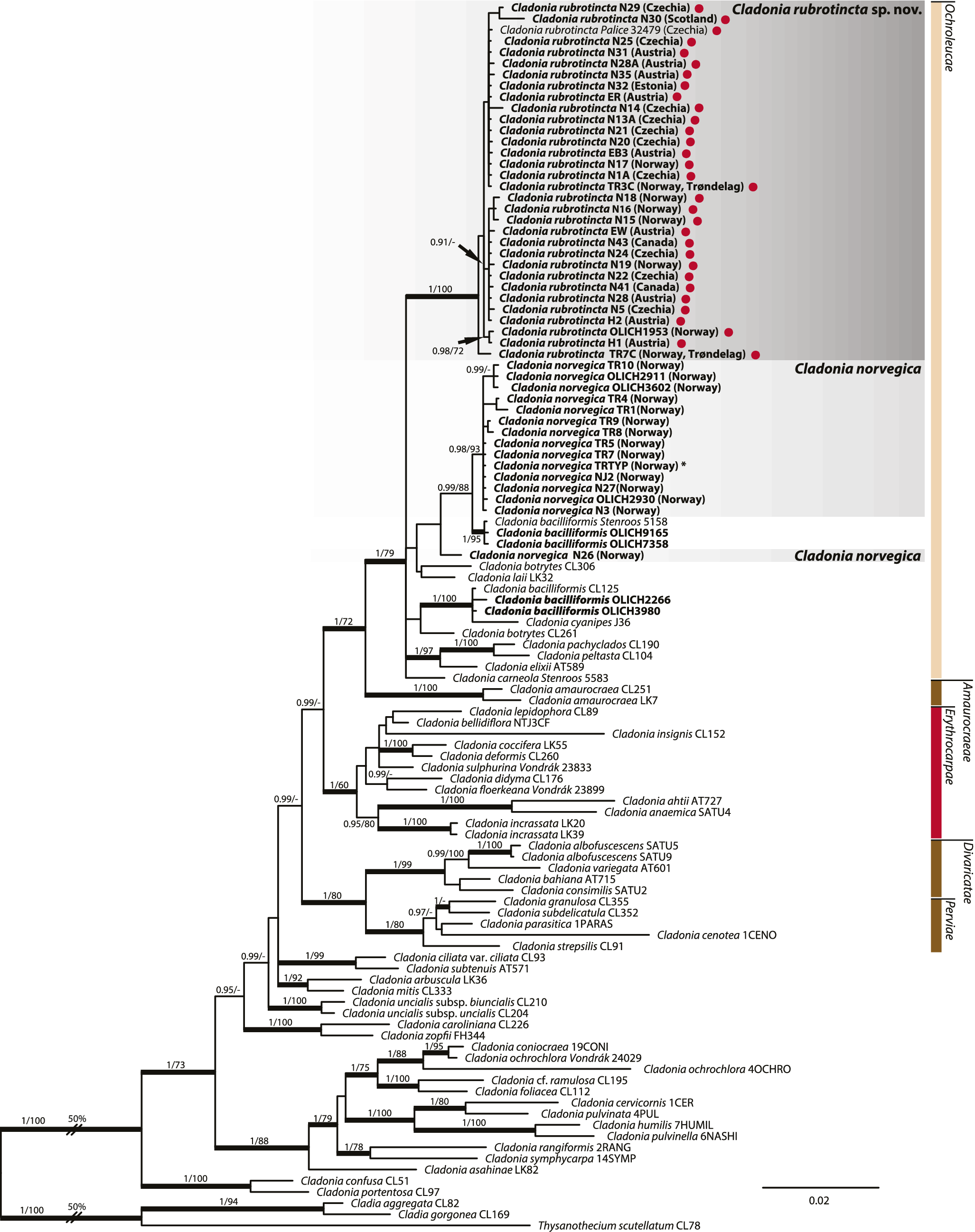
Our Bayesian and maximum-likelihood phylogenetic analyses (Fig. 1) confirmed for the first time that the studied material, including both red-spotted and non-spotted forms, belongs to the Ochroleucae clade. The tree topology revealed that the investigated material is split into two clades. The first clade contains only material without rhodocladonic acid. The second, strongly supported clade (1.00 PP, 100 BS) includes all red-spotted samples, including those from Central Norway (Trøndelag), where only the spotless form had been previously documented. Samples within this clade share several chemical and morphological synapomorphies, including the production of rhodocladonic acid and the generally smaller, more slender and irregular podetia compared to the material without red spots. Based on these findings, we describe the material previously assigned to Cladonia norvegica but containing rhodocladonic acid as the new species Cladonia rubrotincta (see description below).
Phylogenetic tree of Cladonia norvegica s. lat. and related taxa, inferred by Bayesian inference of concatenated ITS rDNA, mtSSU and EF-1α sequences. Node values represent statistical support, given as Bayesian posterior probabilities (PP) and maximum likelihood (ML) bootstrap values. Only values of PP > 0.9 and ML > 70% are shown. Thickened branches indicate full PP support. Newly obtained sequences are highlighted in bold. Red dots mark specimens with red pigment on the thallus, and an asterisk denotes the specimen collected at the type locality of C. norvegica. In colour online.
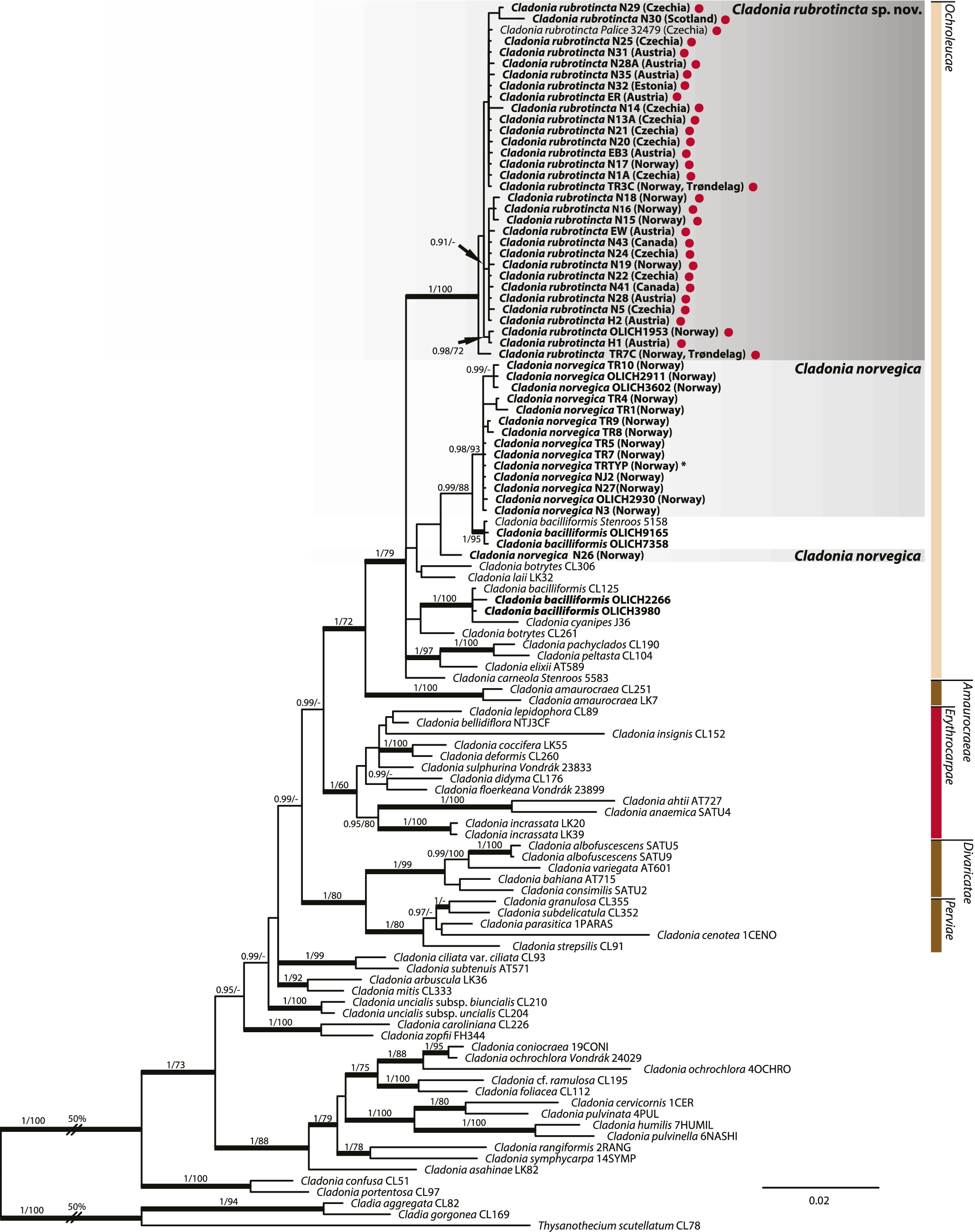
Most specimens identified morphologically and chemically as Cladonia norvegica lacking rhodocladonic acid (including material from the type locality: specimen TRTYP marked with an asterisk in Fig. 1) formed a well-supported clade (0.99 PP, 88 BS). However, one specimen (C. norvegica N26) was recovered outside this clade. While this sample shares the chemical composition (barbatic and 4-O-demethylbarbatic acids) and morphological characteristics of C. norvegica s. str., it is separated by a well-supported clade containing samples identified as C. bacilliformis. Interestingly, C. bacilliformis appears to be polyphyletic, as three additional specimens assigned to this species were recovered in a separate, highly supported clade (1.00 PP, 100 BS) together with C. cyanipes (Sommerf.) Nyl. and as a sister group to C. botrytes (K.G. Hagen) Willd. Furthermore, two samples labelled as C. botrytes (CL306 and CL261) were placed in different parts of the phylogenetic tree, indicating they do not cluster closely in this dataset, although broader multilocus analyses have previously recovered them within the same species clade (Stenroos et al. Reference Stenroos, Pino‐Bodas, Hyvönen, Lumbsch and Ahti2018).
For the photobiont, we obtained 41 new ITS rDNA sequences and seven actin type I sequences. The concatenated photobiont alignment comprised 62 unique sequences and 1115 nucleotide positions. The algal ITS rDNA alignment was 482 bp long and included 88 variable and 52 parsimony-informative positions. In contrast, the actin alignment exhibited much higher variability, comprising 633 bp with 454 variable and 380 parsimony-informative positions.
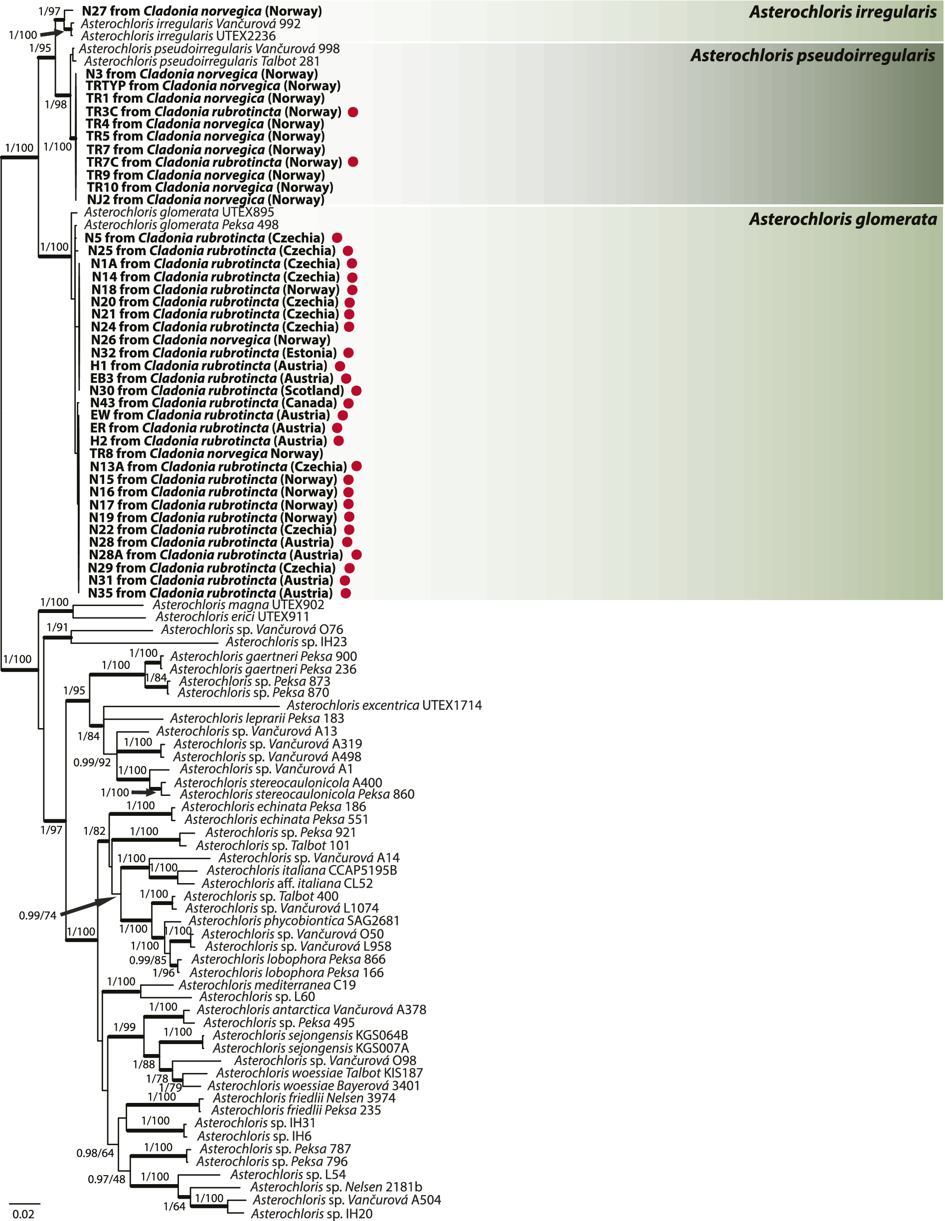
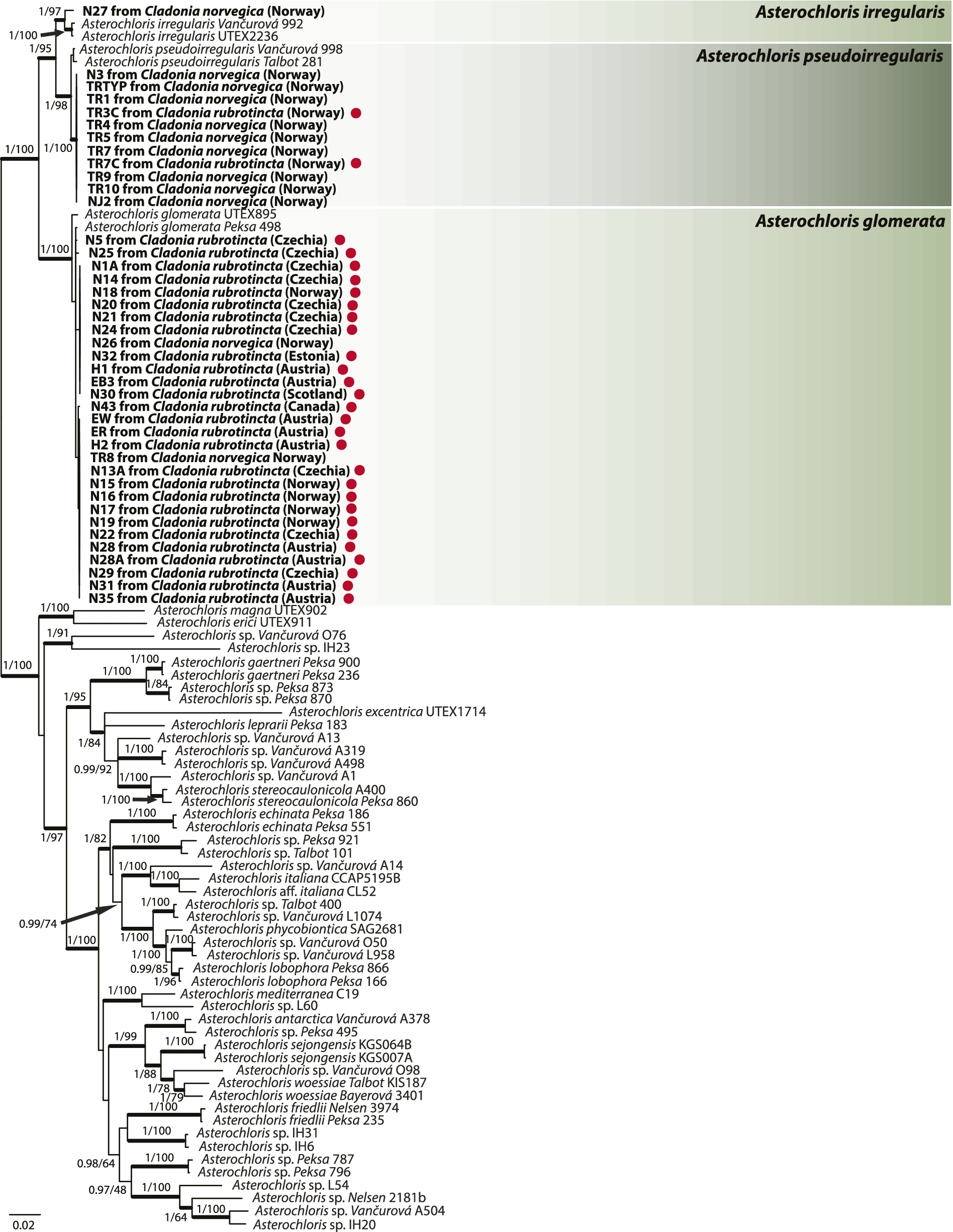
The concatenated photobiont phylogenetic tree (Fig. 2) reveals the presence of three Asterochloris species associated with Cladonia norvegica and C. rubrotincta. Asterochloris glomerata was primarily found in C. rubrotincta samples from Europe and Canada, except in the Trøndelag area of Norway, where A. pseudoirregularis was the dominant photobiont. Additionally, A. glomerata was identified in two samples of C. norvegica. In contrast, A. pseudoirregularis was the most common photobiont for C. norvegica, occurring exclusively in samples (of both lichen species) from the Trøndelag region. Finally, A. irregularis was detected in a single sample of C. norvegica from Trøndelag.
Phylogeny of Asterochloris obtained by Bayesian inference of concatenated ITS rDNA and actin type I. Node values represent statistical support, given as Bayesian posterior probabilities (PP) and maximum likelihood (ML) bootstrap values. Only values of PP > 0.9 and ML > 70% are shown. Thickened branches indicate full PP support. Newly obtained sequences are highlighted in bold. Red dots mark specimens with red pigment on the thallus. In colour online.
Morphological analysis
A significant difference in podetium length was observed between Cladonia norvegica and C. rubrotincta. Specimens of C. rubrotincta had significantly shorter podetia (5.98 ± 2.18 mm) compared to C. norvegica (12.78 ± 4.39 mm), as confirmed by a Welch’s two-sample t-test (t = 6.26, df = 16.58, P < 0.001, 95% CI: 4.50–9.09; see Supplementary Material Fig. S10, available online).
No significant differences were found in squamule length or squamule width between the two species. The mean squamule length was 1.54 ± 0.35 mm in C. norvegica and 1.48 ± 0.41 mm in C. rubrotincta (t = 0.45, df = 17.19, P > 0.05), while the mean squamule width was 1.19 ± 0.27 mm in C. norvegica and 1.13 ± 0.35 mm in C. rubrotincta (t = 0.71, df = 14.47, P > 0.05; see Supplementary Material Fig. S10).
Taxonomy
Cladonia rubrotincta Vtípilová, Timdal, Resl & Steinová sp. nov
MycoBank No.: MB 858583
Distinguished from Cladonia norvegica by the presence of red spots on the thallus and by its shorter podetia, usually not exceeding 15 mm in height and often having a deformed appearance.
Type: Norway, Akershus, Enebakk, Østmarka National Park, 100 m NE of Lake Ålmarktjernet, 59.7878°N, 11.0085°E, 300 m alt., on lying spruce log in spruce forest, 20 September 2014, S. Rui & E. Timdal WG1-0925 (O L-199979—holotype). GenBank Accession no.: PV664558 (ITS).
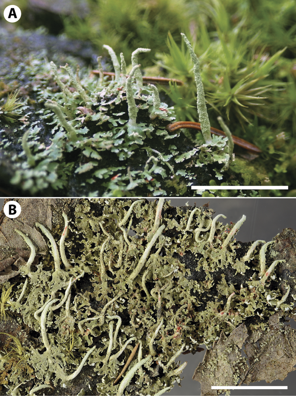
(Fig. 3)
A & B, Cladonia rubrotincta sp. nov. Norway, O L-199979, holotype. A, field photograph before collection. B, the herbarium specimen. Scale = 1 cm. In colour online.
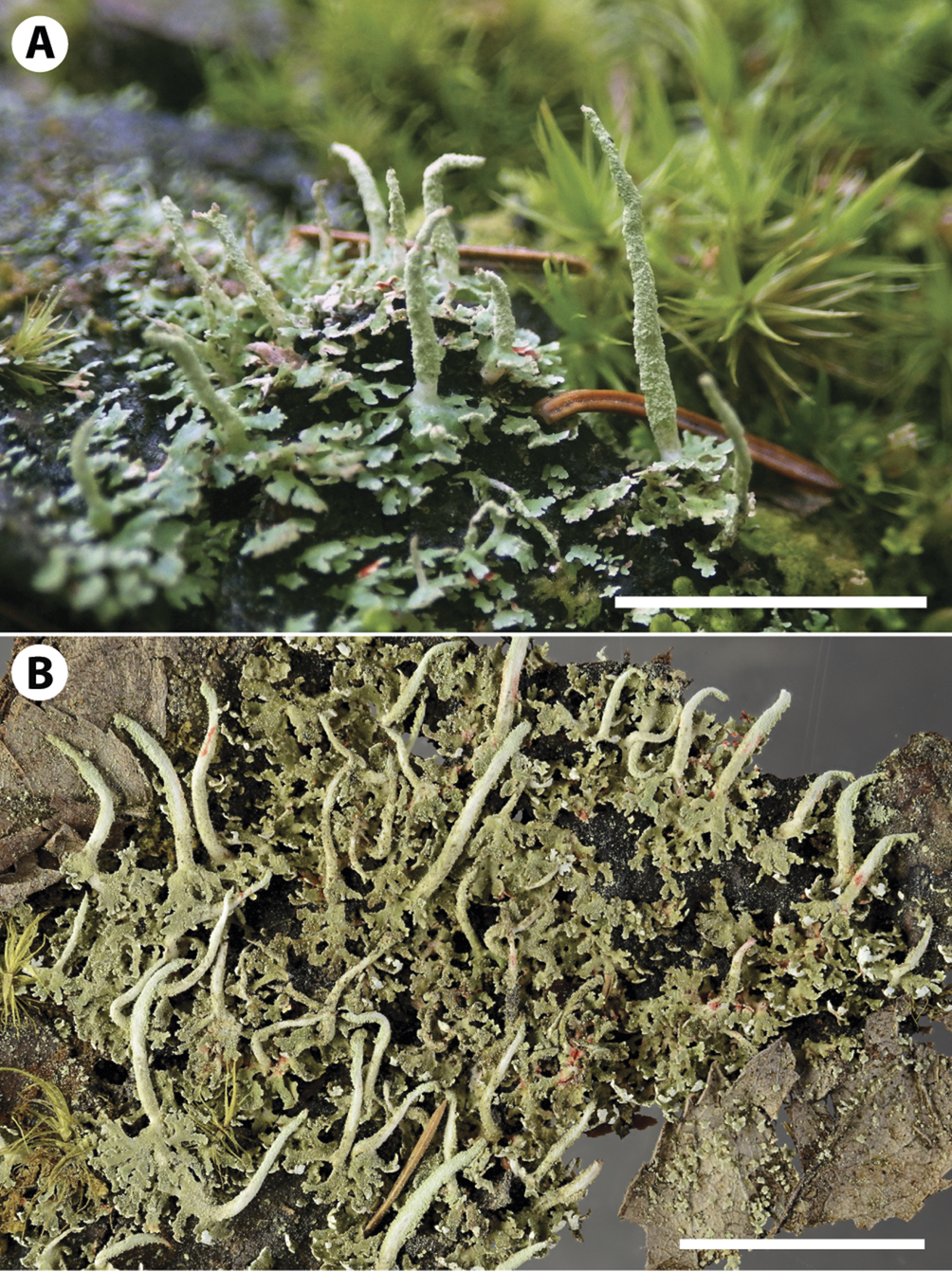
Primary thallus squamulose, persistent, usually well developed, sometimes dominant and forming loose cushions; squamules up to 4 mm long, up to 2.5 mm wide, deeply incised to laciniate, often involute, often with farinose soredia on the underside near the tips, pale green on the upper side, white on the underside, often with red spots. Podetia up to 10(–15) mm tall, up to 1 mm wide, pale green, often with red spots, slender, subulate, straight or curved, unbranched or rarely with one or two branches, ascyphose or more rarely with a narrow scyphus (up to 1 mm wide), farinosely sorediate in upper part, often corticate towards the base, sometimes with a small number of squamules in lower half, often becoming eroded and showing the white or red-spotted stereome.
Apothecia rare, up to 0.4 mm wide, pale brown; ascospores 8 per ascus, ovoid to oblong, 12–15 × 3–4.5 μm.
Pycnidia infrequent, on tips of podetia, indistinctly sessile to shortly stalked, variable, mostly doliiform or cylindrical, brown, with hyaline gelatine; conidia not observed.
Chemistry
P−, K−, C−, KC−, UV+ bluish white. Contains barbatic acid (major) and 4-O-demethylbarbatic acid (minor); rhodocladonic acid and demethyl-rhodocladonic acid in the red spots.
Etymology
The epithet rubrotincta is derived from the Latin words ruber, meaning ‘red’, and tinctus, meaning ‘tinged’ or ‘coloured’. This name refers to the distinctive red spots observed on the thallus, which are a prominent morphological feature.
Habitat and ecology
On rotting wood (primarily on Picea abies), bark and bryophytes; on logs, stumps and at tree bases in shady, humid forests.
Distribution
Genetically confirmed from Austria, Canada, Czechia, Estonia, Great Britain and Norway. Based on morphology, the species is probably more widespread, and morphologically matching specimens (containing red pigment on the thallus) have also been observed in Finland, Germany, Japan, Madeira, Romania, Russia and Sweden.
Remarks
Cladonia rubrotincta s. str. can be easily confused in the field with other Cladonia species, particularly those occurring in similar habitats, such as C. coniocraea, C. macilenta and C. norvegica. The best distinguishing characteristic appears to be the presence of red spots on the thallus. However, as mentioned below, we cannot completely rule out the possibility that specimens lacking rhodocladonic acid also exist.
Compared to C. coniocraea, which has basal squamules 1–6 mm long (Brodo & Ahti Reference Brodo and Ahti1996), C. rubrotincta usually has smaller and more finely divided basal squamules. Additionally, the podetia of C. rubrotincta are narrower, shorter, and often appear deformed, whereas C. coniocraea typically has taller and more robust podetia. The two species also differ in their chemistry: while C. rubrotincta contains barbatic and 4-O-demethylbarbatic acids and therefore reacts P− and UV+ bluish, C. coniocraea contains substances from the fumarprotocetraric acid complex causing its P+ red and UV− reactions.
Cladonia macilenta var. bacillaris has the same chemistry as C. rubrotincta but has red apothecia and pycnidial gelatine. However, both species are often sterile. In such cases, smaller podetia with slender podetial tips are the best features to distinguish the two species.
Similarly, Cladonia norvegica shares the same chemical pattern as C. rubrotincta but, apart from the absence of the red spots, it differs by having larger podetia, usually 1.5–3 cm tall (Tønsberg & Holien Reference Tønsberg and Holien1984), that are at least partly corticate, whereas those of C. rubrotincta are distinctly shorter, reaching only up to 10(–15) mm.
Another species that may be confused with Cladonia rubrotincta is C. bacilliformis, for which occasional occurrences of red spots have been reported (Timdal Reference Timdal1989). Cladonia bacilliformis can be distinguished by its yellowish tint caused by the presence of usnic acid (Timdal Reference Timdal1989). Additionally, the species also typically occurs in different habitats: it prefers relatively dry and open environments (Tønsberg & Holien Reference Tønsberg and Holien1984) and commonly grows on the sawn surfaces or barkless sides of old stumps (Ahti et al. Reference Ahti, Stenroos and Moberg2013).
Selected material studied (paratypes)
Norway: Akershus: Enebakk, south of Tonevann, on decaying stump in a dark, old spruce forest, 59.8225°N, 11.0063°E, 23 viii 1981, E. Timdal (O-L-25377). Aust-Agder: Bygland, west side of Botnfjell, on a stump in moist spruce forest, 58.674°N, 7.8189°E, 18 vii 1982, J. E. Nordnes (O-L-25306). Buskerud: Sigdal, at the road to Skår, on a decaying stump in a dark, old spruce forest, 60.2054°N, 9.3698°E, 26 vi 1981, J. Holtan-Hartwig, E. Timdal (O-L-25370). Hedmark: Sør-Odal, Disenå, west of Sagen, fallen trunk of Picea abies in a herb-rich spruce forest, 60.177°N, 11.6332°E, 13 viii 2015, R. Haugan (O-L-203967). Oppland: Jevnaker, Dal, decaying stump in old spruce forest, 60.3063°N, 10.4024°E, 26 vi 1981, J. Holtan-Hartwig, E. Timdal (O-L-25369). Østfold: Indre Østfold, 700 m west of Stokkstad, 59.6744°N, 10.9664°E, 30 v 1982, E. Timdal (O-L-25382). Oslo: Oslo, Østmarka, between Trollvann and Svartkulp, decaying stump in an old spruce forest, 59.8466°N, 10.8917°E, 14 v 1981, E. Timdal (O-L-25350). Nord-Trøndelag: Grong, east of the railway station, 64.4635°N, 12.3488°E, 15 ix 1981, E. Timdal (O-L-25378). Telemark: Drangedal, Dammen, old spruce forest, 59.0472°N, 9.305°E, 4 ix 1982, E. Timdal (O-L-25399).—Austria: Niederösterreich: Ybbstaler Alpen, Wildnisgebiet Dürrenstein, Lunz am See - Rothwald, Grosser Urwald, primeval beech-silver fir forest in the valley of Rothausbach brook, on lying wood, 47.77689°N, 15.09471°E, 2023, J. Malíček 16204 (hb. J. Malíček). Steiermark: Weinebene, Bad Schwanberg, on a stump of Picea abies, 46.8417563°N, 15.0206204°E, 9 viii 2023, V. Vtípilová, P. Resl, J. Steinová (PRC: Vtípilová 19); ibid., on dead wood, 46.8411546°N, 15.0221733°E, 9 viii 2023, V. Vtípilová, P. Resl, J. Steinová (PRC: Vtípilová 20); Ennstal Alps, Johnsbach, Gesäuse National Park, Hartelsgraben, dead wood by the roadside, 47.58564°N, 14.70632°E, 2022, P. Resl 1164 (GZU); Ennstal Alps, Johnsbach, Gesäuse National Park, Hartelsgraben, dead wood by the roadside, 47.5862575°N, 14.7061034°E, 2022, P. Resl 1163 (GZU); Wielfresen, c. 800 m south of Gasthof Wirtbartl, along the Wirtbartl forest road towards St Oswald, on dead wood, 46.7430118°N, 15.0796671°E, 2022, P. Resl 1166 (GZU); Eibiswald, south-west of Oberfresen, along the forest road Deschlitzweg, near the Kochsimabach brook, 47.4203889°N, 15.4519589°E, 2022, P. Resl 1161 (GZU); Sankt Radegund bei Graz, Schöcklkreuz, mixed forest, on a stump of Picea abies, 47.2059017°N, 15.4819816°E, 2023, V. Vtípilová 22 (PRC); Wielfresen, c. 800 m south of Gasthof Wirtbartl, along the Wirtbartl forest road towards St Oswald, on dead wood, 46.7430118°N, 15.0796671°E, 2022, P. Resl 1165 (GZU).—Canada: Ontario: Heron Bay, 230 m south-west of the road leading to Lafarge and 40 m east of the bay, mixed-wood forest, base of black spruce (Picea mariana), 48.650935°N, 86.305119°W, 14 vii 2021, R. Boisvert (CANL 3A4-S3-BA); Heron Bay, 635 m east of Road 627, 200 m north of the train tracks and 320 m south-west of Pic River, mixed-wood forest, base of black spruce (Picea mariana), 48.661116°N, 86.273823°W, 19 vii 2021, R. Boisvert (CANL 3C4-S1-BA).—Czechia: South Bohemia: Šumava, Černý Kříž, Mrtvý luh State Nature Reserve, on rotten wood of pine/spruce log, 48.8596389°N, 13.8826389°E, 2023, Z. Palice 35719 (PRA); ibid., Volary, wet spruce forest (with occasional pines and birches) near regulated stream of the Hučina brook, c. 0.4 km east of the railway station, dead wood of a coniferous tree, 48.8604167°N, 13.8665833°E, 2021, Z. Palice 32349 (PRA); Šumava Mts, Mrtvý luh State Nature Reserve, on dead wood (pine), 48.8701072°N, 13.8663342°E, 2022, J. Steinová 1221 (PRC); Novohradské hory, edge of the forest Žofínský prales, on a fallen trunk of spruce, 48.66599°N, 14.70066°E, 2022, D. Svoboda 3109 (PRC); Třeboňsko, Nítovice, c. 1 km from Džbán, on a stump, 49.1620000°N, 14.8080000°E, 2022, V. Vtípilová 1 (PRC); Třeboňsko, NPR Žofinka, on decaying wood of Pinus log, 48.8152778°N, 14.8793056°E, 2022, Z. Palice 33812, 33807 (PRA). West Bohemia: Šumava Mts, Vydra River Valley, c. 500 m north-west of the former Hálkova chata cottage, in a spruce forest, on dead wood (spruce), 49.0789300°N, 13.5068644°E, 2022, J. Steinová 1220 (PRC); Šumava, Horská Kvilda, Hamerský potok, on bark at the foot of old Picea abies by the brook, 49.0554167°N, 13.5434722°E, 2022, Z. Palice 33582 (PRA); Šumava Mts, Vydra River Valley, c. 300 m north of the former Hálkova chata cottage, on dead wood (spruce), 49.0788075°N, 13.5101569°E, 2022, J. Steinová 1219 (PRC).—Estonia: Pärnu County: drained coniferous forest, c. 70 years old, on decaying wood of Pinus sylvestris, 58.286081°N, 25.016297°E, 23 x 2023, P. Lõhmus (PRC: Vtípilová 23).—Finland: North Ostrobothnia: Mt Konttainen, on decaying trunk lying on the ground in spruce forest, 7 vii 1981, E. Timdal (O-L-164392).—Germany: Baden-Württemberg: Schwarzwald, Freudenstadt, south of Ober-Zwieselberg, on Abies in a moist, shaded fir forest, 27 ix 1983, H. Schindler (O-L-301240).—Great Britain: Scotland: V.C. 105, Kinlochewe, wet mixed forest (Betula, Fraxinus, Pinus), on a piece of decaying wood covered in mosses, 57.60406°N, 5.29230°W, 22 ix 2023, L. Janošík (PRC: Vtípilová 21).—Japan: Tochigi: Utsunomiya University forest, c. 3 km east-north-east of the Senjogahara marsh, on trunk of Larix kaempferi in plantation, at the tree base, 30 cm trunk diam., 36.7938°N, 139.474°E, 29 ix 2017, R. Haugan, E. Timdal (O-L-209795).—Sweden: Bohuslän: Fjällbacka par., between Hud and Solhem, spruce forest, 21 i 1983, E. Timdal (O-L-301235). Dalsland: Dals-Ed par., at the road between Ed and Nössemark, c. 1 km south-west of Lund, 58.9447°N, 11.894°E, 20 ix 1982, E. Timdal (O-L-301231). Småland: Båraryd par., along Road 27 c. 1.5 km south of Båraryd church, spruce forest, 5 x 1983, E. Timdal (O-L-301236). Södermanland: Mörkön par., on the limestone ridge between Egelsvik and Egelslund (1.5–2.5 km east of the parish church), 27 ix 1981, E. Timdal (O-L-301234). Uppland: Vånge par., Fiby urskog (c. 15 km west of Uppsala), on fallen trunk of Picea in old spruce forest, 29 ix 1981, E. Timdal (O-L-301237). Värmland: Hjällstad, on decaying stump in dark, old spruce forest, 17 vii 1981, E. Timdal (O-L-164380). Västergötland: Billingen, at the road between Skövde and Lerdala, 2.4 km north-west of Skövde, 18 ix 1982, E. Timdal (O-L-301233).
Discussion
Here we described Cladonia rubrotincta (Fig. 3) as a new species. The investigated C. rubrotincta material from Europe and Canada formed a well-supported monophyletic clade (Fig. 1) in all our phylogenetic analyses. We showed that it is genetically distinct from C. norvegica s. str., to which red-spotted material with red pigment was previously assigned based on morphological and chemical characters. We resolved C. rubrotincta within the Ochroleucae clade, together with other species possessing pale ochraceous apothecia (Stenroos et al. Reference Stenroos, Pino‐Bodas, Hyvönen, Lumbsch and Ahti2018). Furthermore, we confirmed the identity of the red pigment as rhodocladonic acid by HPLC and LC-HRMS, consistent with its previous identification by TLC (Timdal Reference Timdal1989), and additionally detected demethyl-rhodocladonic acid.
A quick history of the confusion with Cladonia norvegica
Cladonia rubrotincta material has long been considered conspecific with C. norvegica, mainly due to a lack of more than just subtle morphological differences. Both species share small, deeply incised basal squamules, a characteristic traditionally used to distinguish C. norvegica s. lat. from other Cladonia species. Additionally, apart from the presence of rhodocladonic acid in C. rubrotincta, both species have identical chemical profiles (presence of barbatic and 4-O-demethylbarbatic acids).
Early work on red-spotted material (Timdal Reference Timdal1989), conducted under the assumption of conspecificity with C. norvegica, identified the pigment as rhodocladonic acid. This compound is common in the genus Cladonia and has been reported mainly from the fruiting bodies of red-fruiting Cladonia species (e.g. Baker & Bullock Reference Baker and Bullock1969; Stenroos Reference Stenroos1989b). In addition, it is also produced as a medullary substance in several South American Cladonia representatives of subsection Miniatae, giving their medulla an orange tint or causing red patches (Stenroos Reference Stenroos1989a; Ahti Reference Ahti2000; Stenroos et al. Reference Stenroos, Pino‐Bodas, Hyvönen, Lumbsch and Ahti2018). Our HPLC and LC-HRMS analyses confirmed that the red pigment in the thallus of Cladonia rubrotincta is indeed rhodocladonic acid. Furthermore, by LC-HRMS and UV-VIS analysis we also identified demethyl-rhodocladonic acid as a new substance close to rhodocladonic acid for the first time. In particular, the OCH3 substituent of the rhodocladonic acid is replaced with OH, but further analyses are needed to confirm the final structure of this compound.
Timdal (Reference Timdal1989) suggested that the rhodocladonic acid produced in the squamules and podetia of Cladonia norvegica, which leads to distinct red spots, is a response to the presence of oribatid mites. These red spots were soon proposed to be an important taxonomic character by some authors (Kuusinen et al. Reference Kuusinen, Stenroos and Ahti1989; Lõhmus & Lõhmus Reference Lõhmus and Lõhmus2009; Wirth et al. Reference Wirth, Hauck and Schultz2013) and, until our study, served as the primary distinguishing feature of C. norvegica in some regions, especially for field identification. However, other lichenologists did not consider the presence of the red pigment on the thallus to be taxonomically significant (see e.g. Ahti et al. Reference Ahti, Stenroos and Moberg2013; Burgaz et al. Reference Burgaz, Ahti and Pino-Bodas2020). Our study reveals that red spots containing rhodocladonic acid are characteristic of C. rubrotincta, not C. norvegica s. str. While the exact cause of this pigmentation is not yet fully explained (see the section ‘Cause of red coloration’), all investigated specimens of C. rubrotincta exhibit distinct red coloration on the basal squamules, podetia or both. The consistent presence of this pigment across all sequenced material supports its use as a distinguishing character from C. norvegica s. str. However, we cannot completely rule out the possibility that C. rubrotincta material without this feature also exists. If such material occurs, it may be confused with C. coniocraea or C. norvegica s. str. based on morphology. Cladonia coniocraea can be easily identified by its P− reaction. However, sequencing of the conspicuous material may be necessary to distinguish C. rubrotincta from C. norvegica s. str.
A second set of characters contributing to the confusion between Cladonia norvegica and C. rubrotincta concerns the size and shape of podetia. Timdal (Reference Timdal1989) observed that red-spotted material had smaller and often deformed podetia compared to uninfected C. norvegica. Podetia of C. norvegica s. str. range from 1–3(4) cm in height (Tønsberg & Holien Reference Tønsberg and Holien1984), making them significantly larger than the typical podetia of C. rubrotincta (0.5–1.5 cm). In subsequent studies, this size difference was attributed to mite infection (Timdal Reference Timdal1989; Tønsberg & Goward Reference Tønsberg and Goward1992) rather than recognized as a stable morphological trait. However, our morphological evaluation of the two species revealed that the smaller, irregularly shaped podetia are a consistent morphological feature of C. rubrotincta, distinguishing it from C. norvegica, rather than resulting from impaired podetial development.
Distribution ranges and ecological preferences of Cladonia rubrotincta and C. norvegica s. str.
Before the recognition of Cladonia rubrotincta, C. norvegica was reported from Europe, Asia, and North and South America. Unravelling which of the original C. norvegica records belong to C. rubrotincta is challenging: using genetic data, we now confirm that C. rubrotincta is widely distributed across Europe and also occurs in Canada. Based on previous reports of red-spotted C. norvegica material, we infer that C. rubrotincta probably has an even broader geographical range, as indicated by red-spotted records from Madeira (Timdal Reference Timdal1989) and the Asian part of Russia (Kuznetsova & Dudov Reference Kuznetsova and Dudov2017). However, authors reporting C. norvegica have not always specified whether red spots were present or absent (e.g. Brodo & Ahti Reference Brodo and Ahti1996; Quilhot et al. Reference Quilhot, Cuellar, Díaz, Riquelme and Rubio2011). For example, the specimens on which the first records for Germany (Wirth Reference Wirth1987) and Sweden (Muhr Reference Muhr1986) were based, were only later revealed to exhibit red spots (Timdal Reference Timdal1989). Similarly, red spots remain unmentioned in the first record from the Alps (Pongratz s. n., 7.6.1985, GZU; in Ruoss et al. (Reference Ruoss, Mayrhofer and Pongratz1987)), despite T. Tønsberg confirming the species identity and adding a note to the specimen about red spots being typical for the species (‘Note the red pigment in some of the squamules! Typical for Cl. norv.’) (P. Resl, personal observation).
The ecology of Cladonia rubrotincta is similar to that of C. norvegica s. str. Based on our field observations and earlier literature records of red-spotted C. norvegica material, C. rubrotincta primarily grows on decaying wood, fallen trunks, stumps, and at the base of live conifers (e.g. Picea abies and Pinus sylvestris) or birches (Liška et al. Reference Liška, Palice and Bayerová1999; Lõhmus & Lõhmus Reference Lõhmus and Lõhmus2009; Kuznetsova & Dudov Reference Kuznetsova and Dudov2017; Szczepańska et al. Reference Szczepańska, Kubiak, Ossowska, Kukwa, Jaskólska, Kowalewska, Schiefelbein, Bohdan, Kepel and Sęktas2023), often among bryophytes. It requires humid, primarily coniferous or mixed forests (Liška et al. Reference Liška, Palice and Bayerová1999; Lõhmus & Lõhmus Reference Lõhmus and Lõhmus2009; Malíček et al. Reference Malíček, Bouda, Palice and Peksa2011). In Poland, it is predominantly known from old deciduous forests (Szczepańska et al. Reference Szczepańska, Kubiak, Ossowska, Kukwa, Jaskólska, Kowalewska, Schiefelbein, Bohdan, Kepel and Sęktas2023). It is often found near aquatic habitats (especially along forest streams or near lakes and ponds). Less common occurrences include growth on alder (Malíček Reference Malíček2022) and mossy boulders (Malíček et al. Reference Malíček, Bouda, Palice and Peksa2011).
Similarly to Cladonia rubrotincta, the distribution of C. norvegica is probably not restricted to Norway, from where the species was originally described. Early records of C. norvegica from North America (United States) (Tønsberg & Goward Reference Tønsberg and Goward1992), Japan (Stenroos & Ahti Reference Stenroos and Ahti1994) and South America (Stenroos & Ahti Reference Stenroos and Ahti1990) refer to material that closely matches the original description and lacks red spots. Judging from the known European distribution, C. norvegica seems to be a more oceanic species than C. rubrotincta.
Cladonia norvegica s. str. typically grows on decaying bark or wood of spruce, pine or birch (Tønsberg & Holien Reference Tønsberg and Holien1984). Most commonly, the species occurs in humid spruce forests where it often grows together with C. coniocraea and C. cenotea (Ach.) Schaer. (Tønsberg & Holien Reference Tønsberg and Holien1984).
Future studies should carefully examine fresh material to distinguish Cladonia norvegica from C. rubrotincta. We confirm that both species can co-occur in the same habitat, as we have collected them growing just a few metres apart in Trøndelag, Norway. To our knowledge, this is so far the only documented area where both species coexist, but similar localities may be discovered in the future.
Photobiont diversity and specificity
The analysis of the associated photobionts revealed that both Cladonia norvegica and C. rubrotincta contained photobionts from a strongly supported monophyletic clade comprising Asterochloris glomerata, A. irregularis and A. pseudoirregularis. This finding is consistent with previous results by Pino-Bodas & Stenroos (Reference Pino-Bodas and Stenroos2021), who reported A. glomerata and A. irregularis to be among the most common photobionts in members of the Cladonia clade Ochroleucae. These photobiont lineages have also been associated with sorediate species in the zeorin-containing Cladonia coccifera complex (Steinová et al. Reference Steinová, Škaloud, Yahr, Bestová and Muggia2019). Similarly, a previous study focusing on the genus Cladonia found a positive association between sorediate species and the occurrence of A. glomerata (Škvorová et al. Reference Škvorová, Černajová, Steinová, Peksa, Moya and Škaloud2022).
The fact that both species studied associate with the same set of Asterochloris lineages suggests they share a common photobiont pool. This is probably due in part to the fact that both species are closely related and occur in similar habitats, and it corresponds to the concept of lichen guilds (Rikkinen et al. Reference Rikkinen, Oksanen and Lohtander2002), in which co-occurring lichens share compatible photobionts.
Despite this overlap, we observed specific trends in photobiont diversity in both species. With two exceptions, all sequenced specimens of C. rubrotincta harboured A. glomerata, whereas C. norvegica specimens were predominantly associated with A. pseudoirregularis. This suggests a possible preference for specific photobionts in both lichens, though regional variation may also play a role, since all A. pseudoirregularis sequences originated from Trøndelag County in Norway.
Cause of red coloration
The red-pigmented areas found in Cladonia rubrotincta are hypothesized to result from oribatid mite activity (Timdal Reference Timdal1989). These mites are thought to feed on the lichen, thereby inducing the production of rhodocladonic acid in affected areas. The specific mite species responsible for this phenomenon remain unclear. The only species mentioned in this context is Carabodes marginatus (Pino-Bodas et al. Reference Pino-Bodas, Sanderson, Cannon, Aptroot, Coppins, Orange and Simkin2021).
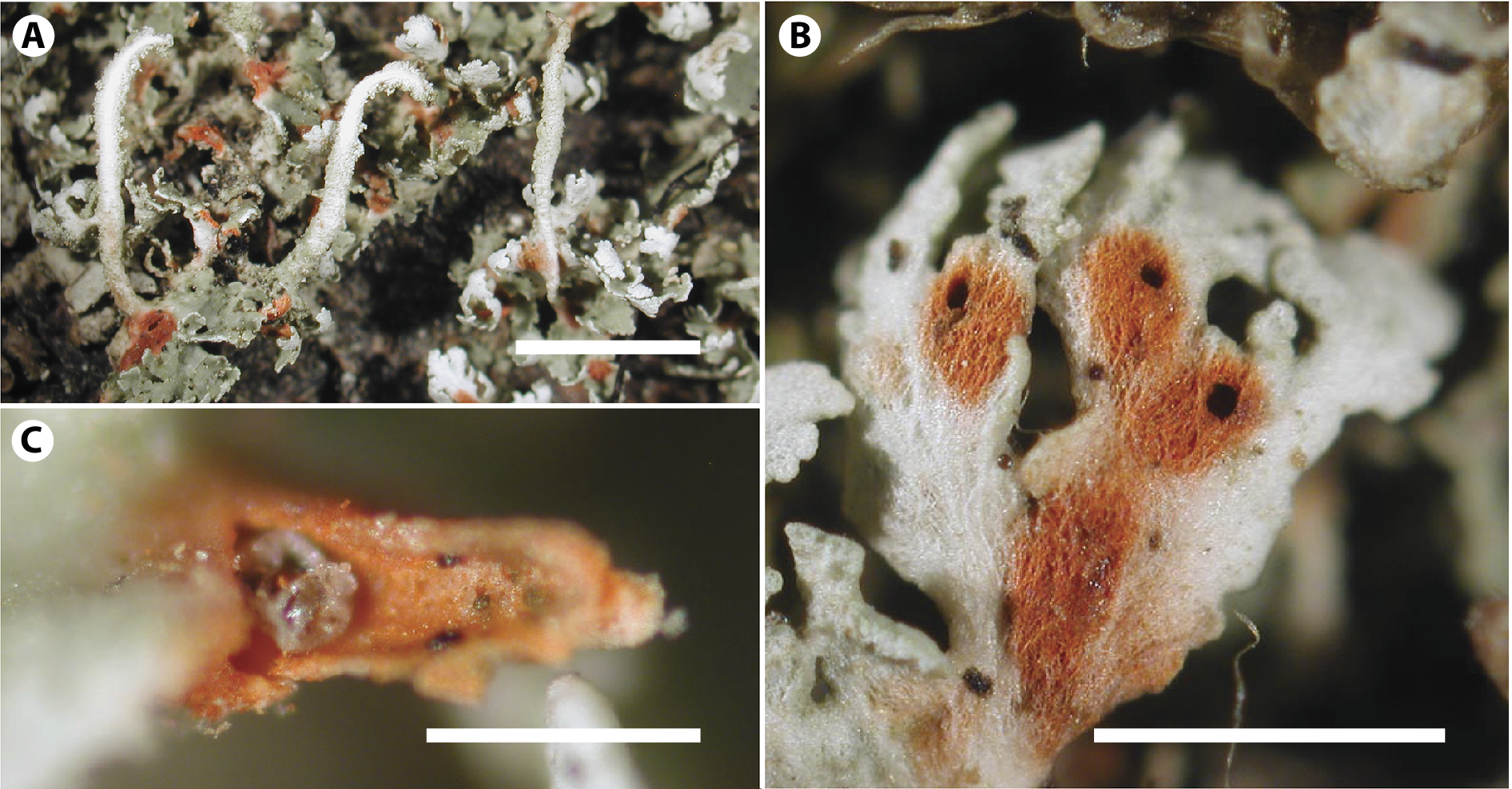
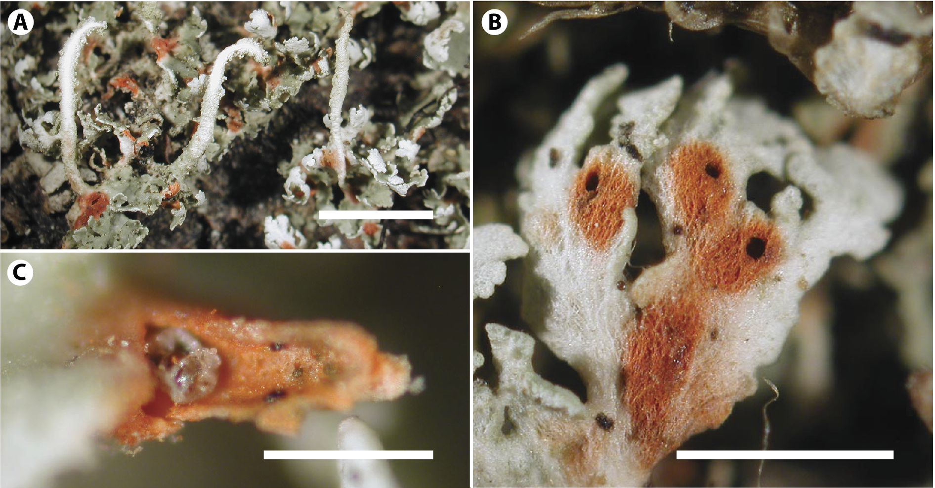
Our own investigation of Central European material performed independently in Austria and Czechia indicates that, apart from Carabodes marginatus, only a small number of other species participate in this feeding relationship (J. Mourek, T. Pfingstl, P. Resl & V. Vtípilová, personal observations). We can also corroborate the observation by Timdal (Reference Timdal1989), that mites found within Cladonia rubrotincta cavities are often colourless (Fig. 4), suggesting that the lichen is inhabited by juvenile nymphs rather than adult mites, although adults also regularly feed on the lichen (Pfingstl et al., Reference Pfingstl, Vtípilová, Ghlimová, Mourek, Steinová, Schäffer and Resl2025).
A–C, mite infection of Cladonia rubrotincta, O L-25383. A, red spots on the thallus caused by mites. B, cavities on the squamules with red coloration. C, view into the inner part of the podetium with the mite nymph. Scales: A = 3 mm; B = 1 mm; C = 0.2 mm. In colour online.
Moreover, the production of rhodocladonic acid in C. rubrotincta can also result from physical damage to the lichen. In our studies, we have regularly noted such damage, although this was less common than mite-induced damage. In some cases, when neither physical nor feeding damage was evident, and no mites were visible, it was impossible to identify the cause of the red pigmentation (Pfingstl et al., Reference Pfingstl, Vtípilová, Ghlimová, Mourek, Steinová, Schäffer and Resl2025).
Reddish or orange spots have also been observed in several other Cladonia species, including C. bacilliformis (Timdal Reference Timdal1989), and several red-fruiting species (Ahti Reference Ahti2000). In the case of C. bacilliformis (which was shown to be polyphyletic in this study), the spots bear a strong resemblance to those observed in C. rubrotincta. In red-fruited Cladonia species, the presence of red or orange spots may be attributable either to the red pigmentation of the medulla (subclade Miniatae, e.g. C. anaemica (Nyl.) Ahti) or to the production of anthraquinones in decaying parts, typically visible as orange spots (T. Ahti, personal communication). However, the origin, chemical basis and ecological significance of these pigmentations in the different species remain unclear and require further investigation.
Conclusion
Red-spotted material previously assigned to Cladonia norvegica belongs to a distinct species, C. rubrotincta, which we describe here. The two species are genetically well separated, with C. rubrotincta forming a well-supported monophyletic clade in our phylogenetic analysis. All analyzed material of C. rubrotincta has red spots containing rhodocladonic acid on basal squamules and/or podetia. Although red pigmentation on the thallus is frequently associated with the presence of mites or mechanical damage, our results show that it is consistently present in genetically verified material of C. rubrotincta, and can therefore be considered a stable morphological feature useful in distinguishing it from C. norvegica s. str. In addition to red pigmentation, the smaller size and irregular shape of the podetia appear to be consistent diagnostic characters. Future studies should re-evaluate the identity of material previously assigned to C. norvegica.
Supplementary Material
The Supplementary Material for this article can be found at http://doi.org/10.1017/S002428292510128X.
Acknowledgements
We would like to thank Rémi Boisvert, Lukáš Jánošík, Piret Lõhmus, Jiří Malíček, Bruce McCune, Troy McMullin, Zdeněk Palice and David Svoboda for providing material. We are grateful to Maria Liebmann-Reidl for collecting material and for her help in the early stages of the project. We would like to thank Jan Mourek and Tobias Pfingstl for valuable discussions about lichen-associated mites. Special thanks to Håkon Holien for his assistance in the field and with TLC, to Miroslav Kolařík for his help with HPLC and LC-HRMS analysis and to Teuvo Ahti for his valuable comments on the first draft of the manuscript. This study was supported by projects N. 96p3 and 98p2 of the AKTION Austria – Czech Republic Initiative. The work of E. Stodůlková and J. Semerád was supported by the Strategie AV21 project ‘VP33 MycoLife – the world of fungi’ of the Czech Academy of Sciences. Jana Steinová was supported by the Czech Science Foundation (GA ČR), project no. 24-10510K.
Author ORCIDs
Věra Vtípilová, 0009-0006-0154-5127; Einar Timdal, 0000-0003-4524-0617; Eva Stodůlková, 0000-0002-1801-5038; Jaroslav Semerád, 0000-0002-6470-183X; Philipp Resl, 0000-0002-7841-6060; Jana Steinová, 0000-0003-0229-4535.
Competing Interests
The authors declare none.