Case report
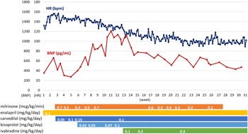
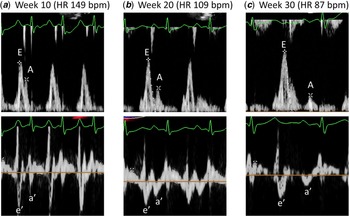
A 10-year-old girl experienced cardiopulmonary arrest with preceding symptoms of fatigue and respiratory distress 1 year and 4 months after receiving a heart transplant for chronic lymphocytic myocarditis. After resuscitation, the patient required extracorporeal membrane oxygenation support and intra-aortic balloon pumping. Endomyocardial biopsy revealed antibody-mediated rejection; thus, she was treated with gamma-globulin, steroid pulse, and plasma exchange therapy. After weaning from extracorporeal membrane oxygenation support and intra-aortic balloon pumping, the patient was transferred to our hospital for additional treatment. The patient received rituximab to prevent recurrence of antibody-mediated rejection, and another endomyocardial biopsy showed improvement in rejection findings. However, soon after transfer, her oedema and fatigue worsened. The patient developed decreased left ventricular contractility (left ventricular ejection fraction, 30%) and sinus tachycardia (heart rate, 130–150 bpm, Fig 1), whereas blood pressure was maintained (systolic and diastolic, 100–120/60–70 mmHg). Continuous intravenous administration of milrinone was initiated; additionally, enalapril and beta-blocker titration (carvedilol 0.15 mg/kg/day and bisoprolol 0.1 mg/kg/day) were initiated to improve heart failure treatment and tachycardia. However, the B-type natriuretic peptide levels remained at 1000–1200 pg/ml, and further titration was difficult due to hypotension. Owing to continued milrinone dependence, the patient was registered for re-transplantation. Because of resistance to conventional heart failure therapy, ivabradine administration was initiated at 0.05 mg/kg twice daily and titrated up to 0.15 mg/kg twice daily. Soon after initiating ivabradine, the heart rate decreased to 100–120 bpm while blood pressure remained 90–100/60–70 mmHg, and left ventricular contractility improved (left ventricular ejection fraction, 50%), with a prolonged left ventricular diastolic filling time (Fig 2). The pulmonary artery wedge pressure reduced from 15 to 12 mmHg, and the B-type natriuretic peptide level decreased to 400–500 pg/ml. Soon after initiating ivabradine administration, the patient complained of a visual abnormality, suspected to be photopsia, but it was tolerable and improved over time. Four months following the initiation of ivabradine, the patient was discharged home after milrinone treatment was discontinued.
Trends in heart rate at rest (dark blue line) and BNP levels (red line) after hospitalisation. Carvedilol and bisoprolol were up titrated under milrinone administration; heart rate decreased slightly, and BNP levels increased. When ivabradine administration was initiated from week 13, BNP levels began to decline as the heart rate dropped, and milrinone was discontinued without worsening the signs of heart failure. HR (heart rate), BNP (B-type natriuretic peptide).

Echocardiography: Pulse-wave Doppler recordings of trans-mitral diastolic inflow filling velocities (upper panels) and tissue Doppler recordings of septal mitral annular velocities (lower panels) 10 (a), 20 (b), and 30 (c) weeks after hospitalization. Before administering ivabradine in week 13, E and A waves and e’ and a’ waves were not separated, and the diastolic time was short. As the heart rate decreased due to ivabradine administration, each waveform became separated and the diastolic time became prolonged.

Discussion
Antibody-mediated rejection, a poor prognostic complication after heart transplantation, is caused by antibodies produced against the transplanted heart. The therapies include enhanced immunosuppression and targeting of antibodies and antibody-producing cells. However, post-treatment deterioration of cardiac function and progression of cardiac allograft vasculopathy often result in the need for a re-transplantation. Reference Colvin, Cook and Chang1
Ivabradine is a relatively new medication for chronic heart failure that acts directly on the sinus node as an If current inhibitor, reducing only heart rate without affecting cardiac contraction or blood pressure. Reference Koruth, Lala, Pinney, Reddy and Dukkipati2 Its use as a treatment for heart failure began with the 2011 SHIFT study, which showed that ivabradine administration significantly reduced cardiovascular death, hospitalisations for adult patients with heart failure, and ejection fraction. Reference Borer, Böhm and Ford3 A randomised trial of ivabradine versus placebo in paediatric patients with dilated cardiomyopathy showed a significant improvement in left ventricular ejection fraction, and a trend towards improvement in NYHA and Ross classifications in the ivabradine group along with a reduction in heart rate. Reference Bonnet, Berger, Jokinen, Kantor and Daubeney4 Moreover, ivabradine administration in patients after heart transplant was associated with a lower left ventricular end-diastolic pressure and NT-proBNP level and an improved 5-year survival rate than metoprolol administration in a registry study of adult patients with inappropriate sinus tachycardia after heart transplantation. Reference Rivinius, Helmschrott and Rahm5 This report highlights that ivabradine, which acts directly on the sinus node, is more effective than beta-blockers, which act via the autonomous nervous system, in a denervated transplanted heart. To our knowledge, no studies have reported the efficacy of ivabradine in paediatric patients after heart transplant.
The mechanism by which ivabradine improves heart failure is thought to be a decrease in heart rate, which leads to sufficient left ventricular filling time, resulting in improved left ventricular diastolic capacity Reference Fang, Debunne and Vercauteren6 and contractility Reference Colvin, Cook and Chang1 and utilisation of atrial contraction, as indicated by the separation of the E and A waves in the left ventricular inflow. Reference Izumida, Imamura, Nakamura, Fukuda and Kinugawa7 In the present case, we speculated that ivabradine corrected inappropriate sinus tachycardia, resulting in improved left ventricular contractility and utilisation of atrial contraction (Fig 2), ultimately contributing to improvement in heart failure, as reported in adults. Ivabradine may be a useful option for the treatment of heart failure in patients with denervated transplanted hearts, irrespective of age.
In conclusion, we encountered a heart transplant rejection case that was refractory to heart failure therapy and in which ivabradine administration avoided retransplantation. Ivabradine may be more effective than beta-blockers for the treatment of denervated transplanted hearts in children and adults.
Acknowledgements
We would like to thank the pediatric ICU team, pediatric cardiology team, and cardiac surgeons (especially Dr Yasutaka Hirata and Dr Minoru Ono), who contributed significantly to the treatment of the patient.
Financial support
This research received no specific grant from any funding agency, commercial, or not-for-profit sectors.
Conflicts of interest
None
Ethical standards
The authors assert that all procedures contributing to this work comply with the ethical standards of the relevant national guidelines on human experimentation and with the Helsinki Declaration of 1975, as revised in 2008. Informed consent was obtained from the patient’s family for submission and publication of this paper.